. 2003 Sep 30;100(20):11469-73.
doi: 10.1073/pnas.1634991100. Epub 2003 Sep 18.

Evolutionary origins of the vertebrate heart: Specification of the cardiac lineage in Ciona intestinalis

Affiliations

Evolutionary origins of the vertebrate heart: Specification of the cardiac lineage in Ciona intestinalis

Brad Davidson et al. Proc Natl Acad Sci U S A. .

Abstract

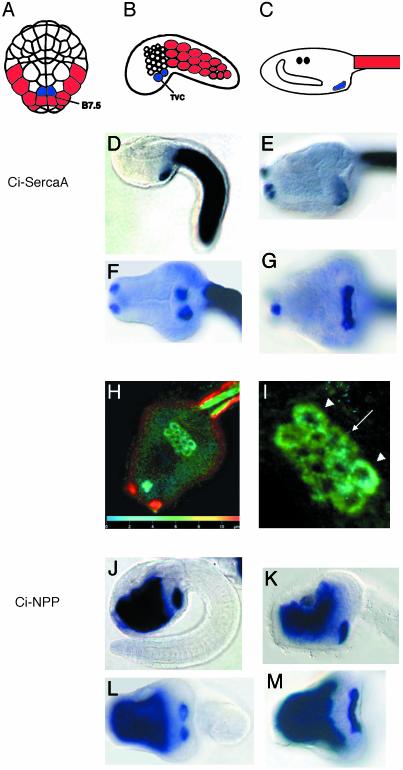
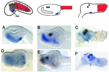
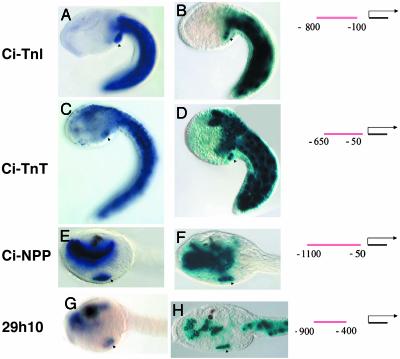
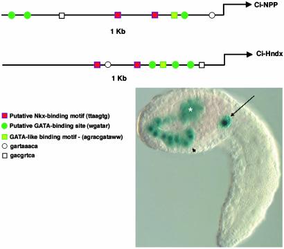
Here we exploit the extensive cell lineage information and streamlined genome of the ascidian, Ciona intestinalis, to investigate heart development in a basal chordate. Several cardiac genes were analyzed, including the sole Ciona ortholog of the Drosophila tinman gene, and tissue-specific enhancers were isolated for some of the genes. Conserved sequence motifs within these enhancers facilitated the isolation of a heart enhancer for the Ciona Hand-like gene. Altogether, these studies provide a regulatory framework for the differentiation of the cardiac mesoderm, beginning at the 110-cell stage, and extending through the fusion of cardiac progenitors during tail elongation. The cardiac lineage shares a common origin with the germ line, and zygotic transcription is first detected in the heart progenitors only after its separation from the germ line at the 64-cell stage. We propose that germ-line determinants influence the specification of the cardiac mesoderm, both by inhibiting inductive signals required for the development of noncardiac mesoderm lineages, and by providing a localized source of Wnt-5 and other signals required for heart development. We discuss the possibility that the germ line also influences the specification of the vertebrate heart.


Figures

Fig. 1.
Fig. 1.
Gene expression in the heart lineage. (A–C) Diagrams of Ciona embryos and larvae. Tail muscle myoblasts are red, and heart lineage cells (TVCs) are blue. The anterior is up (A) to the left (B and C). (D–M) Ciona larvae stained with antisense RNA probes for Ci-SercaA (ci0100137099, clone ID no. GC29o11) (D–I) and Ci-NPP (ci0100144439, clone ID no. GC29e18) (J–M). The top rows are side views of tailbud embryos (D and J) and larval heads (E and K). The bottom rows are ventral views of tailbud embryos (F and L) and larval heads (G and M). (H) Ventral view of larva hybridized to Ci-SercaA and stained with Fast red. Projections of confocal z series false colored to represent depth are shown (scale on the z axis is indicated on bar at the bottom of the image). (I) Closeup (from H) of emerging heart field. Arrowheads point to four large outer cells, and the arrow points to inner cluster of eight smaller cells. To obtain sequence information, use the gene numbers provided above to search within the annotation section (http://genome.jgi-psf.org/ciona4/ciona4.home.html), or use the clone ID no. to search the EST site (http://ghost.zool.kyoto-u.ac.jp/indexr1.html).
Fig. 2.
Fig. 2.
Ci-Nkx expression. The top row displays diagrams of representative stages as in Fig. 1. (Right Upper) The diagram portrays a juvenile ≈36 h after metamorphosis, with the resorbed tail muscle in red. The anterior is to the left in all diagrams and pictures. (A–E) Ciona larvae stained with antisense RNA probes against Ci-Nkx (ci0100140552, clone ID no.GC29k02). (AC) Side views of a tailbud head (A), larval head (B), and a juvenile (C) ≈36 h after metamorphosis. Note staining of the differentiating heart (arrowhead). Contractile activity in the heart can first be detected ≈60 h after metamorphosis. Also shown are ventral views of a tailbud head (D) and a larval head (E). Note staining of the TVCs in A and D (arrow). We have also included a side view of an ≈36-h-old juvenile stained with Ci-TnI (F). Note staining of the siphon muscles (arrow), along with the heart (arrowhead).
Fig. 3.
Fig. 3.
Enhancers for Ciona heart genes. (A, C, E, and G) In situ staining for four probes, as indicated to the left, Ci-TnI (ci0100137955, clone ID no. GC29b15), Ci-TnT (ci0100146672, clone ID no. GC26c17), Ci-NPP, and 29h10 (ci0100135212, named after clone ID no. GC29h10). (B, D, F, and H) Corresponding lacZ reporter assays for regulatory DNA located upstream of the relevant genes. Diagrams to the right indicate the position of the tested fragment relative to the predicted transcriptional start site of the relevant gene. Note staining of the TVC progeny in all images (arrowheads). Note staining in the tail muscle in A–D, staining in the endoderm in E–H, and staining of the CNS in C–D. Also note that the 29h10 enhancer drives expression in the tail muscle (H), although this is not seen in the endogenous expression pattern (G). (A–D) Side view of tailbud embryos. (E–H) Side view of larval heads. The anterior is to the left in all images.
Fig. 4.
Fig. 4.
Prediction of the Ci-Hndx enhancer. (Upper) Alignment of proximal 1 kb of upstream sequence for Ci-NPP and Ci-Hndx (ci0100140298, clone ID no. GC31k05) showing conservation of clustered motifs. (Left Lower) Sequence and tentative designation of motifs is shown. (Right Lower) A typical staining pattern for the 5′ region upstream of Ci-Hndx attached to Fkh-lacZ. Note staining in the endoderm (arrowhead), trunk lateral cells (asterisk), and TVCs (arrow).
Fig. 5.
Fig. 5.
Model of ascidian heart specification. (A and B) Diagrams of 32- and 64-cell embryos seen from the vegetal side. In these diagrams, the PP is purple and the PVC is outlined in red. Gray represents the expression patterns of Ci-Sna and Cs-ZicL. Note that when expression is limited to the nuclei (as in the B7.5 cells at the 64-cell stage), this indicates recently initiated transcription (29). (C) Antisense Ci-Wnt5 (ci0100135747, clone ID no. GC12l09) staining of a 110-cell embryo, on the vegetal side. (D) A 110-cell embryo in which descendents of the posterior vegetal quadrant have been color-coded to correspond with the cell fates listed in the diagrams to the right. (E–G) Diagrammatic representations of the posterior vegetal quadrant of the Ciona embryo. (E) At the 32-cell stage, the posteriormost blastomere (B6.3) still contains PP, repressing transcription, and thereby inhibiting a response to PVC-mediated myogenesis, as well as inductive Fgf signaling from the endoderm (yellow arrows). (F) At the 64-cell stage, the newly born B7.5 cells have now emerged from the PP and are therefore newly competent to respond to inductive signals. Possible signaling, which may occur after the 64-cell stage, is indicated by arrows, including the expression of BMP by the B7.5 cells at the 110-cell stage (red arrow) (34), FGF-8 signaling from the mesenchyme (MES) (green arrow; unpublished data), Wnt-5 signaling from the germ cell lineage (purple), or signaling from the endoderm (ENDO) (yellow). (G) By the 180-cell stage, the TVC lineage has separated from the tail muscle (MUS) lineage (35). It is unclear to what extent the TVCs are prespecified to from the heart at this point; later positional signaling may play a significant role in heart lineage specification.

References

    1. Andree, B., Duprez, D., Vorbusch, B., Arnold, H. H. & Brand, T. (1998) Mech. Dev. 70, 119–131. - PubMed
    1. Cripps, R. M. & Olson, E. N. (2002) Dev. Biol. 246, 14–28. - PubMed
    1. Pandur, P., Lasche, M., Eisenberg, L. M. & Kuhl, M. (2002) Nature 418, 636–641. - PubMed
    1. Reifers, F., Walsh, E. C., Leger, S., Stainier, D. Y. & Brand, M. (2000) Development (Cambridge, U.K.) 127, 225–235. - PubMed
    1. Satoh, N. (1994) Developmental Biology of Ascidians (Cambridge Univ. Press, New York).

Publication types

LinkOut - more resources