. 2003 Sep 30;100(20):11594-9.
doi: 10.1073/pnas.1930781100. Epub 2003 Sep 18.

A natural mutation in the Tyk2 pseudokinase domain underlies altered susceptibility of B10.Q/J mice to infection and autoimmunity

Affiliations

A natural mutation in the Tyk2 pseudokinase domain underlies altered susceptibility of B10.Q/J mice to infection and autoimmunity

Michael H Shaw et al. Proc Natl Acad Sci U S A. .

Abstract

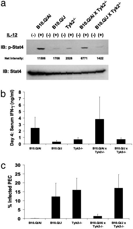
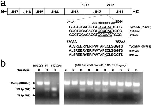
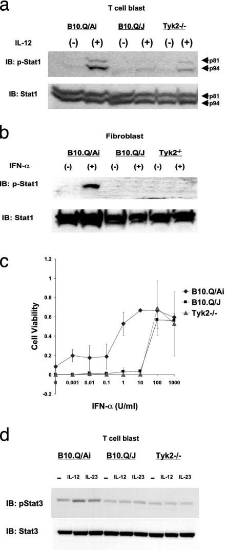
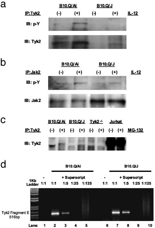
The B10.Q/J strain of mice was serendipitously discovered to be highly susceptible to infection by the intracellular protozoan parasite, Toxoplasma gondii but markedly resistant to induction of autoimmune arthritis. We have previously shown that the B10.Q/J phenotype is controlled by a single recessive locus and is associated with lymphocyte hyporesponsiveness to IL-12. Using genetic approaches, we have now localized the B10.Q/J locus to chromosome 9 and established its identity as Tyk2, a Janus kinase essential for IL-12 and IFN-alpha/beta cytokine signaling. The B10.Q/J Tyk2 gene contained a single missense mutation resulting in a nonconservative amino acid substitution (E775K) in an invariant motif of the pseudokinase (Janus kinase homology 2) domain. This mutation appeared to result in the absence of the B10.Q/J-encoded Tyk2 protein, despite presence of Tyk2-specific transcripts. Phenotypically, B10.Q/J cells were indistinguishable from Tyk2-deficient cells, showing impaired signaling and biologic responses to IL-12, IL-23, and type I IFNs. The analogous E782K mutant of human Tyk2 failed to restore IFN-alpha responsiveness in Tyk2 null 11,1 cells. Our results indicate a crucial role for Tyk2 in T helper 1-mediated protective and pathogenic immune responses. An additional implication of our findings is that naturally occurring mutations in the Tyk2 gene may underlie altered susceptibilities to infectious or autoimmune diseases in human and animal populations.


Figures

Fig. 1.
Fig. 1.
B10.Q/J mice fail to genetically complement Tyk2-deficient mice. (a) T cell blasts from wild-type, B10.Q/J, Tyk2/, B10.Q/Ai × Tyk2/ F1, and B10.Q/J × Tyk2/ F1 mice were assayed for IL-12-induced phosphorylation of Stat4. (b) IFN-γ levels were determined in sera obtained 4 days after T. gondii infection in parental strains, B10.Q/Ai × Tyk2/ F1, and B10.Q/J × Tyk2/ F1 mice. (c) The percentage of infected peritoneal cells of the same mice described in b were determined on day 5 postinfection. Results shown are representative of one of two experiments. In each experiment, three to four age- and sex-matched mice were used.
Fig. 2.
Fig. 2.
B10.Q/J Tyk2 contains a single G→ A mutation in the JH2 domain. (a) Comparison of B10.Q/Ai and B10.Q/J Tyk2 cDNA sequences. Splenic cDNA from B10.Q/Ai and B10.Q/J mice were amplified by PCR by using primers (as described in Materials and Methods) spanning the entire coding region of Tyk2. B10.Q/J mice exhibit a G→ A base substitution at position 2538. An E775K amino acid substitution is predicted in the JH2 domain. Note: The G→ A polymorphism resulted in the loss of an AvaI restriction site present in the wild-type Tyk2 sequence. (b) A 204-bp Tyk2 fragment was amplified from genomic DNA of parental, F1, and F1 backcross progeny (as described in Materials and Methods) and subjected to AvaI restriction typing. The presence of the wild-type Tyk2 allele is indicated by the appearance of 126- and 78-bp AvaI digested fragments. The AvaI-resistant 204-bp PCR product corresponds to the mutant B10.Q/J Tyk2 allele. AvaI digestion patterns for individual parental and F1 mice (Left) and representative backcross mice (Right) are shown. The resistance or susceptibility phenotype of individual mice to T. gondii infection is indicated by R or S, respectively.
Fig. 3.
Fig. 3.
B10.Q/J cells exhibit impaired cellular responses to IL-12, IFN-α, and IL-23. Impaired Stat1 phosphorylation in B10.Q/J cells after IL-12 (a) or IFN-α (b) exposure is shown. (a) T cell blasts derived from B10.Q/Ai, B10.Q/J, and Tyk2/ mice were cultured with (+) or without (–) IL-12 (50 ng/ml) for 15 min. (b) Lung-derived fibroblasts from B10.Q/Ai, B10.Q/J, and Tyk2/ mice were cultured with (+) or without (–) IFN-α (1,000 units/ml) for 15 min. Cells were analyzed for activation of Stat1 by direct Western blot with antiphosphotyrosine-Stat1 (p-Stat1) Ab. Protein loading was controlled by reprobing with normal Stat1 Ab. (c) Antiviral response of B10.Q/J fibroblasts. Fibroblasts from B10.Q/Ai, B10.Q/J, and Tyk2/ mice were stimulated with IFN-α and cultured with vesicular stomatitis virus. After 48 h, surviving cells were quantified by crystal violet staining. (d) Lymphocytes from wild-type, B10.Q/J, and Tyk2/ mice were analyzed for Stat3 activation after 30-min stimulation with IL-12 (50 ng/ml) or IL-23 (1 μg/ml) with p-Stat3 Ab. Protein loading was controlled by reprobing with normal Stat3 Ab. a and b are representative of one of three experiments, and c and d are representative of two experiments.
Fig. 4.
Fig. 4.
Lack of immunoreactive Tyk2 expression in B10.Q/J cells despite the presence of Tyk2-specific transcript. (a and b) T cell blasts from B10.Q/Ai and B10.Q/J mice were cultured with (+) or without (–) IL-12 (50 ng/ml) and analyzed for Tyk2 activation (a) or Jak2 activation (b) by immunoprecipitation with Tyk2- or Jak2-specific Abs and subsequent immunoblotting of precipitates with phosphotyrosine-specific (p-Y) Ab. The same blots were reprobed with Tyk2- and Jak2-specific Abs to control for protein loading. (c) T cell blasts from B10.Q/Ai, B10.Q/J, and Tyk2/ mice were cultured for 8 h in the presence (+) or absence (–) of proteasome inhibitor MG-132 (10 ng/ml). Total protein extracts were then immunoprecipitated with Tyk2-specific Ab followed by immunoblotting with the same Tyk2 Ab. Jurkat T cells were included to serve as positive control for immunoprecipitation and immunoblotting. (d) B10.Q/J cells have equivalent amounts of Tyk2-specific transcripts compared with wild-type cells. cDNA was prepared from the spleens of B10.Q/Ai or B10.Q/J mice followed by PCR with primers specific for the 3′ terminus of Tyk2 (Fragment E: 2143–2580). cDNA primed with random hexamers in the presence of reverse transcriptase (RT) from both strain of mice was diluted serially (1:5) (lanes 2–5 and 7–10). cDNA primed in the absence of RT lack Tyk2-specific transcripts (lanes 1 and 6).

References

    1. Scharton-Kersten, T. M., Yap, G., Magram, J. & Sher, A. (1997) J. Exp. Med. 185, 1261–1273. - PMC - PubMed
    1. Yap, G., Pesin, M. & Sher, A. (2000) J. Immunol. 165, 628–631. - PubMed
    1. Yap, G. S., Ortmann, R., Shevach, E. & Sher, A. (2001) J. Immunol. 166, 5720–5725. - PubMed
    1. Cooper, A. M., Magram, J., Ferrante, J. & Orme, I. M. (1997) J. Exp. Med. 186, 39–45. - PMC - PubMed
    1. Mattner, F., Magram, J., Ferrante, J., Launois, P., Di Padova, K., Behin, R., Gately, M. K., Louis, J. A. & Alber, G. (1996) Eur. J. Immunol. 26, 1553–1559. - PubMed

Publication types

LinkOut - more resources