. 2003 Oct 1;31(19):5526-33.
doi: 10.1093/nar/gkg761.

A requirement for PARP-1 for the assembly or stability of XRCC1 nuclear foci at sites of oxidative DNA damage

Affiliations

A requirement for PARP-1 for the assembly or stability of XRCC1 nuclear foci at sites of oxidative DNA damage

Sherif F El-Khamisy et al. Nucleic Acids Res. .

Abstract

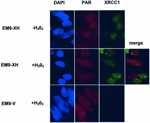
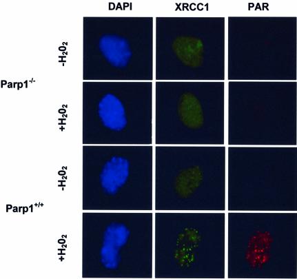
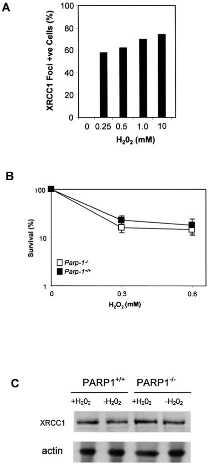
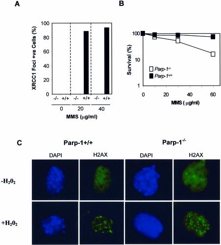
The molecular role of poly (ADP-ribose) polymerase-1 in DNA repair is unclear. Here, we show that the single-strand break repair protein XRCC1 is rapidly assembled into discrete nuclear foci after oxidative DNA damage at sites of poly (ADP-ribose) synthesis. Poly (ADP-ribose) synthesis peaks during a 10 min treatment with H2O2 and the appearance of XRCC1 foci peaks shortly afterwards. Both sites of poly (ADP-ribose) and XRCC1 foci decrease to background levels during subsequent incubation in drug-free medium, consistent with the rapidity of the single-strand break repair process. The formation of XRCC1 foci at sites of poly (ADP-ribose) was greatly reduced by mutation of the XRCC1 BRCT I domain that physically interacts with PARP-1. Moreover, we failed to detect XRCC1 foci in Adprt1-/- MEFs after treatment with H2O2. These data demonstrate that PARP-1 is required for the assembly or stability of XRCC1 nuclear foci after oxidative DNA damage and suggest that the formation of these foci is mediated via interaction with poly (ADP-ribose). These results support a model in which the rapid activation of PARP-1 at sites of DNA strand breakage facilitates DNA repair by recruiting the molecular scaffold protein, XRCC1.


Figures

Figure 1
Figure 1
Co-localization of XRCC1 foci at sites of poly (ADP-ribose) synthesis after oxidative DNA damage. EM9 CHO cells expressing either human XRCC1 (EM9-XH) or harbouring empty vector (EM9-V) were mock-treated (–H2O2) or treated with 10 mM H2O2 for 20 min and then incubated in drug-free medium for 10 min. Untreated and treated cells were then fixed with methanol/acetone and immunostained with anti-poly (ADP-ribose) (‘PAR’) monoclonal antibody 10H and anti-XRCC1 polyclonal antibody (AHP428, Serotec) for analysis by indirect immunofluorescence. Representative images (×100 magnification) were photographed and coloured using an RTI/CCD-1300/Y digital camera and Metamorph software.
Figure 2
Figure 2
Temporal assembly of PAR foci and XRCC1 foci in EM9 CHO cells expressing wild-type human XRCC1 or XRCC1C389A. (A) EM9-XH cells (squares) or EM9-XHC389A cells (circles) were mock-treated (‘no damage’) or treated with 10 mM H2O2 for 20 min and then incubated for 0, 10, 30, 60 or 120 min as indicated (‘R0-R120’) in drug-free medium to allow time for repair. Cells were then fixed and immunostained with anti-PAR antibodies (solid lines) and anti-XRCC1 antibodies (dotted lines) as described for Figure 1. For each time point, 100 cells were scored for the presence of six or more foci per cell. Data points are the mean of two or three experiments and error bars represent the data range or 1 S.D. from the mean (R10), respectively. Where not visible, error bars are smaller than the symbols. (B) 250 cells of the indicated cell lines were plated in 10 cm dishes in duplicate and either mock-treated or treated with the indicated concentrations of H2O2 for 10 min or MMS for 1 h. Cells were then rinsed in PBS and incubated in drug-free medium for 7–10 days to allow formation of macroscopic colonies. Survival was calculated by dividing the average number of colonies on treated plates by the average number on untreated plates. Data are the mean of three independent experiments for each drug and error bars represent ±1 S.D. Where not visible, error bars are smaller than the symbols.
Figure 3
Figure 3
The BRCT I domain is required for appearance of XRCC1 nuclear foci after oxidative DNA damage. (A) Schematic of full-length XRCC1 (top) and XRCC1242–533 (bottom) depicting the proposed PAR- binding motif (dotted box) located within BRCT I domain. The position of the nuclear localization signal (NLS) and of the mutations employed in this study are shown. (B) Mutation of the BRCT I domain inhibits assembly of XRCC1 nuclear foci after H2O2 treatment. The indicated cell lines were mock-treated (‘–’) or treated (‘+’) with H2O2 for 20 min, incubated in drug-free medium for 10 min, and then processed for anti-XRCC1 immunofluorescence as described for Figure 1. 100 cells from each cell line were scored for the presence or absence of six or more foci. Each data point represents the mean of two independent experiments and error bars reflect the data range. EM9-XH5 is a single transfectant clone and is the wild-type control for EM9-XHLI360/361DD and EM9-XHW385D, with which it expresses similar levels of recombinant human XRCC1 (25). EM9-XH is a pooled population of more than 50 transfectants and is the control cell line for EM9-XH242–533 and EM9-XHR399Q (25). (C) XRCC1 protein levels in EM9-V, EM9-XH, EM9-XH242–533 and EM9-XHC389A cells. Protein extracts from the indicated cell lines were fractionated by SDS–PAGE, transferred to nitrocellulose and immunoblotted with anti-XRCC1 polyclonal antibody.
Figure 4
Figure 4
PARP-1 is required for appearance of XRCC1 nuclear foci at sites of oxidative DNA damage. Parp1+/+ or Parp1–/– MEFs were mock-treated or treated for 20 min with 10 mM H2O2 and then incubated in drug-free medium for 10 min. Cells were then fixed in methanol/acetone and processed for indirect immunofluorescence using anti-PAR monoclonal antibody and anti-XRCC1 polyclonal antibody and counterstained with DAPI. Representative cells are shown (×100 magnification).
Figure 5
Figure 5
XRCC1 protein levels, nuclear foci and survival to H2O2 in Parp1+/+ or Parp1–/– MEFs. (A) Parp1+/+ MEFs were treated for 20 min with the indicated concentrations of H2O2 and then incubated in drug-free medium for 10 min. Cells were immunostained for Xrcc1 and PAR as described in Figure 4 and the fraction of cells with >6 Xrcc1 foci was determined. The absence of visible bars for untreated cells indicates the absence of detectable foci-positive cells. Data are the mean of duplicate samples from a single experiment, and the data range for each duplicate was less than five. (B) The clonogenic survival of Parp1+/+ or Parp1–/– MEFs treated with the indicated concentrations of H2O2 was determined as described in Figure 2B. (C) Protein extracts from mock-treated or H2O2-treated Parp1+/+ or Parp1–/– MEFs were fractionated by SDS–PAGE, transferred to nitrocellulose and immunoblotted with anti-XRCC1 polyclonal, or anti-actin monoclonal, antibody.
Figure 6
Figure 6
XRCC1 and H2AX nuclear foci in Parp1+/+ and Parp1–/– MEFs following exposure to MMS and H2O2. (A) Parp1+/+ (‘+/+’) and Parp1–/– (‘–/–’) MEFs were treated for 20 min with the indicated concentrations of MMS and then incubated in drug-free medium for 10 min. Cells were immunostained for Xrcc1 and PAR as described in Figure 4 and the fraction of cells with >6 Xrcc1 foci was determined. The absence of visible bars for untreated cells and for treated Parp1–/– MEFs indicates the absence of detectable foci-positive cells. Data are the mean of duplicate samples from a single experiment, and the data range for each duplicate was less than five. (B) The clonogenic survival of Parp1+/+ or Parp1–/– MEFs treated with the indicated concentrations of MMS for 1 h was determined as described in Figure 2B. Where not visible, error bars are smaller than the symbols. (C) Parp1+/+ or Parp1–/– MEFs were mock-treated or treated for 20 min with 10 mM H2O2 as indicated and then incubated in drug-free medium for 10 min. Cells were then fixed in methanol/acetone and processed for indirect immunofluorescence using anti-phospho H2AX monoclonal antibody (Upstate; clone JBW301) and counterstained with DAPI. Representative cells are shown (×100 magnification).

Similar articles

Cited by

References

    1. Xu Y.J., Kim,E.Y. and Demple,B. (1998) Excision of C-4′-oxidized deoxyribose lesions from double-stranded DNA by human apurinic/apyrimidinic endonuclease (Ape1 protein) and DNA polymerase beta. J. Biol. Chem., 273, 28837–28844. - PubMed
    1. Caldecott K.W. (2001) Mammalian DNA single-strand break repair: an X-ra(y)ted affair. Bioessays, 23, 447–455. - PubMed
    1. Ochs K., Sobol,R.W., Wilson,S.H. and Kaina,B. (1999) Cells deficient in DNA polymerase beta are hypersensitive to alkylating agent-induced apoptosis and chromosomal breakage. Cancer Res., 59, 1544–1551. - PubMed
    1. Thompson L.H., Brookman,K.W., Dillehay,L.E., Carrano,A.V., Mazrimas,J.A., Mooney,C.L. and Minkler,J.L. (1982) A CHO-cell strain having hypersensitivity to mutagens, a defect in DNA strand-break repair, and an extraordinary baseline frequency of sister-chromatid exchange. Mutat. Res., 95, 427–440. - PubMed
    1. Dominguez I., Daza,P., Natarajan,A.T. and Cortes,F. (1998) A high yield of translocations parallels the high yield of sister chromatid exchanges in the CHO mutant EM9. Mutat. Res., 398, 67–73. - PubMed

Publication types

MeSH terms