. 2003 Oct 1;31(19):5635-43.
doi: 10.1093/nar/gkg786.

Discovery of novel splice forms and functional analysis of cancer-specific alternative splicing in human expressed sequences

Affiliations

Discovery of novel splice forms and functional analysis of cancer-specific alternative splicing in human expressed sequences

Qiang Xu et al. Nucleic Acids Res. .

Abstract

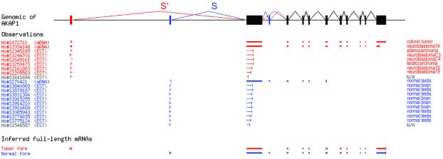
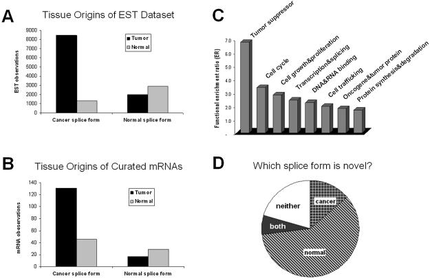
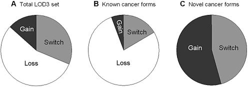
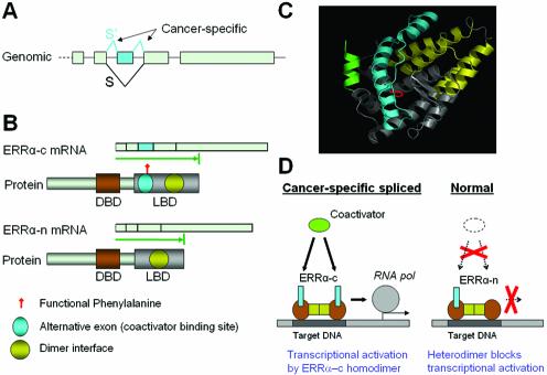
We report here a genome-wide analysis of alternative splicing in 2 million human expressed sequence tags (ESTs), to identify splice forms that are up-regulated in tumors relative to normal tissues. We found strong evidence (P < 0.01) of cancer-specific splice variants in 316 human genes. In total, 78% of the cancer-specific splice forms we detected are confirmed by human-curated mRNA sequences, indicating that our results are not due to random mis-splicing in tumors; 73% of the genes showed the same cancer-specific splicing changes in tissue-matched tumor versus normal datasets, indicating that the vast majority of these changes are associated with tumorigenesis, not tissue specificity. We have confirmed our EST results in an independent set of experimental data provided by human-curated mRNAs (P-value 10(-5.7)). Moreover, the majority of the genes we detected have functions associated with cancer (P-value 0.0007), suggesting that their altered splicing may play a functional role in cancer. Analysis of the types of cancer-specific splicing shifts suggests that many of these shifts act by disrupting a tumor suppressor function. Sur prisingly, our data show that for a large number (190 in this study) of cancer-associated genes cloned originally from tumors, there exists a previously uncharacterized splice form of the gene that appears to be predominant in normal tissue.


Figures

Figure 1
Figure 1
Detection of cancer-specific alternative splicing in AKAP1. Raw data showing the schematic alignment of all 17 ESTs aligning to the S/S′ region of the gene structure (including two ESTs of unclassified origin, excluded from our calculation) and 3 mRNAs. In normal tissues, splice form S was strongly preferred (9/9 ESTs and 1/1 mRNA), but in tumors, splice form S′ replaced it entirely (6/6 ESTs and 2/2 mRNA). The odds ratio for the null hypothesis that no shift from S to S′ occurs between normal and tumor samples is less than 10–4. The UniGene sequence identifier is shown for each sequence.
Figure 2
Figure 2
Independent validation, functional enrichment and novelty of cancer-specific alternative splice forms. (A) The tissue distribution of EST observations for both cancer and normal splice forms. (B) The tissue distribution of mRNA observations for both cancer and normal splice forms. (C) Functional categories that were enriched among the cancer-specific alternatively spliced genes, expressed as the ratio of genes observed in the LOD3 set relative to a random sample of 100 alternative spliced genes. (D) Classification of which splice forms within 316 cancer-specific alternatively spliced genes were novel. Splice forms were classified as known if they matched an existing mRNA sequence in GenBank, otherwise as novel.
Figure 3
Figure 3
Types of cancer-specific splicing shifts. (A) The relative proportions of LOD3 cancer-specific splicing that constituted a complete shift from one splice form in normal to another form in tumors (Switch); increase in one splice form in tumors (Gain); or loss of one form in tumors (Loss). (B) The same proportions, measured in genes for which the cancer-specific splice form was previously known; (C) in genes for which the cancer- specific form is novel.
Figure 4
Figure 4
Cancer-specific alternative splicing of ERRα. (A) Genomic structure of the last 5 exons of ERRα gene. Exons are shown as gray boxes and the cancer-specific splices (S′) and exon are colored cyan. (B) The two alternative mRNA and protein isoforms of ERRα. The protein-coding region for each isoform is represented as a green arrow. The functional protein domains are marked on protein isoforms (DBD, DNA binding domain; LBD, ligand binding domain). (C) 3-D structure model of ERRα ligand-binding domain (LBD). The cancer-specific exon is colored cyan. The dimer interface is colored yellow. The functional phenylalanine is colored red. The coactivator peptide is colored green. (D) Proposed model of functional regulation of alternative splicing on ERRα. The cancer-specific spliced form ERRα-c contains complete coactivator-binding site and is a fully functional transcription factor in cancer tissue. The truncated form ERRα-n misses part of the coactivator binding site and is likely to heterodimerize with the full-length form, suppressing its activity (dominant negative effect) in normal tissue.

References

    1. Ross D.T., Scherf,U., Eisen,M.B., Perou,C.M., Rees,C., Spellman,P., Iyer,V., Jeffrey,S.S., Van de Rijn,M., Waltham,M. et al. (2000) Systematic variation in gene expression patterns in human cancer cell lines. Nature Genet., 24, 227–235. - PubMed
    1. Mironov A.A., Fickett,J.W. and Gelfand,M.S. (1999) Frequent alternative splicing of human genes. Genome Res., 9, 1288–1293. - PMC - PubMed
    1. Brett D., Hanke,J., Lehmann,G., Haase,S., Delbruck,S., Krueger,S., Reich,J. and Bork,P. (2000) EST comparison indicates 38% of human mRNAs contain possible alternative splice forms. FEBS Lett., 474, 83–86. - PubMed
    1. Croft L., Schandorff,S., Clark,F., Burrage,K., Arctander,P. and Mattick,J.S. (2000) ISIS, the intron information system, reveals the high frequency of alternative splicing in the human genome. Nature Genet., 24, 340–341. - PubMed
    1. IHGS Consortium (2001) Initial sequencing and analysis of the human genome. Nature, 409, 860–921. - PubMed

Publication types