. 2003 Oct;12(10):2337-47.
doi: 10.1110/ps.03110703.

Stabilization of a metastable state of Torpedo californica acetylcholinesterase by chemical chaperones

Affiliations

Stabilization of a metastable state of Torpedo californica acetylcholinesterase by chemical chaperones

Charles B Millard et al. Protein Sci. 2003 Oct.

Abstract

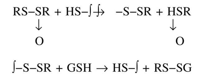
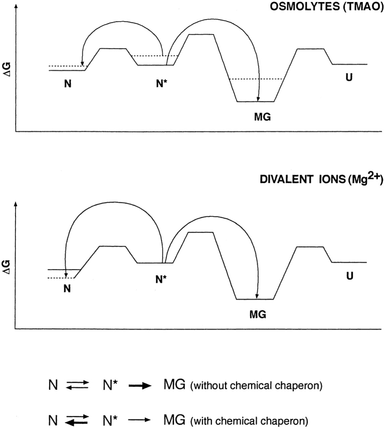
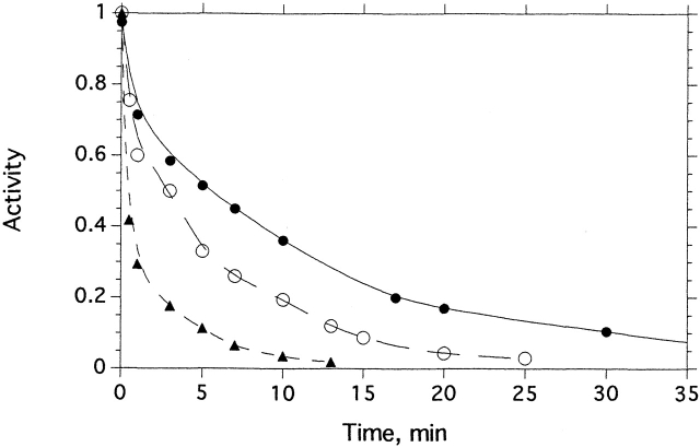
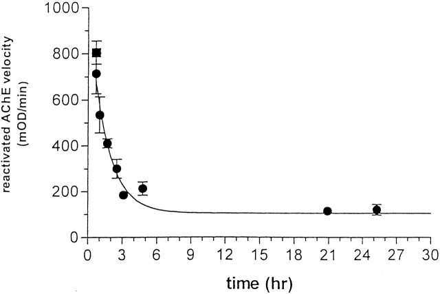
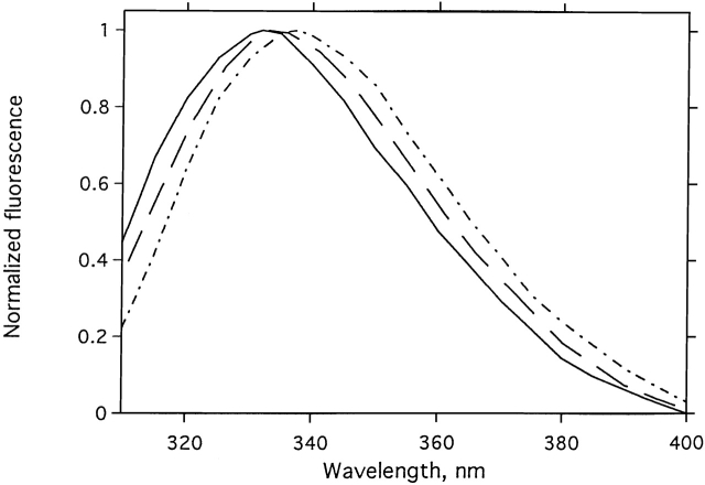
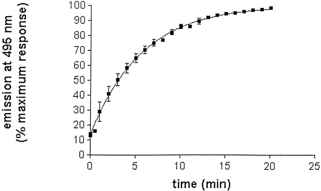
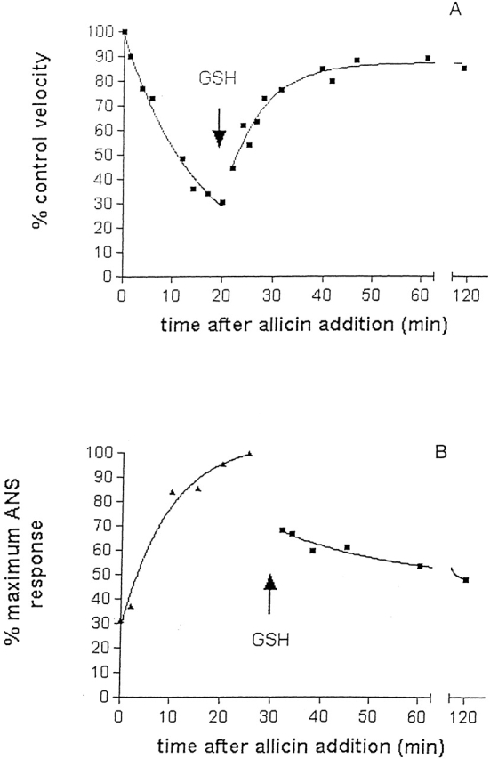
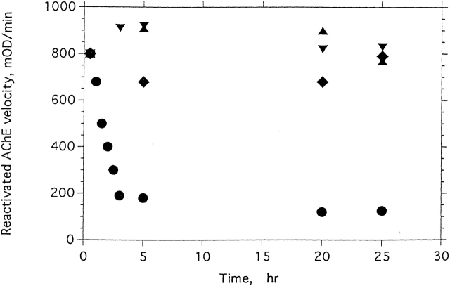
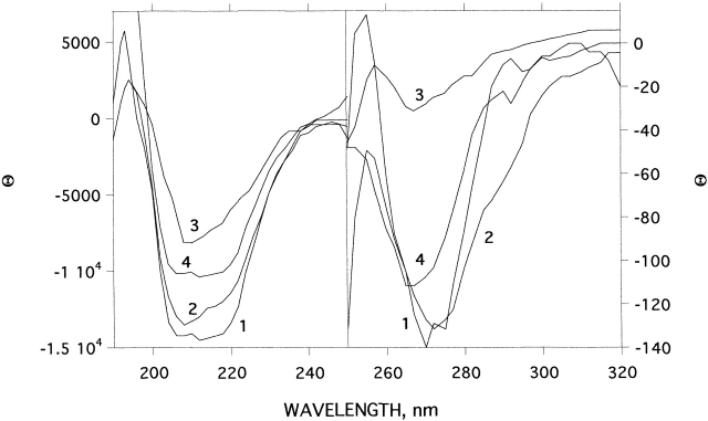
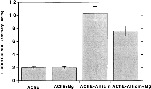
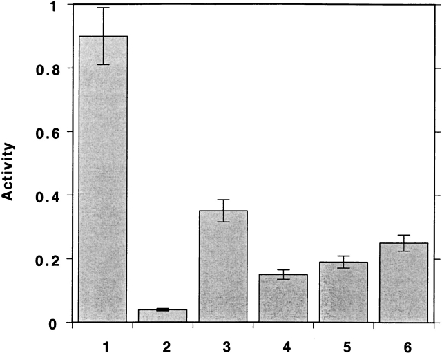
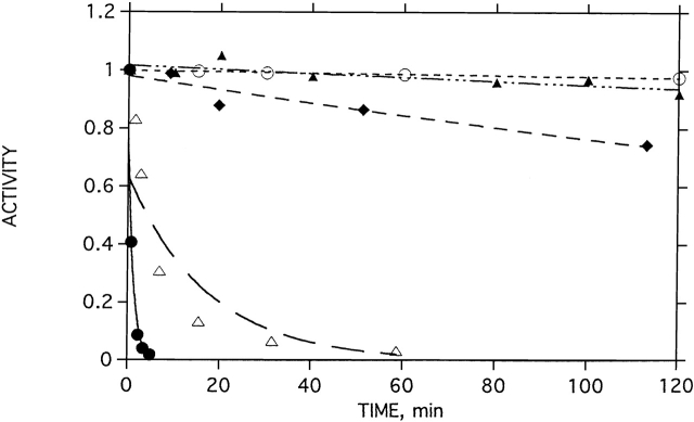
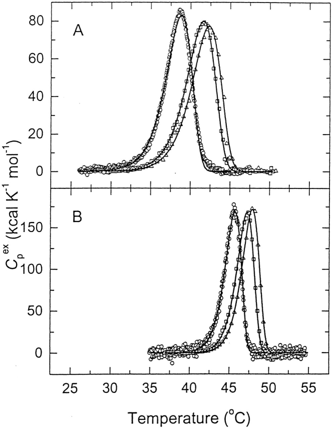
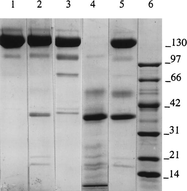
Chemical modification of Torpedo californica acetylcholinesterase by the natural thiosulfinate allicin produces an inactive enzyme through reaction with the buried cysteine Cys 231. Optical spectroscopy shows that the modified enzyme is "native-like," and inactivation can be reversed by exposure to reduced glutathione. The allicin-modified enzyme is, however, metastable, and is converted spontaneously and irreversibly, at room temperature, with t(1/2) approximately 100 min, to a stable, partially unfolded state with the physicochemical characteristics of a molten globule. Osmolytes, including trimethylamine-N-oxide, glycerol, and sucrose, and the divalent cations, Ca(2+), Mg(2+), and Mn(2+) can prevent this transition of the native-like state for >24 h at room temperature. Trimethylamine-N-oxide and Mg(2+) can also stabilize the native enzyme, with only slight inactivation being observed over several hours at 39 degrees C, whereas in their absence it is totally inactivated within 5 min. The stabilizing effects of the osmolytes can be explained by their differential interaction with the native and native-like states, resulting in a shift of equilibrium toward the native state. The stabilizing effects of the divalent cations can be ascribed to direct stabilization of the native state, as supported by differential scanning calorimetry.


Figures

Scheme 1
Scheme 1
Scheme 2
Scheme 2
Scheme 3.
Scheme 3.
Proposed profile of relative free energy levels of conformational states of TcAChE in the presence and absence of chemical chaperones. The solid lines show energy levels in the absence of osmolytes and Mg2+, and the dashed lines, the levels in their presence.
Figure 1.
Figure 1.
Kinetics of inhibition of TcAChE by allicin. The enzyme (3.6 μM) was incubated with the appropriate concentration of allicin at 23°C. The decrease in enzymic activity as a function of time is shown at three allicin concentrations: (•) 0.5 mM; (○) 3 mM; and (▴) 8 mM.
Figure 2.
Figure 2.
Time-dependent loss by allicin-modified TcAChE of capacity for reactivation by GSH. TcAChE (1 μM) was inhibited >80% by incubation with 1 mM allicin at room temperature for 25 min. The sample was maintained at room temperature for >24 h; at appropriate intervals, 10-μL aliquots were withdrawn, mixed with 5 μL of freshly reduced GSH, and incubated at 23°C for 30 min, prior to assay using 2 mM PNPA in Sorenson buffer.
Figure 3.
Figure 3.
Effect of modification by allicin on the CD spectra of TcAChE in the near and far UV. (1) Native TcAChE; (2) allicin-modified TcAChE after 22°C for 1 h, subsequent to modification as described in Materials and Methods; (3) as in 2, but 24 h after modification; (4) MG produced by heating of TcAChE at 39°C for 1 h.
Figure 4.
Figure 4.
Effects of modification by allicin or by DTP on the intrinsic fluorescence spectrum of TcAChE. (Solid line) Native TcAChE; (dashed line) allicin-modified TcAChE; (dotted/dashed line) DTP-modified TcAChE.
Figure 5.
Figure 5.
Allicin-induced increase in ANS fluorescence of TcAChE. The enzyme (2.5 μM), was incubated with 3 mM allicin in the presence of 0.1 mM ANS. The points show the averages of triplicate values. In some cases, the error bars are smaller than the symbols. The t1/2 for transition from the state that binds ANS poorly, the N state, to the state that binds ANS maximally, the quasinative state, N*, is 3.9 min.
Figure 6.
Figure 6.
Reversal by GSH of inactivation of TcAChE, and concomitant capacity to bind ANS, produced by modification with allicin. Native enzyme (2.5 μM) was incubated at 23°C for 20 min with 1 mM allicin, in the absence (A) and presence (B) of 0.1 mM ANS. Aliquots from A were taken for enzymic assay, and from B for estimation of ANS binding by fluorescence measurements. After 20 min (A) and 30 min (B), 11 mM GSH (final concentration) was added, and recovery of activity and decrease in ANS binding in A and B, respectively, were monitored as prior to addition of GSH.
Figure 7.
Figure 7.
Cross-linking by hypericin of N* TcAChE, produced by modification with allicin, and of MG TcAChE, produced by treatment with 1.2 M Gdn.HCl. Allicin modification was carried out with 1 mM allicin at 23°C for 30 min. Treatment with 1.2 M Gdn.HCl was in Sorenson buffer at 22°C for 2 h, followed by removal of the Gdn.HCl by washing three times in a YM-30 Centricon centrifugal filter. Irradiation in the presence of 1 mM hypericin for 1 h was performed as described in Materials and Methods. SDS-PAGE was as described previously (Weiner et al. 1999). (Lane 1) Allicin-modified (N*); (lane 2) allicin-modified enzyme after irradiation for 1 h in the presence of hypericin; (lane 3) Gdn.HCl-treated (MG) TcAChE; (lane 4) Gdn.HCl-treated enzyme after irradiation for 1 h in the presence of hypericin; (lane 5) molecular weight markers.
Figure 8.
Figure 8.
Effects of glycerol, TMAO, and Mg2+ on the degree of reactivation by GSH of N* TcAChE produced by allicin. The native enzyme (1.5 μM) was inhibited by incubation with 1 mM allicin at 23°C for 25 min, yielding 90% inhibition. Samples were then treated with (♦) 1.1 M glycerol, (▾) 3 M TMAO, (▴) 10 mM MgCl2, or (•) an equivalent volume of the incubation buffer. At appropriate times, 10-μL aliquots were mixed with 5 μL of fresh 30 mM aqueous GSH and incubated at 23°C for 30 min. The amount of reactivation was assayed spectrophotometrically on 2 mM PNPA in Sorenson buffer. The velocity measured was corrected for GSH-catalyzed hydrolysis of PNPA.
Figure 9.
Figure 9.
Effect of Mg2+ on the CD spectra of allicin-modified TcAChE in the near and far UV. (1) Native TcAChE + 10 mM MgCl2; (2) allicin-modified TcAChE at 23°C for 1 h, after modification as described in Materials and Methods; (3) as in 2, but 24 h after modification; (4) allicin-modified TcAChE + 10 mM MgCl2 at 22°C for 24 h, after modification.
Figure 10.
Figure 10.
Effect of Mg2+ on the ANS-binding capacity of native and allicin-modified TcAChE. ANS, at a final concentration of 0.2 mM, was added to either native or freshly prepared allicin-modified TcAChE (1.5 μM), in the presence or absence of 10 mM MgCl2.
Figure 11.
Figure 11.
Effects of TMAO and Mg2+ on the activity of N* TcAChE. The enzyme (4 μM) was incubated with 1 mM allicin at 23°C for 20 min. The inhibited enzyme was either demodified by addition of GSH to a final concentration of 2 mM, to determine maximum reactivation capacity; or incubated with TMAO, at a final concentration of 4 M; or of MgCl2, at a final concentration of 10 mM; or both together, before dilution 102–103-fold into the assay mixture, followed by immediate assay. (1) Control; (2) allicin treated; (3) allicin treatment followed by GSH; (4) allicin treatment followed by TMAO; (5) allicin treatment followed by Mg2+; (6) allicin treatment followed by TMAO + Mg2+.
Figure 12.
Figure 12.
Effects of Mg2+ and TMAO on thermal denaturation of TcAChE. The native enzyme was incubated at 39°C in the presence and absence of MgCl2. At appropriate times, aliquots were withdrawn and assayed for enzymic activity. (•) No MgCl2; (▵) 0.1 mM MgCl2; (♦) 1 mM MgCl2; (○) 5 mM MgCl2; (▴) 3 M TMAO.
Figure 13.
Figure 13.
Differential scanning calorimetry of native TcAChE in the absence and presence of Mg2+. The curves show the temperature dependence of the excess molar heat capacity at three scan rates: (○) 0.21 K/min; (□) 1.0 K/min; (▵) 1.45 K/min. The buffer was 0.1 M NaCl/10 mM HEPES (pH 7.5), and the concentration of enzyme was 9.2 μM for the scans at 0.21 K/min; 6.2 μM for those at 1.0 K/min, and 5.6 μM for those at 1.45 K/min. (A) Control; (B) in the presence of 10 mM MgCl2. The solid lines represent the best fits of the experimental data to equation 1.
Figure 14.
Figure 14.
SDS-PAGE analysis of susceptibility to tryptic digestion of native and allicin-modified TcAChE. SDS-PAGE was carried out under nonreducing conditions on 5%–15% acrylamide gradient gels. Enzyme samples (1 mg/mL) were digested with trypsin (1% [w/w]) at 23°C for 1 h. (Lane 1) Control, native TcAChE; (lane 2) trypsin-digested native TcAChE; (lane 3) TcAChE modified by treatment with 1 mM allicin at 22°C for 30 min; (lane 4) allicin-modified enzyme digested with trypsin; (lane 5) allicin-modified enzyme digested with trypsin in the presence of 1 mM MgCl2; (lane 6) molecular weight markers.

References

    1. Ananthan, J., Goldberg, A.L., and Voellmy, R. 1986. Abnormal proteins serve as eukaryotic stress signals and trigger the activation of heat shock genes. Science 232 522–524. - PubMed
    1. Arai, M. and Kuwajima, K. 2000. Role of the molten globule state in protein folding. Adv. Protein Chem. 53 209–282. - PubMed
    1. Baskakov, I.V., Legname, G., Prusiner, S.B., and Cohen, F.E. 2001. Folding of prion protein to its native α-helical conformation is under kinetic control. J. Biol. Chem. 276 19687–19690. - PubMed
    1. Bolen, D.W. and Baskakov, I.V. 2001. The osmophobic effect: Natural selection of a thermodynamic force in protein folding. J. Mol. Biol. 310 955–963. - PubMed
    1. Carr, C.M., Chaudhry, C., and Kim, P.S. 1997. Influenza hemagglutinin is spring-loaded by a metastable native conformation. Proc. Natl. Acad. Sci. 94 14306–14313. - PMC - PubMed

Publication types

MeSH terms

LinkOut - more resources