

doi: 10.1110/ps.03152903.
The role of protein stability, solubility, and net charge in amyloid fibril formation
Affiliations
- PMID: 14500896
- PMCID: PMC2366926
- DOI: 10.1110/ps.03152903
Item in Clipboard
The role of protein stability, solubility, and net charge in amyloid fibril formation
Protein Sci.
2003 Oct.
Abstract
Ribonuclease Sa and two charge-reversal variants can be converted into amyloid in vitro by the addition of 2,2,2-triflouroethanol (TFE). We report here amyloid fibril formation for these proteins as a function of pH. The pH at maximal fibril formation correlates with the pH dependence of protein solubility, but not with stability, for these variants. Additionally, we show that the pH at maximal fibril formation for a number of well-characterized proteins is near the pI, where the protein is expected to be the least soluble. This suggests that protein solubility is an important determinant of fibril formation.
Figures
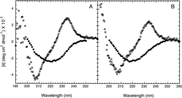
Far-UV CD spectra for RNase Sa. Representative spectra are shown for the protein in 0% TFE (open circles) or 30% TFE (filled circles) at pH 7 (A) and pH 3.5 (B). The spectra were obtained after 5-h incubation at 25°C in 10 mM (citrate, phosphate, and borate) buffer. The presence of TFE clearly induces a substantial amount of β-structure in RNase Sa at both pH values.
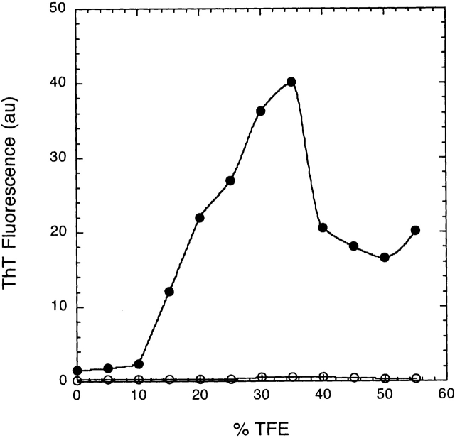
Fibril formation (monitored by ThT fluorescence) as a function of TFE concentration. The curves represent the changes of fluorescence with TFE concentrations at pH 3.5 (filled circles) and pH 5.5 (open circles) after 18 h of incubation. ThT binding was monitored after removing an aliquot of the aggregation mixture (10 μL) and diluting it with 990 μL of 50 mM sodium phosphate, pH 6.0, containing 3 μM ThT. Fluorescence intensity was determined at 485 nm after excitation at 440 nm using a 96-well plate reader equipped with cutoff filters as previously described (Wood et al. 1996).
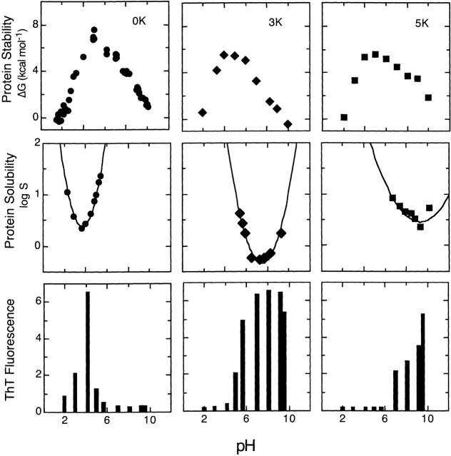
The conformational stability, solubility, and fibril formation are shown as a function of pH for wild-type RNase Sa, and the 3K and 5K variants. The proteins were purified as described previously (Hebert et al. 1997), and the pH dependence of stability, the pI determination, and solubility have been described in more detail in an earlier report (Shaw et al. 2001). The solubility data are expressed as logS, where S is the solubility in mg mL−1. The lines through the data are meant only to guide the eye. The amyloid formation data were obtained from the ThT fluorescence assay as described in Figure 2 ▶. The samples were incubated at room temperature in 30% TFE at the indicated pH before measuring ThT fluorescence.
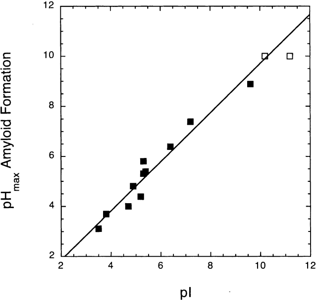
The correlation between the pH of maximal amyloid formation and the pI of the proteins listed in Table 1. The open squares represent the 5K variant of RNase Sa and hen egg white lysozyme (HEWL). These two proteins have pHmax values greater than 10, and could not be determined. The correlation coefficient for the 11 proteins shown as solid squares is 0.98.
References
Publication types
MeSH terms
Substances
Grants and funding
LinkOut - more resources
Full Text Sources
Other Literature Sources
Research Materials
Miscellaneous
