. 2003 Sep 30;100(20):11795-800.
doi: 10.1073/pnas.2032704100. Epub 2003 Sep 19.

Blue light regulates an auxin-induced K+-channel gene in the maize coleoptile

Affiliations

Blue light regulates an auxin-induced K+-channel gene in the maize coleoptile

I Fuchs et al. Proc Natl Acad Sci U S A. .

Abstract

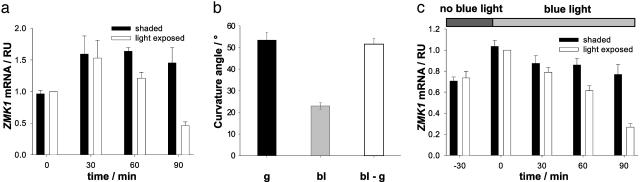
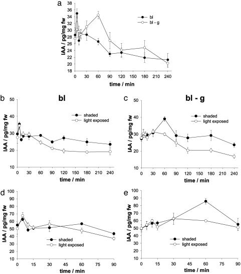
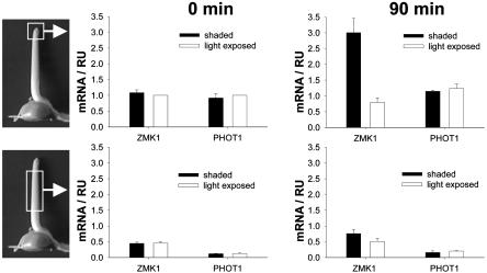
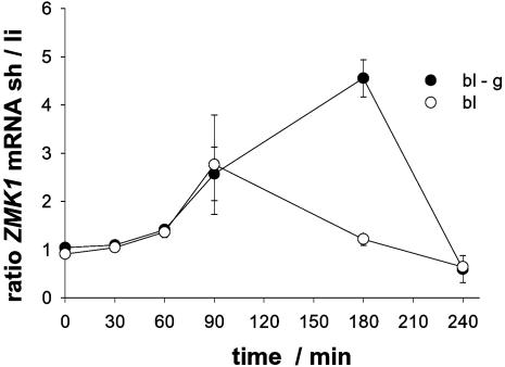
Auxin redistribution along gravistimulated maize coleoptiles causes differential expression of the auxin-induced K+-channel gene ZMK1 (Zea mays K+ channel 1) and precedes the curvature response. To evaluate the role of ZMK1 during phototropism, we here investigated blue light-stimulated coleoptiles. Four hours of blue light stimulation resulted in phototropic bending (23 degrees ). Rotation on a clinostat, at nominally "zero" gravity, and simultaneous stimulation with unidirectional blue light, however, resulted in up to 51 degrees bending toward the light. Differential ZMK1 transcription reached a maximum after 90 min of blue light stimulation under gravity, whereas ZMK1 expression remained asymmetric for at least 180 min in photostimulated coleoptiles on a clinostat. We therefore conclude that the stronger phototropic bending under nominally "zero" gravity results from prolonged differential expression of ZMK1. Under both conditions, asymmetric expression of ZMK1 could be superimposed on the lateral auxin gradient across the coleoptile tip, whereas the gene for the blue light receptor phototropin 1 (PHOT1), expressed in the tip only, was not differentially regulated in response to blue light. The activation of the two different receptors eliciting the photo- and gravitropic response of the coleoptile thus feeds into a common signaling pathway, resulting in auxin redistribution in the coleoptile tip and finally in differential transcription of ZMK1. In the process of signal integration, gravity transduction restricts the magnitude of the blue light-inducible ZMK1 gradient. The spatial and temporal distribution of ZMK1 transcripts and thus differential K+ uptake in both flanks of the coleoptile seem to limit the stimulus-induced bending of this sensory organ.


Figures

Fig. 1.
Fig. 1.
Differential expression of ZMK1 and coleoptile bending angles during gravi- and phototropism of the maize coleoptile. (a) Relative transcript level (RU, relative units) of ZMK1 in light-exposed (open bars) and shaded (solid bars) coleoptile halves after 0, 30, 60, and 90 min of stimulation with unilateral blue light. The mRNA content (n = 3 ± SE) was quantified as described in Materials and Methods. The level of ZMK1 mRNA in the light-exposed half at t = 0 min was set to 1.0. (b) Curvature angle of coleoptiles after 4 hr of gravistimulation (g, black bar) and stimulation with unilateral blue light (bl, gray bar) and of photostimulated seedlings rotating on the BLC (bl-g, open bar). Bending angles (mean of n ≥ 30 ± SE) were measured as described in Materials and Methods. (c) Relative transcript level (RU) of ZMK1 in light-exposed (open bars) and shaded (closed bars) coleoptile halves of seedlings rotating on the BLC after 0, 30, 60, and 90 min of simultaneous stimulation with unilateral blue light. Blue light illumination started 30 min after the onset of clinostat rotation (–30). The mRNA content (n = 3 ± SE) was quantified as described in Materials and Methods. The level of ZMK1 mRNA in the light-exposed half at t = 0 min was set to 1.0.
Fig. 2.
Fig. 2.
Redistribution of endogenous IAA in response to stimulation of the coleoptile with blue light. (a) IAA concentration in the entire coleoptile, stimulated with unilateral blue light under normal gravity conditions (bl, filled circles) and on the rotating BLC (bl-g, open circles). (b) IAA concentration in shaded (filled circles) and light-exposed (open circles) coleoptile halves after photostimulation (bl). (c) IAA concentration in shaded (filled circles) and light-exposed (open circles) coleoptile halves after photostimulation on the rotating BLC (bl-g). (d) IAA concentration in shaded (filled circles) and light-exposed (open circles) sides of the coleoptile tip after photostimulation (bl). (e) IAA concentration in shaded (filled circles) and light-exposed (open circles) sides of the coleoptile tip after photostimulation on the rotating BLC (bl-g). Stimulation with blue light occurred for 240 min (a–c) or 90 min (d and e). IAA concentrations are given in picograms of IAA/milligram of fresh weight (fw), mean ± SE, n = 3.
Fig. 3.
Fig. 3.
Localization of ZMK1 and PHOT1 expression in the photostimulated coleoptile. Relative transcript levels (RU, relative units) of ZMK1 and PHOT1 in light-exposed (open bars) and shaded (solid bars) halves of the coleoptile tip (Upper) and base (Lower) after 0 (Left) and 90 (Right) min of stimulation with unilateral blue light. The mRNA content (n = 3 ± SE) was quantified as described in Materials and Methods. The level of ZMK1 and PHOT1 mRNA in the light-exposed half at t = 0 min was set to 1.0.
Fig. 4.
Fig. 4.
Time-dependent differential expression of ZMK1 in photostimulated coleoptiles under different gravity conditions. Ratio of ZMK1 transcript level between shaded (sh) and light-exposed (li) sides of phototropically stimulated maize coleoptiles under control conditions (bl, open circles) and on a rotating BLC (bl-g, filled circles) after 0, 30, 60, 90, 180, and 240 min of the corresponding treatment. The mRNA content (n = 3 ± SE) was quantified as described in Materials and Methods.

References

    1. Darwin, C. (1880) The Power of Movements in Plants (Murray, London).
    1. Went, F. W. (1928) Receuil des Travaux Botaniques Neerlandais 25, 1–116.
    1. Cholodny, N. (1928) Planta 6, 118–134.
    1. Parker, K. E. & Briggs, W. R. (1990) Plant Physiol. 94, 1763–1769. - PMC - PubMed
    1. Iino, M. (1991) Plant Cell Environ. 14, 279–286.

Publication types

MeSH terms

Associated data