. 2003 Oct;4(10):964-8.
doi: 10.1038/sj.embor.embor939. Epub 2003 Sep 19.

An essential function of the mitogen-activated protein kinase Erk2 in mouse trophoblast development

Affiliations

An essential function of the mitogen-activated protein kinase Erk2 in mouse trophoblast development

Marc K Saba-El-Leil et al. EMBO Rep. 2003 Oct.

Abstract

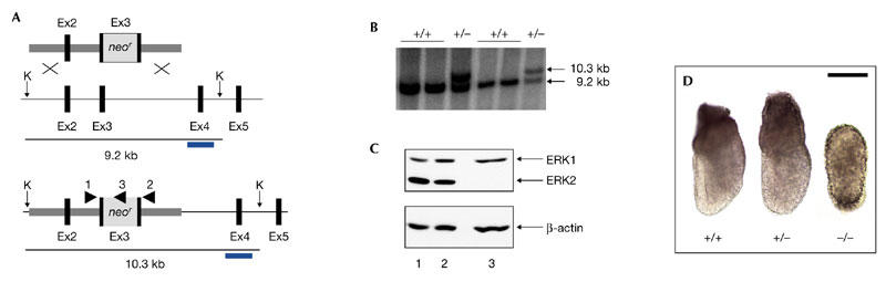
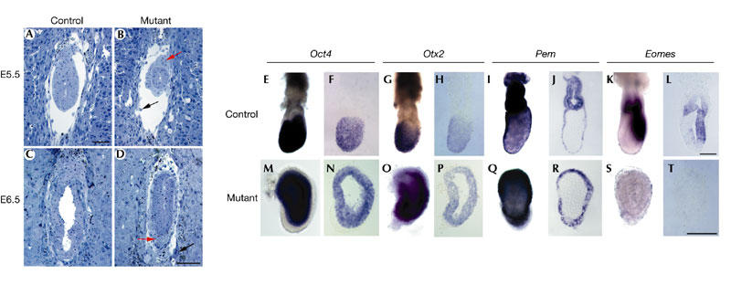
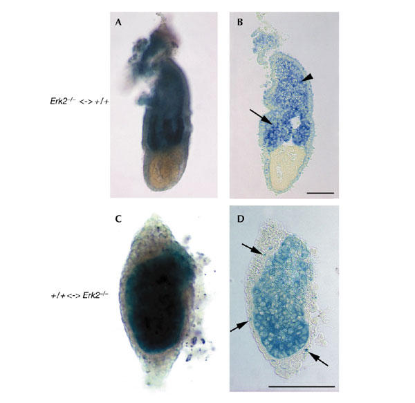
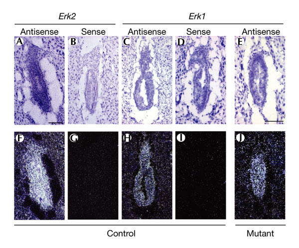
The closely related mitogen-activated protein kinase isoforms extracellular signal-regulated kinase 1 (ERK1) and ERK2 have been implicated in the control of cell proliferation, differentiation and survival. However, the specific in vivo functions of the two ERK isoforms remain to be analysed. Here, we show that disruption of the Erk2 locus leads to embryonic lethality early in mouse development after the implantation stage. Erk2 mutant embryos fail to form the ectoplacental cone and extra-embryonic ectoderm, which give rise to mature trophoblast derivatives in the fetus. Analysis of chimeric embryos showed that Erk2 functions in a cell-autonomous manner during the development of extra-embryonic cell lineages. We also found that both Erk2 and Erk1 are widely expressed throughout early-stage embryos. The inability of Erk1 to compensate for Erk2 function suggests a specific function for Erk2 in normal trophoblast development in the mouse, probably in regulating the proliferation of polar trophectoderm cells.


Figures

Figure 1
Figure 1
Targeted disruption of the mouse Erk2 gene. (A) The targeting vector (top), the wild-type Erk2 locus (middle) and the mutant Erk2 locus (bottom) are shown. Black boxes represent Erk2 exons (Ex2–Ex5) and the grey box represents the neor (neomycin resistance gene)–poly(A) cassette. The arrowheads indicate the positions and orientations of PCR primers that were used for genotyping analysis. The 3′ external probe used for Southern analysis is shown as blue bars. KpnI restriction sites (K) are indicated. (B) Southern blot analysis of wild-type and heterozygous embryonic stem cell DNA. DNA samples were digested with KpnI and hybridized with the 3′ external probe. The positions and sizes of wild-type and mutant fragments are indicated. (C) Immunoblot analysis of whole extracts from embryos obtained from Erk2 intercrosses. Top panel, blotting using the α1cp44 antibody, which recognizes both Erk1 and Erk2 isoforms. Lower panel, control blot using anti-β-actin. (D) Gross morphology of wild-type, heterozygous and homozygous Erk2 mutant embryos at embryonic day 6.5. Scale bar, 100 μm. Erk2, extracellular signal-regulated kinase 2.
Figure 2
Figure 2
Impairment of extra-embryonic ectoderm development in Erk2−/− embryos. (AD) Histological sections of control (A,C) and mutant embryos (B,D) at early post-implantation stages (embryonic days (E) 5.5 and 6.5) show the absence of ectoplacental cones and reduced size of the mutant embryo. (ET) Marker gene expression in control (EL) and mutant (MT) embryos by whole-mount in situ hybridization followed by section analyses reveal that the extra-embryonic ectoderm domains of Pem (n = 2) (I,J,Q,R) and eomesodermin (Eomes; n = 2) (K,L,S,T) are not expressed in the mutant embryos, whereas the embryonic domains of Oct4 (n = 2) (E,F,M,N) and Otx2 (n = 2) (G,H,O,P) are still present. (AD) are shown at the same magnification, as are (EL) and (MT). Scale bars, 100 μm. Erk2, extracellular signal-regulated kinase 2.
Figure 3
Figure 3
Erk2 is required in extra-embryonic tissues for the development of the extra-embryonic ectoderm and the ectoplacental cone. Chimeric embryos at embryonic day (E) 6.5 stained for β-galactosidase, and the corresponding sagittal sections, are shown. (A,B) Chimeric embryo with wild-type extra-embryonic tissues (blue) and a predominantly Erk2−/− epiblast (white), showing rescue of extra-embryonic ectoderm (arrow in (B)) and ectoplacental cone (arrowhead in (B)) development. (C,D) By contrast, a chimeric embryo with Erk2−/− extra-embryonic tissues (white) and a predominanly wild-type (blue) epiblast phenocopies Erk2−/− embryos. In the latter chimaera, a significant number of wild-type cells also contributed to the visceral endoderm (arrows in (D)). Magnifications are the same in (A,B) and in (C,D). Scale bars, 100 μm. Erk2, extracellular signal-regulated kinase 2.
Figure 4
Figure 4
Gene expression patterns for Erk1 and Erk2 at E6.5. Brightfield (AE) and corresponding darkfield images (FJ) are shown. Erk2 (A,B,F,G) and Erk1 (CE,HJ) are ubiquitously expressed throughout the whole embryo in control embryos ((A,F) and (C,H), respectively) at E6.5, as assessed by 35S radioactive in situ hybridization. Erk1 is expressed in mutant embryos (E,J). 3′ UTR sense and antisense probes were used. Magnifications are the same in (AD,FI) and (E,J). Scale bars, 100 μm. Erk2, extracellular signal-regulated kinase

References

    1. Ang S.L, Conlon R.A., Jin O. & Rossant J. ( 1994) Positive and negative signals from mesoderm regulate the expression of mouse Otx2 in ectoderm explants. Development, 120, 2979–2989. - PubMed
    1. Ballif B.A. & Blenis J. ( 2001) Molecular mechanisms mediating mammalian mitogen-activated protein kinase (MAPK) kinase (MEK)–MAPK cell survival signals. Cell Growth Differ., 12, 397–408. - PubMed
    1. Beddington R.S. & Robertson E.J. ( 1989) An assessment of the developmental potential of embryonic stem cells in the midgestation mouse embryo. Development, 105, 733–737. - PubMed
    1. Boulton T.G. et al. . ( 1990) An insulin-stimulated protein kinase similar to yeast kinases involved in cell cycle control. Science, 249, 64–67. - PubMed
    1. Boulton T.G. et al. . ( 1991) ERKs: a family of protein-serine/threonine kinases that are activated and tyrosine phosphorylated in response to insulin and NGF. Cell, 65, 663–675. - PubMed

Publication types

MeSH terms

Substances