

Review
doi: 10.1038/nri1200.
Macrophage migration inhibitory factor: a regulator of innate immunity
Affiliations
- PMID: 14502271
- PMCID: PMC7097468
- DOI: 10.1038/nri1200
Item in Clipboard
Review
Macrophage migration inhibitory factor: a regulator of innate immunity
Nat Rev Immunol.
2003 Oct.
Abstract
For more than a quarter of a century, macrophage migration inhibitory factor (MIF) has been a mysterious cytokine. In recent years, MIF has assumed an important role as a pivotal regulator of innate immunity. MIF is an integral component of the host antimicrobial alarm system and stress response that promotes the pro-inflammatory functions of immune cells. A rapidly increasing amount of literature indicates that MIF is implicated in the pathogenesis of sepsis, and inflammatory and autoimmune diseases, suggesting that MIF-directed therapies might offer new treatment opportunities for human diseases in the future.
Figures
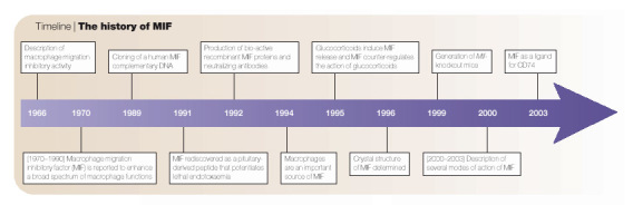
The history of MIF
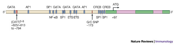
The human macrophage migration inhibitory factor (MIF) gene is composed of three short exons (green boxes) of 107, 172 and 66 base pairs, and two introns (pink boxes) of 188 and 94 base pairs. Its 5′ regulatory region contains several consensus DNA-binding sequences for transcription factors, notably activator protein 1 (AP1), nuclear factor-κB (NF-κB), ETS, GATA, SP1 and cAMP response element binding protein (CREB). However, little is known about the relevance of these putative DNA-binding sites in the regulation of expression of the human MIF gene. Two polymorphisms of the human MIF gene (arrows) — a CATT-tetranucleotide sequence repeated five to eight times at position −794 and a G-to-C single nucleotide polymorphism (SNP) at position −173 — have been associated with the severity of rheumatoid arthritis and systemic-onset juvenile idiopathic arthritis.
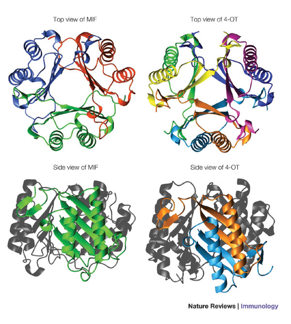
Macrophage migration inhibitory factor (MIF; top and bottom left panels) is a trimer with structural homology with the bacterial isomerases 4-oxalocrotonate tautomerase (4-OT; top and bottom right panels) and 5-carboxymethyl-2-hydroxymuconate isomerase (CHMI; not shown).
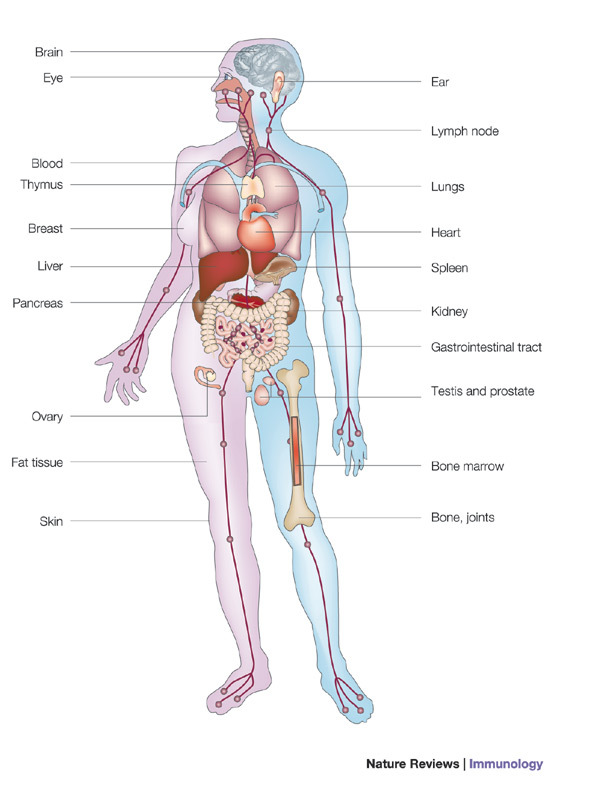
The tissue distribution and cellular sources of macrophage migration inhibitory factor (MIF) are shown. MIF is expressed in the brain (by the cortex, hypothalamus and cerebellum neurons, hippocampus, pons, glial cells, ependyma and astrocytes); in the eye (by the lens and epithelial cells of the cornea, iris and ciliary body, endothelial cells, and cells of the retina including epithelial cells, Muller cells and astrocytes); in the ear (by middle ear effusion); in the immune system (in the thymus, spleen, lymph nodes, blood and bone marrow, by monocytes/macrophages, T cells, B cells, dendritic cells, eosinophils, basophils, neutrophils and mast cells); in the lungs (by macrophages and epithelial cells); in the heart and vasculature (by endothelial cells); in the breast; in the endocrine system (by the pituitary gland, adrenal cortex and β-islet cells of the pancreas); in the liver (by Kuppfer cells, hepatocytes and endothelial cells); in the testis, prostate and ovaries (by Leydig cells, epithelial cells and granulosa cells of the follicles); in the gastrointestinal tract (by epithelial cells of the oesophagus, stomach, small and large intestines, and neurons); in the kidney (by epithelial cells, endothelial cells and mesangial cells); in fat tissue (by adipocytes); in the skin (by keratinocytes, sebaceous glands, hair follicles, endothelial cells and fibroblasts); and in bone and joints (by osteoblasts, fibroblasts and synoviocytes).
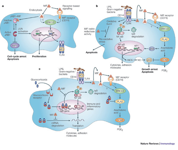
a | Macrophage migration inhibitory factor (MIF) might mediate its biological activities either through a classical receptor-mediated pathway or through a non-classical endocytic pathway. MIF has been shown to bind to CD74 and to phosphorylate the extracellular signal-regulated kinase 1 (ERK1)/ERK2 proteins. MIF promotes cell growth and activates transcription factors of the ETS family known to be crucial for expression of the Toll-like receptor 4 (TLR4) gene that encodes the signal-transducing molecule of the lipopolysaccharide (LPS) receptor complex. MIF binds to JUN-activation domain-binding protein 1 (JAB1), preventing JAB1-induced activation of JUN and JAB1-induced degradation of the cell-proliferation inhibitor KIP1, thereby leading to cell-cycle arrest and apoptosis. b | The induction and regulation of inflammatory responses of innate immune cells by MIF. MIF upregulates the expression of TLR4 by macrophages allowing rapid recognition of endotoxin-containing bacteria, which promotes the production of cytokines (including MIF), nitric oxide (NO) and other mediators. After it is released, MIF activates a cascade of events consisting of the phosphorylation of ERK1/ERK2, the induction of cytoplasmic phospholipase A2 (PLA2), arachidonic acid, JUN N-terminal kinase (JNK) activity and prostaglandin E2 (PGE2). Through the generation of oxidoreductase activity and cyclooxygenase 2 (COX2), MIF prevents activation-induced apoptosis mediated by the oxidative burst and by p53. c | MIF counter-regulates the immunosuppressive effects of glucocorticoids at transcriptional and post-transcriptional levels. MIF inhibits the glucocorticoid-mediated induction of inhibitor of nuclear factor-κB (IκB) synthesis and messenger RNA destabilization, and overrides the glucocorticoid-mediated inhibition of PLA2 activity and arachidonic acid production. iNOS, inducible nitric oxide synthase.
References
-
- Janeway CA, Jr., Medzhitov R. Innate immune recognition. Annu. Rev. Immunol. 2002;20:197–216. - PubMed
-
- Hoffmann JA, Kafatos FC, Janeway CA, Ezekowitz RA. Phylogenetic perspectives in innate immunity. Science. 1999;284:1313–1318. - PubMed
-
- Bloom BR, Bennett B. Mechanism of a reaction in vitro associated with delayed-type hypersensitivity. Science. 1966;153:80–82. - PubMed
Publication types
MeSH terms
Substances
LinkOut - more resources
Full Text Sources
Other Literature Sources
Molecular Biology Databases
Miscellaneous
