. 2003 Sep 29;162(7):1223-32.
doi: 10.1083/jcb.200303113. Epub 2003 Sep 22.

A carboxyl-terminal interaction of lamin B1 is dependent on the CAAX endoprotease Rce1 and carboxymethylation

Affiliations

A carboxyl-terminal interaction of lamin B1 is dependent on the CAAX endoprotease Rce1 and carboxymethylation

Christopher P Maske et al. J Cell Biol. .

Abstract

The mammalian nuclear lamina protein lamin B1 is posttranslationally modified by farnesylation, endoproteolysis, and carboxymethylation at a carboxyl-terminal CAAX motif. In this work, we demonstrate that the CAAX endoprotease Rce1 is required for lamin B1 endoproteolysis, demonstrate an independent pool of proteolyzed but nonmethylated lamin B1, as well as fully processed lamin B1, in interphase nuclei, and show a role for methylation in the organization of lamin B1 into domains of the nuclear lamina. Deficiency in the endoproteolysis or methylation of lamin B1 results in loss of integrity and deformity of the nuclear lamina. These data show that the organization of the nuclear envelope and lamina is dependent on a mechanism involving the methylation of lamin B1, and they identify a potential mechanism of laminopathy involving a B-type lamin.


Figures
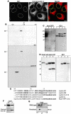
Figure 1.
Figure 1.
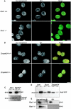
Monoclonal antibody 8D1 reacts with farnesylated lamin B1. (A) Immunofluorescence with 8D1 (green in merge) and the endoplasmic reticulum marker rhodamine– concanavalin A (red in merge) show partial colocalization at the nuclear envelope of HeLa cells. (B) HeLa nuclear envelope proteins were separated by two-dimensional electrophoresis on 13-cm, pH 4–7, gradients and immunoblotted with 8D1 and commercial antilamin antibodies. 8D1 reactivity co-migrated with the reactivity of a commercial anti–lamin B1 antibody, but shows differential reactivity with isoforms of lamin B1, suggesting a posttranslational modification is required for formation of the epitope. No cross reactivity with lamin B2 is present. (C) Replicate Western blots with polyclonal anti-GFP and 8D1. YFP–lamin C (lane 1) was fused upstream of 13 (lane 2) or 25 (lane 3) residues of the carboxyl terminus of lamin B1 and expressed in HeLa cells. GFP–lamin B1 is expressed as a control (lane 4). 8D1 reacted with lamin C fusion proteins containing the last 13 residues of lamin B1 and, therefore, binds near the CAAX motif. (D) 8D1 reacts with a eukaryotic posttranslational modification of lamin B1. Western blot with polyclonal anti–lamin B1 and 8D1; (lane 1) His6-Xpress-lamin B1 expressed in bacteria; (lane 2) HeLa lysate; and (lane 3) His6-Xpress-lamin B1 expressed in HeLa and (lane 4) in the presence of 100 μM FTI III. Lower molecular mass band represents a carboxyl- terminal apoptotic fragment of lamin B1. (E) Sequence alignment of the carboxyl terminal 40 residues of human lamin B1, lamin B2, construct lamin C-C13, human lamin C, and construct NLS-YFP-C13 (last 13 residues of lamin B1 fused directly to YFP). (F) Western blot of whole cell lysates from Icmt+/+ and Icmt−/− mouse embryonic fibroblasts with 8D1 and polyclonal anti–lamin B1 antibodies. 8D1 reacts with lamin B1 that is not methylated. (G) Two-dimensional electrophoresis on 13-cm, pH 4–7, gradients of total protein from Icmt+/+ (wild type) and Icmt−/− (knockout) mouse embryonic fibroblasts followed by immunolabeling for total lamin B1 shows loss of the most neutral isoform of lamin B1 in Icmt-deficient cells. Bars, 10 μm.
Figure 2.
Figure 2.
Rce1 is the lamin B1 CAAX endoprotease. Mouse embryonic fibroblasts from control mice and animals with homozygous deficiency of either Rce1 (A) or Zmpste24 (B) were double labeled with goat anti–lamin B1 (green in merge) and 8D1 (red in merge). Images represent single confocal sections. Rce1-deficient cells do not label with 8D1. (C) Western blot of whole cell lysates from wild-type and Rce1- or Zmpste24-deficient mouse embryonic fibroblasts with 8D1 and polyclonal anti–lamin B1 antibodies confirms the lack of an 8D1 reactivity in Rce1−/− cells. (D) A heterologous reporter containing the last 40 residues of lamin B1 including the CAAX motif, NLS-YFP-C40, was expressed in wild-type and CAAX processing–deficient cells and analyzed by SDS-PAGE and Western blot with an anti-GFP antibody. Primary antibody was detected with an alkaline phosphatase–conjugated secondary antibody and chromogenic substrate on the membrane. The protein migrates slower when expressed in Rce1−/− cells compared with wild- type cells or cells deficient in Icmt or Zmpste24, indicating a defect in proteolysis. The doublet in Icmt−/− cells is due to partial endoproteolysis. (E) Two-dimensional electrophoresis of total protein from wild-type and Rce1 deficient fibroblasts immunolabeled with polyclonal anti–lamin B1 and 8D1. Rce1 deficiency results in the loss of the methylated isoform of lamin B1. No isoforms of lamin B1 are reactive with 8D1 in Rce1−/− cells. Bars, 10 μm.
Figure 3.
Figure 3.
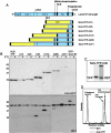
The lamin B1 CAAX endoprotease requires 40 residues of upstream sequence. (A) Schematic representation of full-length lamin B1 and carboxyl-terminal tail constructs. Increasing lengths of the lamin B1 carboxyl terminus were fused to the carboxyl terminus of NLS-YFP. (B) Constructs were expressed in HeLa cells in the absence and presence of 50 μM FTI III and analyzed by Western blot with anti-GFP and 8D1. Constructs are indicated above lanes; the numbers represent the number of residues from the carboxyl terminus of full-length lamin B1. 8D1 reactivity is acquired when 40 residues of lamin B1 sequence are present and is dependent on farnesylation. (C) Construct NLS-YFP-C40 is shown as an inset with 8D1 and anti-YFP signals aligned, indicating that 8D1 recognizes a lower molecular weight band corresponding to proteolyzed lamin B1 carboxyl terminus. The data are derived from a single nitrocellulose membrane that was reprobed to ensure perfect alignment of bands from the two antibodies. (D) The relative mobility of bands recognized by the antibodies is represented. 8D1 (solid line) recognizes endogenous lamin B1 and the reporter, which co-migrates with the shoulder present in the anti-YFP label (dotted line), corresponding to the lower molecular weight, endoproteolyzed species.
Figure 4.
Figure 4.
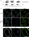
Differential CAAX processing of lamin B1 in interphase cells. (A) Two-dimensional electrophoresis of proteins from purified HeLa nuclei and immunolabeling with goat anti–lamin B1 and 8D1 antibodies demonstrates differential CAAX processing of lamin B1. Relative molecular mass and pI are shown. Nuclei purified from cells treated with cycloheximide (CHX) or farnesyltransferase inhibitor (FTI) demonstrate stability of two isoforms of lamin B1, representing methylated (arrow) and proteolyzed (arrowhead) isoforms. An acidic isoform of lamin B1 (asterisk), depleted by CHX treatment and increased by FTI treatment, may represent a nonfarnesylated pool of lamin B1. (B) Single confocal sections of untreated and FTI-treated HeLa cells labeled with goat anti–lamin B1 (green in merge) and 8D1 (red in merge) antibodies. FTI treatment increased the intranuclear pool of lamin B1 and decreased the mature pool of lamin B1 at the nuclear periphery even though total lamin B1 was maintained. The 8D1 signal was quantitatively lower in the nuclear lamina of FTI-treated cells and showed a discontinuous pattern. (C) FTI-treated cells were labeled with dilutions of polyclonal antibody to achieve similar signal intensities for both antibodies. Subdomains of CAAX-processed lamin B1 identified by 8D1 are present in the lamina (shown at a higher magnification in the inset), compared with a more continuous pattern seen with the polyclonal antibody. Bars, 10 μm.
Figure 5.
Figure 5.
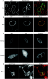
A carboxymethyl-dependent lamin receptor in the nuclear envelope. (A) NLS-YFP-C13 (green in merge), representing the terminal 13 residues of lamin B1, was expressed in HeLa cells; the cells were fixed and labeled with 8D1 (red in merge). A single confocal section is shown. NLS-YFP-C13 localized throughout the endomembrane system and plasma membrane. The absence of 8D1 reactivity confirms that the construct was not proteolyzed despite membrane association. (B) NLS-YFP-C40 (green in merge) expressed in HeLa cells localized to the nuclear envelope and was 8D1 positive (red in merge), confirming that CAAX processing took place. A single confocal section is shown. (C) Single confocal sections through the mid-nucleus of wild-type and knockout cell lines, deficient in individual CAAX-processing enzymes, expressing NLS-YFP-C40, show failure to localize to the nuclear envelope in Rce1−/− and Icmt−/− cells. The nuclear envelope localization of NLS-YFP-C40 was not affected in Zmpste24−/− cells. Intranuclear levels of the expressed protein were more pronounced in Icmt−/−cells compared with Rce1−/− cells. (D) Farnesylation and membrane association was not impaired in Rce1- or Icmt-deficient cells. NLS-YFP-C40 is partially colocalized (merge) with rhodamine–concanavalin A in Icmt−/− cells (not depicted) and Rce1−/− cells. Bars, 10 μm.
Figure 6.
Figure 6.
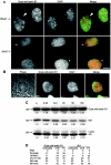
Defective CAAX processing of lamin B1 leads to morphological changes in the nuclear lamina. (A) Epifluorescence images of Rce1−/− and Icmt−/− cells labeled with goat anti–lamin B1 (green in merge) and DAPI (red in merge) for DNA. Defects in the lamina included abnormal distribution of lamin B1 in the periphery (arrows), often localized to one pole of the nucleus; peripheral lamina defects with no lamin B1 present (large arrowheads); and herniation of chromatin through peripheral defects (small arrowheads). (B) An epifluorescence image of an Icmt−/− cell labeled with a polyclonal anti–lamin B1 antibody (green in merge) and DAPI (red in merge) shows a large herniation of chromatin (DAPI) and relative paucity of lamin staining overlying the herniation. The herniation is visible by phase-contrast microscopy of the same cell as an irregularity and protrusion of the nuclear envelope. (C) HeLa cells treated with doubling doses of an FTI were analyzed by Western blot with goat anti–lamin B1 and 8D1 antibodies. Over the period of a cell cycle, mature, 8D1-positive lamin B1 decreased to ∼60% of control levels. Total lamin B1 expression was up-regulated. Pre–lamin A accumulated in a band of higher molecular weight. Lanes were loaded with equal amounts of total protein. (D) HeLa cells were treated with 50 μM farnesyltransferase inhibitor (FTI) III for 24 h or with 20 μg/ml cycloheximide (CHX) for 4 h, or were untreated. Nuclei were purified and subjected to differential extraction. Samples were analyzed by Western blot with goat anti–lamin B1 and 8D1 antibodies and quantitated by densitometry. The amounts of total lamin B1 and mature (8D1 positive) lamin B1 in each extracted fraction are shown as a percentage of the total amount released. PNS, postnuclear supernatant. Bars, 10 μm.
Figure 7.
Figure 7.
Two-step recruitment of lamin B1 at the end of mitosis. Single confocal sections of fluorescent labels and transmission images of mitotic HeLa cells double labeled with goat anti–lamin B1 (green in merge) and 8D1 (red in merge) antibodies demonstrate an unprocessed pool of lamin B1 throughout mitosis that localizes to the spindle region in metaphase and anaphase. In a telophase/early G1 cell, 8D1-positive mature lamin B1 is localized in the reforming envelope; a pool of 8D1-negative, nonproteolyzed lamin B1 was seen in the cytoplasm. Lamin B1 was recruited to the reforming nuclear envelope in two steps, dependent on the degree of carboxyl-terminal modification. Bars, 10 μm.

Similar articles

Cited by

References

    1. Adjei, A.A., J.N. Davis, C. Erlichman, P.A. Svingen, and S.H. Kaufmann. 2000. Comparison of potential markers of farnesyltransferase inhibition. Clin. Cancer Res. 6:2318–2325. - PubMed
    1. Belmont, A.S., Y. Zhai, and A. Thilenius. 1993. Lamin B distribution and association with peripheral chromatin revealed by optical sectioning and electron microscopy tomography. J. Cell Biol. 123:1671–1685. - PMC - PubMed
    1. Bergo, M.O., G.K. Leung, P. Ambroziak, J.C. Otto, P.J. Casey, and S.G. Young. 2000. Targeted inactivation of the isoprenylcysteine carboxyl methyltransferase gene causes mislocalization of K-Ras in mammalian cells. J. Biol. Chem. 275:17605–17610. - PubMed
    1. Bergo, M.O., G.K. Leung, P. Ambroziak, J.C. Otto, P.J. Casey, A.Q. Gomes, M.C. Seabra, and S.G. Young. 2001. Isoprenylcysteine carboxyl methyltransferase deficiency in mice. J. Biol. Chem. 276:5841–5845. - PubMed
    1. Bergo, M.O., P. Ambroziak, C. Gregory, A. George, J.C. Otto, E. Kim, H. Nagase, P.J. Casey, A. Balmain, and S.G. Young. 2002. a. Absence of the CAAX endoprotease Rce1: effects on cell growth and transformation. Mol. Cell. Biol. 22:171–181. - PMC - PubMed

Publication types