. 2003 Nov 28;278(48):48445-52.
doi: 10.1074/jbc.M309238200. Epub 2003 Sep 22.

Atg23 is essential for the cytoplasm to vacuole targeting pathway and efficient autophagy but not pexophagy

Affiliations

Atg23 is essential for the cytoplasm to vacuole targeting pathway and efficient autophagy but not pexophagy

Katherine A Tucker et al. J Biol Chem. .

Abstract

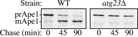
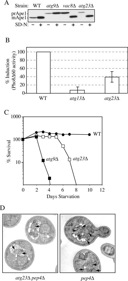
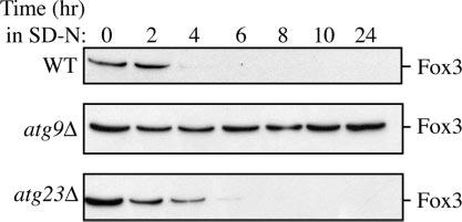
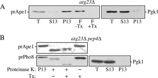
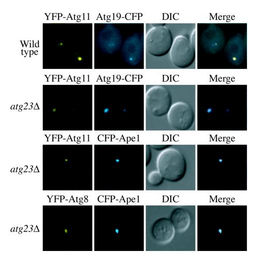
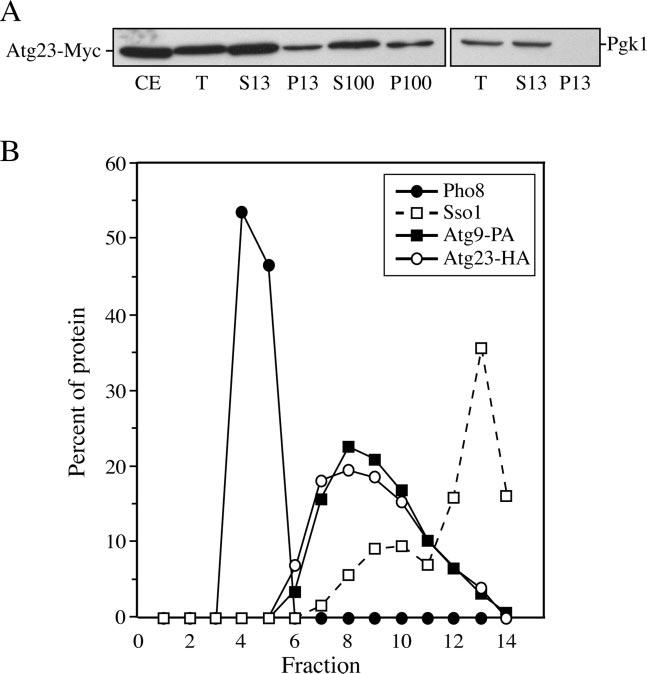
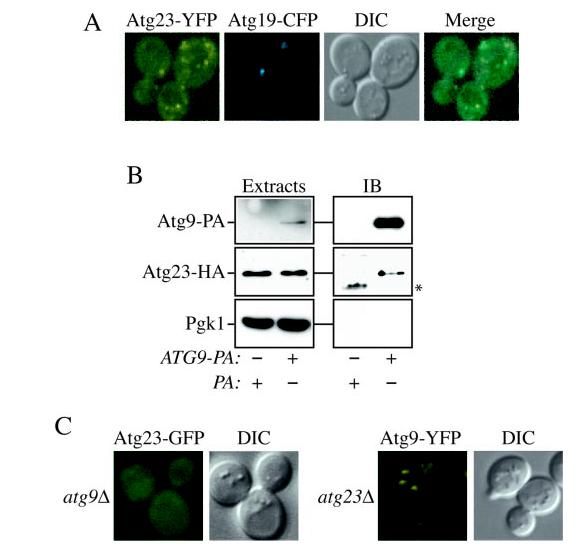
Cells must regulate both biosynthesis and degradation to ensure proper homeostasis of cellular organelles and proteins. This balance is demonstrated in a unique way in the yeast Saccharomyces cerevisiae, which possesses two distinct, yet mechanistically related trafficking routes mediating the delivery of proteins from the cytoplasm to the vacuole: the biosynthetic cytoplasm to vacuole targeting (Cvt) and the degradative autophagy pathways. Several components employed by these two transport routes have been identified, but their mechanistic interactions remain largely unknown. Here we report a novel gene involved in these pathways, which we have named ATG23. Atg23 localizes to the pre-auto-phagosomal structure but also to other cytosolic punctate compartments. Our characterization of the Atg23 protein indicates that it is required for the Cvt pathway and efficient autophagy but not pexophagy. In the absence of Atg23, cargo molecules such as prApe1 are correctly recruited to a pre-autophagosomal structure that is unable to give rise to Cvt vesicles. We also demonstrate that Atg23 is a peripheral membrane protein that requires the presence of Atg9/Apg9 to be specifically targeted to lipid bilayers. Atg9 transiently interacts with Atg23 suggesting that it participates in the recruitment of this protein.


Figures

Fig. 1
Fig. 1
atg23Δ cells are defective in the Cvt pathway. Wild type (WT) (SEY6210) and atg23Δ (KTY14) cells were pulse-labeled for 10 min and subjected to a non-radioactive chase for 90 min. Ape1 was immunoprecipitated from cell lysates and resolved by SDS-PAGE at the indicated time points. The positions of prApe1 and mApe1 are indicated.
Fig.2
Fig.2
Atg23 is required for autophagy. A, precursor Ape1 matures in atg23Δ cells during starvation conditions. Wild type (WT) (SEY6210), atg23Δ (KTY14), atg9Δ (JKY007), and vac8Δ (D3Y102) cells were grown to A600 = 1.0 in SMD and then kept in SMD or starved in SD-N medium for 2 h. Protein extracts were prepared and subjected to immunoblot analysis with anti-Ape1 antiserum. B, autophagy is partially induced in atg23Δ cells. Wild type (TN124), atg23Δ (KTY9), and atg13Δ (D3Y103) cells were shifted from SMD to SD-N medium for 4 h. Autophagy was measured by the levels of Pho8Δ60 activity in whole cell protein extracts. Activity in the wild type strain was set to 100% and activity in the other strains normalized relative to wild type. Error bars represent the S.D. from three separate experiments. C, the atg23Δ cells exhibit intermediate starvation resistance. Wild type (SEY6210), atg23Δ (KTY14), and atg9Δ (JKY007) cells were grown in SMD to A600 = 1.0 and then shifted to SD-N. At the indicated day, viability was determined by removing aliquots, plating in triplicate, and counting the number of colonies per plate after 2–3 days growth. D, fewer autophagic bodies accumulate in atg23Δ pep4Δ cells during starvation conditions. Cells from the pep4Δ (TVY1) and pep4Δ atg23Δ (KTY22) strains were grown in SMD, then shifted to SD-N for 4 h, fixed in potassium permanganate, and processed for electron microscopy as described under “Experimental Procedures.” Arrows indicate autophagic bodies.
Fig.3
Fig.3
Pexophagy is normal in atg23Δ cells. Cells from wild type (WT), atg23Δ, and atg9Δ mutants in the BY4742 background were grown under peroxisome-inducing conditions and shifted to SD-N for 24 h. At the indicated times, protein extracts were prepared and subjected to immunoblot analysis using anti-Fox3 antiserum.
Fig.4
Fig.4
Atg23 functions at the stage of Cvt vesicle formation. A, precursor Ape1 is membrane-associated in atg23Δ cells. The P13 fraction from atg23Δ (KTY14) spheroplasts was collected and loaded on the bottom of a Ficoll step gradient in the presence or absence of the detergent Triton X-100 (Tx). After centrifugation, the float (F) fractions were collected and analyzed by immunoblot using anti-Ape1 antiserum as described under “Experimental Procedures.” Lysis conditions were verified by immunoblot analysis using anti-Pgk1. B, precursor Ape1 is protease-sensitive in atg23Δ pep4Δ cells. The 13,000 × g pellet (P13) fraction was collected from atg23Δ pep4Δ (KTY22) lysed spheroplasts, subjected to treatment with proteinase K in the presence or absence of Triton X-100 (Tx), and analyzed by immunoblot using anti-Ape1 antiserum as described under “Experimental Procedures.” Lysis conditions were verified by immunoblot analysis using anti-Pgk1 and anti-Pho8 antisera. T, total; S13, 13,000 × g supernatant fraction.
Fig.5
Fig.5
Atg23 is not required for the organization of proteins at the PAS. Wild type and atg23Δ (KTY14) cells were co-transformed with plasmids expressing YFP-Atg11 or YFP-Atg8 and Atg19-CFP or CFPApe1, grown in selective SMD medium to mid-log phase and visualized by fluorescence microscopy. DIC, differential interference contrast.
Fig.6
Fig.6
Atg23 exists in both soluble and membrane-associated pools. A, Atg23-Myc is present in both soluble and pelletable fractions by velocity gradient sedimentation. Atg23-Myc (KTY7) cells were converted to spheroplasts, lysed, and subjected to differential centrifugations. All fractions were separated by SDS-PAGE and membranes probed with monoclonal anti-Myc antibody. Lysis conditions were monitored by analysis of Pgk1. CE, whole cell protein extract; T, total spheroplast protein extract; S13, low speed supernatant; P13, low speed pellet; S100, high speed supernatant; P100, high speed pellet. B, Atg23-HA and Atg9 cofractionate at the PAS. Spheroplasts from cells expressing both Atg23-HA and Atg9-PA (KTY74) were osmotically lysed, resulting in total protein extracts, and the membranes were fractionated on a sucrose density gradient as described under “Experimental Procedures.” A total of 14 fractions was collected from the top of the gradient and resolved by SDS-PAGE and membranes probed with antisera to HA, PA, Pho8, and Sso1. Percent of protein per fraction was determined relative to the total protein extracts used as the starting material.
Fig .7
Fig .7
Atg23-GFP and Atg9-YFP exhibit similar localization as well as physical and functional interactions. A, Atg9 and Atg23 localize to several punctate structures dispersed in the cytoplasm. Cells containing Atg9-YFP and CFP-Atg8 integrated at their chromosomal loci (FRY134) or those containing both Atg23-YFP integrated at the chromosomal ATG23 locus (KTY26) and the plasmid bearing Atg19-CFP were grown in selective medium to A600 = 0.8–1.2 and imaged with a fluorescent microscope. B, Atg23 interacts with Atg9. Atg23-HA (KTY8) cells transformed with Atg23-HA and either Atg9-PA (KTY74) or pRS416-CuProtA as a control were used to prepare detergent-solubilized extracts as described under “Experimental Procedures.” IgG-Sepharose beads were used to affinity-purify the PA fusions together with the associated proteins. Eluted polypeptides were separated by SDS-PAGE and then visualized by immunoblotting (IB) with antiserum to PA, HA, and Pgk1. For each experiment, 0.013% of the total lysate or 4.45% of the total eluate was loaded per gel lane. The asterisk marks a contaminating band. C, Atg9 is required to recruit Atg23 to membranes. The atg9Δ mutant bearing an integrated Atg23-GFP (KTY32) or Atg9-YFP cells deleted for the ATG23 ORF (KTY51) were grown and imaged as in A. DIC, differential interference contrast.

Similar articles

Cited by

References

    1. Bryant NJ, Stevens TH. Microbiol. Mol. Biol. Rev. 1998;62:230–247. - PMC - PubMed
    1. Khalfan WA, Klionsky DJ. Curr. Opin. Cell Biol. 2002;14:468–475. - PubMed
    1. Klionsky DJ, Ohsumi Y. Annu. Rev. Cell Dev. Biol. 1999;15:1–32. - PubMed
    1. Stromhaug PE, Klionsky DJ. Traffic. 2001;2:524–531. - PubMed
    1. Klionsky DJ, editor. Autophagy. Landes Bioscience; Georgetown: 2004. pp. 1–327. TX.

Publication types

MeSH terms