. 2003 Sep 30;100 Suppl 1(Suppl 1):11911-6.
doi: 10.1073/pnas.2034195100. Epub 2003 Sep 22.

Nonhuman primate parthenogenetic stem cells

Affiliations

Nonhuman primate parthenogenetic stem cells

Kent E Vrana et al. Proc Natl Acad Sci U S A. .

Erratum in

  • Proc Natl Acad Sci U S A. 2004 Jan 13;101(2):693

Abstract

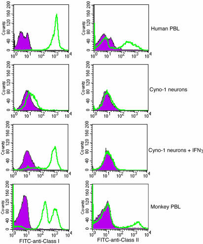
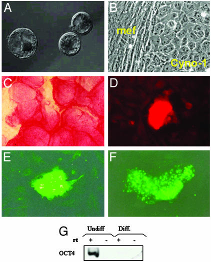
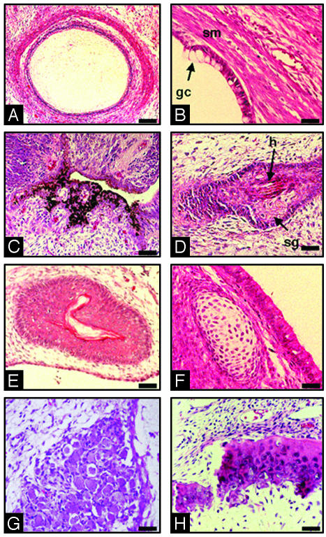
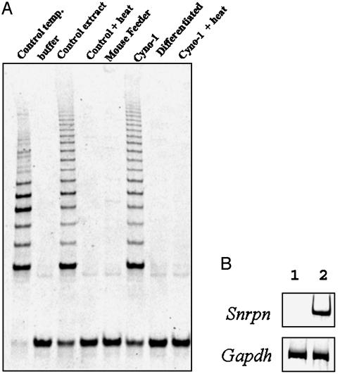
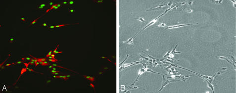
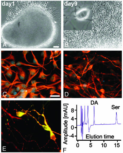
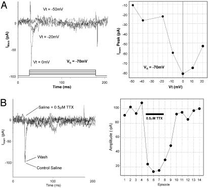
Parthenogenesis is the biological phenomenon by which embryonic development is initiated without male contribution. Whereas parthenogenesis is a common mode of reproduction in lower organisms, the mammalian parthenote fails to produce a successful pregnancy. We herein describe in vitro parthenogenetic development of monkey (Macaca fascicularis) eggs to the blastocyst stage, and their use to create a pluripotent line of stem cells. These monkey stem cells (Cyno-1 cells) are positive for telomerase activity and are immunoreactive for alkaline phosphatase, octamer-binding transcription factor 4 (Oct-4), stage-specific embryonic antigen 4 (SSEA-4), tumor rejection antigen 1-60 (TRA 1-60), and tumor rejection antigen 1-81 (TRA 1-81) (traditional markers of human embryonic stem cells). They have a normal chromosome karyotype (40 + 2) and can be maintained in vitro in an undifferentiated state for extended periods of time. Cyno-1 cells can be differentiated in vitro into dopaminergic and serotonergic neurons, contractile cardiomyocyte-like cells, smooth muscle, ciliated epithelia, and adipocytes. When Cyno-1 cells were injected into severe combined immunodeficient mice, teratomas with derivatives from all three embryonic germ layers were obtained. When grown on fibronectin/laminin-coated plates and in neural progenitor medium, Cyno-1 cells assume a neural precursor phenotype (immunoreactive for nestin). However, these cells remain proliferative and express no functional ion channels. When transferred to differentiation conditions, the nestin-positive precursors assume neuronal and epithelial morphologies. Over time, these cells acquire electrophysiological characteristics of functional neurons (appearance of tetrodotoxin-sensitive, voltage-dependent sodium channels). These results suggest that stem cells derived from the parthenogenetically activated nonhuman primate egg provide a potential source for autologous cell therapy in the female and bypass the need for creating a competent embryo.


Figures

Fig. 7.
Fig. 7.
Immunological profile of Cyno-1 cells. PBLs and Cyno-1-derived neural cells were analyzed by flow cytometry to quantitate expression of M. fasicularis class I (anti-HLA-A,-B,-C) and class II (anti-HLA-DR) antigens and compared with cells stained with isotype-matched control antibodies (shaded curves). PBLs express both class I and class II (DR) antigens whereas differentiated Cyno-1-derived neurons do not express either class of antigen unless treated with IFN-γ.
Fig. 1.
Fig. 1.
Characterization of parthenogenetic embryos and derived cell lines. (A) Parthenogenetically activated eggs at day 8 of development before ICM isolation. (B) Phase contrast of Cyno-1 stem cells growing on top of mitotically inactivated mouse feeder layer (mef). (C) Alkaline phosphatase staining. (D) Stage-specific embryonic antigen 4. (E) Tumor rejection antigen 1-60. (F) Tumor rejection antigen 1-81 staining. (G) RT-PCR octamer-binding transcription factor 4 expression in undifferentiated Cyno-1 cells. (Scale bars = 50 μm in A, 10 μm in B and D-F, and 4 mm in C.)
Fig. 2.
Fig. 2.
In vivo differentiation of Cyno-1 cells. Cells were injected i.p. in severe combined immunodeficient mice. Eight and 15 weeks after injection, teratomas 12 and 30 mm in diameter, respectively, were isolated, fixed with 10% paraformaldehyde, and paraffin-embedded. Sections were stained with hematoxylin/eosin. The following complex structures were observed: gut (A), intestinal epithelium with typical goblet cells (gc) and smooth muscle (sm) (B), neuronal tissue with melanocytes (C), hair follicle complex with evident hair (h) and sebaceous gland (sg) (D), skin (E), cartilage (F), ganglion cells (G), and bone (H). (Scale bars = 40 μmin A,10 μmin B and D-H, and 20 μmin C.)
Fig. 3.
Fig. 3.
Telomerase activity. (A) Cyno-1 cells, maintained in the undifferentiated state on mouse feeder layers, express telomerase activity that diminishes to undetectable levels in differentiated Cyno-1 cells. (B) RT-PCR to detect expression of the paternally expressed imprinted gene Snrpn in Cyno-1 cells (lane 1) and in adult fibroblasts (lane 2) from the same species. The housekeeping gene Gapdh is used as a control to demonstrate that equal amounts of mRNA were used.
Fig. 4.
Fig. 4.
Nestin-positive neural precursors derived from Cyno-1 cells. (A) Neural precursors stained for nestin (green). The nuclei were stained with 4′,6-diamidino-2-phenylindole (DAPI) (blue). (B) Phase contrast of nestin precursors.
Fig. 5.
Fig. 5.
Neural differentiation of Cyno ES cells in vitro. (A) Phase contrast microscopy of proliferating Cyno1-derived neural precursors at day 1 in vitro (DIV1). (B) Same cluster of precursors shown at DIV9. The total cell number has increased by 5- to 8-fold over a 9-day period. (Inset) One of many mitotic figures. Immunohistochemical analyses after 5 days of neural differentiation in the absence of bFGF and epidermal growth factor and the presence of ascorbic acid revealed positive staining for glial fibrillary acidic protein (GFAP), an astrocytic marker seen in C, and TUJ1, a neuronal marker seen in D. (E) Sequential exposure to sonic hedgehog, FGF8b, and ascorbic acid yielded an average of 25% TUJ1+ neurons coexpressing tyrosine-hydroxylase (TH), a marker for dopamine neurons. (F) HPLC revealing the release of dopamine (DA) and serotonin (Ser).
Fig. 6.
Fig. 6.
Single-cell electrophysiology. (A) Neurons derived from Cyno-1 express voltage-dependent inward currents that are blocked by tetrodotoxin. Currents were elicited by membrane depolarizations to 0 mV every 15 s from a holding potential of -70 mV. Application of 0.5 μM tetrodotoxin inhibited >90% of these currents. (B) Inhibition was complete within 30 s of tetrodotoxin application and washed completely in <1 min.

References

    1. Thomson, J. A., Itskovitz-Eldor, J., Shapiro, S. S., Waknitz, M. A., Swiergiel, J. J., Marshall, V. S. & Jones, J. M. (1998) Science 282, 1145-1147. - PubMed
    1. Thomson, J. A., Kalishman, J., Golos, T. G., Durning, M., Harris, C. P., Becker, R. A. & Hearn, J. P. (1995) Proc. Natl. Acad. Sci. USA 92, 7844-7848. - PMC - PubMed
    1. Thomson, J. A., Kalishman, J., Golos, T. G., Durning, M., Harris, C. P. & Hearn, J. P. (1996) Biol. Reprod. 55, 254-259. - PubMed
    1. Suemori, H., Tada, T., Torii, R., Hosoi, Y., Kobayashi, K., Imahie, H., Kondo, Y., Iritani, A. & Nakatsuji, N. (2001) Dev. Dyn. 222, 273-279. - PubMed
    1. Cibelli, J. B., Grant, K. A., Chapman, K. B., Cunniff, K., Worst, T., Green, H. L., Walker, S. J., Gutin, P. H., Vilner, L., Tabar, V., et al. (2002) Science 295, 819. - PubMed

MeSH terms