. 2003 Sep 30;100(20):11583-8.
doi: 10.1073/pnas.2034960100. Epub 2003 Sep 22.

Interference with T cell receptor-HLA-DR interactions by Epstein-Barr virus gp42 results in reduced T helper cell recognition

Affiliations

Interference with T cell receptor-HLA-DR interactions by Epstein-Barr virus gp42 results in reduced T helper cell recognition

Maaike E Ressing et al. Proc Natl Acad Sci U S A. .

Abstract

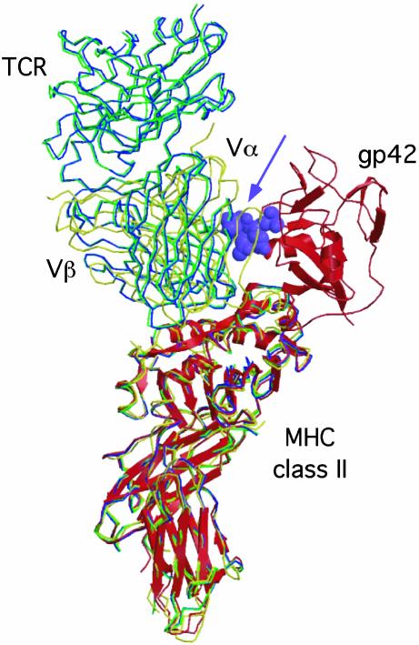
Epstein-Barr virus (EBV) persists lifelong in infected hosts despite the presence of antiviral immunity. Many viral antigens are expressed during lytic infection. Thus, for EBV to spread, it must have evolved effective ways to evade immune recognition. Here, we report that HLA class II-restricted antigen presentation to T helper cells is hampered in the presence of the lytic-phase protein gp42. This interference with T cell activation involves association of gp42 with class II peptide complexes. Using HLA-DR tetramers, we identify a block in T cell receptor (TCR)-class II interactions imposed by gp42 as the underlying mechanism. EBV gp42 sterically clashes with TCR Valpha-domains as visualized by superimposing the crystal structures for gp42-HLA-DR1 and TCR-MHC class II complexes. Blocking TCR recognition provides a previously undescribed strategy for viral immune evasion.


Figures

Fig. 1.
Fig. 1.
Endogenously expressed EBV gp42 associates with HLA class II molecules. (a) Surface-exposed proteins on MJS/gp42 (lanes 4–6 and 8) and control MJS/gfp (lanes 1–3 and 7) cells were labeled with 125I for 1 h. Specific proteins were isolated from cell lysates by immunoprecipitation (IP) with mAbs against gp42 (F-2-1), HLA-DR (Tü36), HLA class I (W6/32), and TfR (66IG10); Ii (VicY1) served as a negative control. Precipitated proteins were separated under nonreducing conditions by SDS/12% PAGE. (b) Total lysates of MJS/gp42 (lanes 2, 4, and 6) and control MJS/gfp (lanes 1, 3, and 5) cells were directly loaded (lanes 1 and 2) or were subjected to immunoprecipitations with rabbit serum no. 32 against gp42 (lanes 3 and 4) and mAb Tü36 against HLA-DR complexes (lanes 5 and 6). Total lysates and immune complexes were boiled in reducing (α) or nonreducing (β) sample buffer, separated by SDS/12% PAGE, and blotted onto poly(vinylidene difluoride) membranes. Western blots were stained with mAbs specific for HLA-DRα (DA6.147) and β (HB10A) chains and visualized by enhanced chemiluminescence.
Fig. 2.
Fig. 2.
Inhibition of T helper responses on antigen presentation by EBV gp42-expressing cells. MJS/gp42 and control cells were assayed for HLA-DR3 presentation of Ad5 (formula image
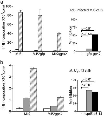
) to R30.95 T cells (a) or the M. tuberculosis hsp65 protein (formula image) and the related p3-15 peptide (formula image) to Rp15.1.1 T cells (b). [3H]Thymidine incorporation is depicted with error bars for triplicates. (Right) Percentage inhibition of antigen-specific T cell proliferation in response to MJS/gp42 or MJS/gfp cells is depicted compared with (uninfected) MJS cells. Statistical analysis on triplicate wells was performed by the Student's t test, and P values are indicated.
Fig. 3.
Fig. 3.
Surface expression and maturation of HLA-DR αβ-peptide complexes is not altered in MJS/gp42 cells. (a) Flow-cytometry histograms are depicted for surface staining of MJS/gfp and MJS/gp42 cells with the following mAbs: CerCLIP for HLA-DR/CLIP complexes (peak 2), Bü45 for Ii (peak 3), 16.23 for an HLA-DM-induced conformation of DR3 (peak 4), and L243 for HLA-DR (peak 5). As a negative control, no primary Ab was added (peak 1). (b) MJS/gp42 (lanes 4–6) and control MJS/gfp (lanes 1–3) cells were metabolically labeled for 1 h with [35S]methionine and chased for 0, 4, and 20 h, followed by cell lysis. HLA-DR complexes were isolated by immunoprecipitation (IP) with mAb L243, were incubated in nonreducing sample buffer with SDS at 37°C, which reveals the SDS-stable class II αβ dimers (αβ), and were analyzed on SDS/12% PAGE. As a control protein, TfR was precipitated by mAb 66IG10 and separated under reducing conditions by SDS/10% PAGE.
Fig. 4.
Fig. 4.
TCR interactions with complexes of peptide–HLA class II, but not class I, are abolished in the presence of soluble EBV gp42. Peripheral blood mononuclear cells from a healthy HLA-A2+ DR4+ donor were labeled with CFSE and stimulated in vitro with DR4/HA307–319 (Upper) or A2/M58–66 (Lower) or without peptides (control). Responding T cells were incubated with specific allophycocyanin-conjugated tetramers in the presence or absence of rgp42. rgp42 concentrations of 0.05, 0.25, and 1.25 μM correspond to gp42:class II ratios of 0.5:1, 3:1, and 12:1, respectively. Cells were stained with phycoerythrin-conjugated mAbs to CD4 (Upper) or CD8 (Lower). Each flow-cytometry dot plot represents ≈45,000 propidium iodide, CFSE cells; percent values refer to the percentage of CD4+ or CD8+ T cells that stained with the tetramers (top right quadrant).
Fig. 5.
Fig. 5.
EBV gp42 sterically clashes with the TCR in known TCR–MHC class II structures. Superposition of the gp42–DR1 structure (shown as red ribbons) with the crystal structures of the HA1.7-TCR–DR1 complex (green), the HA1.7-TCR–DR4 complex (blue), and the D10-TCR–I-Ak complex (yellow). The gp42 loop including residues 157–161 clashes with the TCR Vα domains and is shown as purple Corey–Pauling–Koltun model atoms (arrow). The D10-TCR has the greatest overlap with gp42, whereas the HA1.7 structures are rotated slightly away from gp42.

References

    1. Tortorella, D., Gewurz, B. E., Furman, M. H., Schust, D. J. & Ploegh, H. L. (2000) Annu. Rev. Immunol. 18, 861–926. - PubMed
    1. Cresswell, P. (1994) Annu. Rev. Immunol. 12, 259–293. - PubMed
    1. Rickinson, A. B. & Kieff, E. (2001) in Field's Virology, eds. Knipe, D. M. & Howley, P. M. (Lippincott Williams & Wilkins, Philadelphia), pp. 2575–2627.
    1. Rickinson, A. B. & Moss, D. J. (1997) Annu. Rev. Immunol. 15, 405–431. - PubMed
    1. Levitsky, V. & Masucci, M. G. (2002) Virus Res. 88, 71–86. - PubMed

Publication types