

Lung pathology of severe acute respiratory syndrome (SARS): a study of 8 autopsy cases from Singapore
- PMID: 14506633
- PMCID: PMC7119137
- DOI: 10.1016/s0046-8177(03)00367-8
Lung pathology of severe acute respiratory syndrome (SARS): a study of 8 autopsy cases from Singapore
Erratum in
- Hum Pathol. 2004 Jan;35(1):138
Abstract
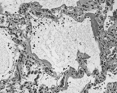
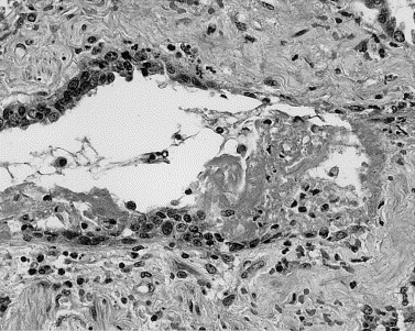
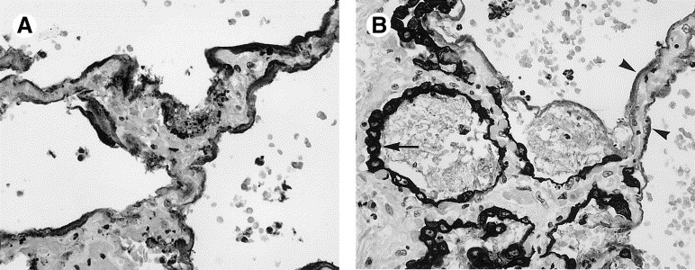
Severe acute respiratory syndrome (SARS) is an infectious condition caused by the SARS-associated coronavirus (SARS-CoV), a new member in the family Coronaviridae. To evaluate the lung pathology in this life-threatening respiratory illness, we studied postmortem lung sections from 8 patients who died from SARS during the spring 2003 Singapore outbreak. The predominant pattern of lung injury in all 8 cases was diffuse alveolar damage. The histology varied according to the duration of illness. Cases of 10 or fewer days' duration demonstrated acute-phase diffuse alveolar damage (DAD), airspace edema, and bronchiolar fibrin. Cases of more than 10 days' duration exhibited organizing-phase DAD, type II pneumocyte hyperplasia, squamous metaplasia, multinucleated giant cells, and acute bronchopneumonia. In acute-phase DAD, pancytokeratin staining was positive in hyaline membranes along alveolar walls and highlighted the absence of pneumocytes. Multinucleated cells were shown to be both type II pneumocytes and macrophages by pancytokeratin, thyroid transcription factor-1, and CD68 staining. SARS-CoV RNA was identified by reverse transcriptase-polymerase chain reaction in 7 of 8 cases in fresh autopsy tissue and in 8 of 8 cases in formalin-fixed, paraffin-embedded lung tissue, including the 1 negative case in fresh tissue. Understanding the pathology of DAD in SARS patients may provide the basis for therapeutic strategies. Further studies of the pathogenesis of SARS may reveal new insight into the mechanisms of DAD.
Figures


References
-
- Wenzel R.P., Hendley J.O., Davies J.A. Coronavirus infections in military recruits. Three-year study with coronavirus strains OC43 and 229E. Am Rev Respir Dis. 1974;109:621–624. - PubMed
-
- Krafft A.E., Duncan B.W., Bijwaard K.E. Optimization of the isolation and amplification of RNA from formalin-fixed, paraffin-embedded tissue: The Armed Forces Institute of Pathology experience and literature review. Mol Diagn. 1997;2:217–230. - PubMed
MeSH terms
Substances
LinkOut - more resources
Full Text Sources
Other Literature Sources
Miscellaneous
