. 2003 Oct;163(4):1233-43.
doi: 10.1016/S0002-9440(10)63483-9.

Hypoxia-induced gene expression in human macrophages: implications for ischemic tissues and hypoxia-regulated gene therapy

Affiliations

Hypoxia-induced gene expression in human macrophages: implications for ischemic tissues and hypoxia-regulated gene therapy

Bernard Burke et al. Am J Pathol. 2003 Oct.

Abstract

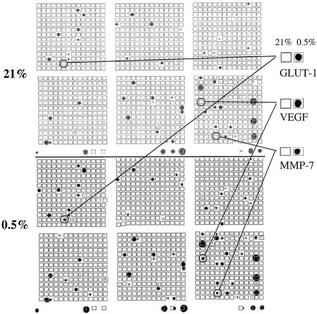
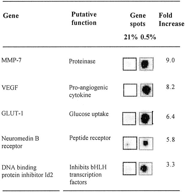
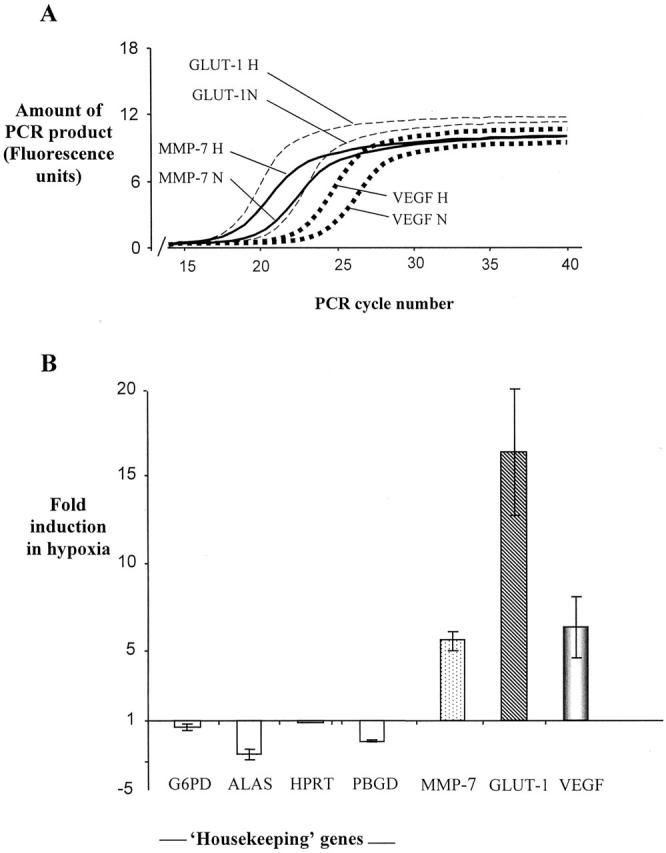
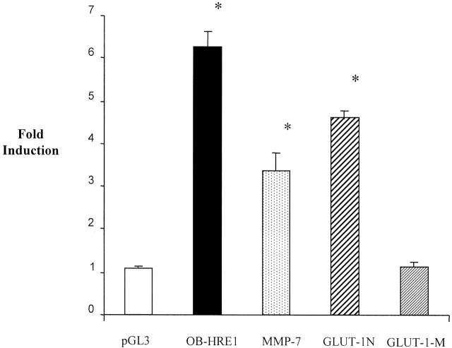
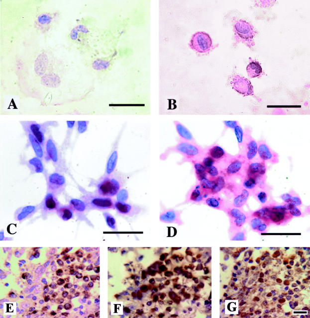
Macrophages accumulate in ischemic areas of such pathological tissues as solid tumors, atherosclerotic plaques and arthritic joints. Studies have suggested that hypoxia alters the phenotype of macrophages in a way that promotes these lesions. However, the genes up-regulated by macrophages in such hypoxic tissues are poorly characterized. Here, we have used cDNA array hybridization to investigate the effects of hypoxia on the mRNAs of 1185 genes in primary human monocyte-derived macrophages. As shown previously in other cell types, mRNA levels for vascular endothelial growth factor (VEGF) and glucose transporter 1 (GLUT-1) were up-regulated by hypoxia. However, the mRNAs of other genes were also up-regulated including matrix metalloproteinase-7 (MMP-7), neuromedin B receptor, and the DNA-binding protein inhibitor, Id2. The promoters of GLUT-1 and MMP-7 confer hypoxic inducibility on a reporter gene in RAW 264.7 macrophages, indicating that the hypoxic up-regulation of these mRNAs may occur, at least in part, at the transcriptional level. GLUT-1 and MMP-7 mRNA were also shown to be up-regulated in hypoxic macrophages in vitro by real-time RT-PCR, and these proteins were elevated in hypoxic macrophages in vitro and in hypoxic areas of human breast tumors. The hypoxia up-regulated genes identified could be important for the survival and functioning of macrophages in hypoxic diseased tissues, and their promoters could prove useful in macrophage-delivered gene therapy.


Figures

Figure 1.
Figure 1.
Genes expressed by human macrophages as detected by cDNA array analysis of mRNA levels. Two representative cDNA array autoradiographs are shown, after hybridization with radiolabeled probes from human MDMs exposed to normoxia (21% oxygen) or hypoxia (0.5% oxygen), respectively, for 16 hours. Enlarged versions of the array spots of the three most highly up-regulated genes are shown. Essentially similar results were different preparations of macrophage mRNA were used on separate arrays.
Figure 2.
Figure 2.
Genes regulated by hypoxia in human macrophages as detected by cDNA array analysis of mRNA levels. Insets show the matched array spots of the five most highly up-regulated genes from a representative cDNA array autoradiograph after hybridization with radiolabeled cDNA probes derived from human MDMs exposed to normoxia (21% oxygen) or hypoxia (0.5% oxygen) for 16 hours. Fold induction values were calculated as the hypoxic values divided by normoxic values for the same gene.
Figure 3.
Figure 3.
Effect of hypoxia on mRNA level determined using real-time RT-PCR. A: A representative experiment illustrating PCR curves for amplifications of GLUT-1, MMP-7, and VEGF, which are used by the Lightcycler software to calculate fold induction data, based on the PCR cycle at which amplification becomes exponential for each individual test gene. B: Fold induction in hypoxia relative to normoxia for each test gene and four housekeeping genes. Negative values indicate a level of mRNA under hypoxia lower than that under normoxia. Data shown is the mean of three independent experiments carried out on three different RNA preparations. Bars represent standard errors of the mean. MMP-7, matrix metalloproteinase 7; GLUT-1, glucose transporter 1; VEGF, vascular endothelial growth factor; β2 mol/L, β2 microglobulin; G6PDH, glucose-6-phosphate dehydrogenase; ALAS, 5-aminolevulinate synthase gene; HPRT, hypoxanthine phosphoribosyltransferase; PBGD, porphobilinogen deaminase.
Figure 4.
Figure 4.
Effect of hypoxia on luciferase reporter constructs after transfection into the murine macrophage-like cell line, RAW 264.7. Cells were exposed to 20.9% (normoxia) or 0.5% O2 (hypoxia) for 16 hours. pGL3, the luciferase reporter construct pGL3 promoter containing the minimal, enhancerless SV40 promoter; OB-HRE1, a luciferase reporter construct controlled by a trimer of the HRE monomer described in Table 1 ▶ (cloned into the multiple cloning site of pGL3 promoter); MMP-7 promoter, a 2.3-kb sequence containing the promoter of the human MMP-7 gene, cloned into the multiple cloning site of pGL2 basic (Promega), a promoterless luciferase reporter construct. GLUT-1-N contains a 184-bp sequence from the rat GLUT-1 promoter cloned into the multiple cloning site of pGL3 promoter (which contains the viral SV40 promoter), and GLUT-1 mol/L is identical apart from a 4-bp mutation (CGTG to ACAT) resulting in ablation of the HRE (HIF binding site). Fold induction values refer to luciferase values obtained under hypoxia divided by those seen under normoxia. Fold inductions (hypoxic luciferase values divided by normoxic values) from four independent transfections are shown. Means ± SEM. *, P < 0.05 compared to pGL3 promoter (Mann Whitney U-test).
Figure 5.
Figure 5.
Immunodetection of GLUT-1 and MMP-7 in human macrophages in vitro and in vivo. These proteins were immunolocalized in/on human MDM grown on coverslips (GLUT-1, A and B; MMP-7, C and D) following exposure to normoxia (20.9% oxygen, A and C) or hypoxia (0.5% oxygen, B and D) for 16 hours in vitro. Although low-level expression of immunoreactive GLUT-1 (brown) and MMP-7 (red) can just be seen in the cytoplasm of these cells in normoxia, this was markedly increased following exposure to hypoxia. Immunoreactive CD68 (E), MMP-7 (F), and HIF-1α (G) proteins were also detectable in macrophages in focal areas of malignant human breast carcinomas (brown color reaction). CD68 and MMP-7 staining was cytoplasmic, whereas HIF-1α staining was seen in both the cytoplasm and nuclei of cells. Sections exposed to a negative control antibody showed no staining reaction (not shown). Bar, 20 μm.

References

    1. Vaupel P, Kallinowski F, Okunieff P: Blood flow, oxygen and nutrient supply, and metabolic environment of human tumors: a review. Cancer Res 1989, 49:6449-6465 - PubMed
    1. Kivisaari J: Oxygen and carbon dioxide tensions in healing tissue. Acta Chir Scand 1975, 141:693-696 - PubMed
    1. Stevens CR, Williams RB, Farrell AJ, Blake DR: Hypoxia and inflammatory synovitis: observations and speculation. Ann Rheum Dis 1991, 50:124-132 - PMC - PubMed
    1. Bjornheden T, Levin M, Evaldsson M, Wiklund O: Evidence of hypoxic areas within the arterial wall in vivo. Arterioscler Thromb Vasc Biol 1999, 19:870-876 - PubMed
    1. Lewis JS, Lee J, Underwood JCE, Harris AL, Lewis CE: Macrophage responses to hypoxia: implications for disease mechanisms. J Leukoc Biol 1999, 66:889-900 - PubMed

Publication types

MeSH terms