. 2003 Oct;163(4):1261-73.
doi: 10.1016/s0002-9440(10)63486-4.

Transforming growth factor-beta-dependent and -independent pathways of induction of tubulointerstitial fibrosis in beta6(-/-) mice

Affiliations

Transforming growth factor-beta-dependent and -independent pathways of induction of tubulointerstitial fibrosis in beta6(-/-) mice

Li-Jun Ma et al. Am J Pathol. 2003 Oct.

Abstract

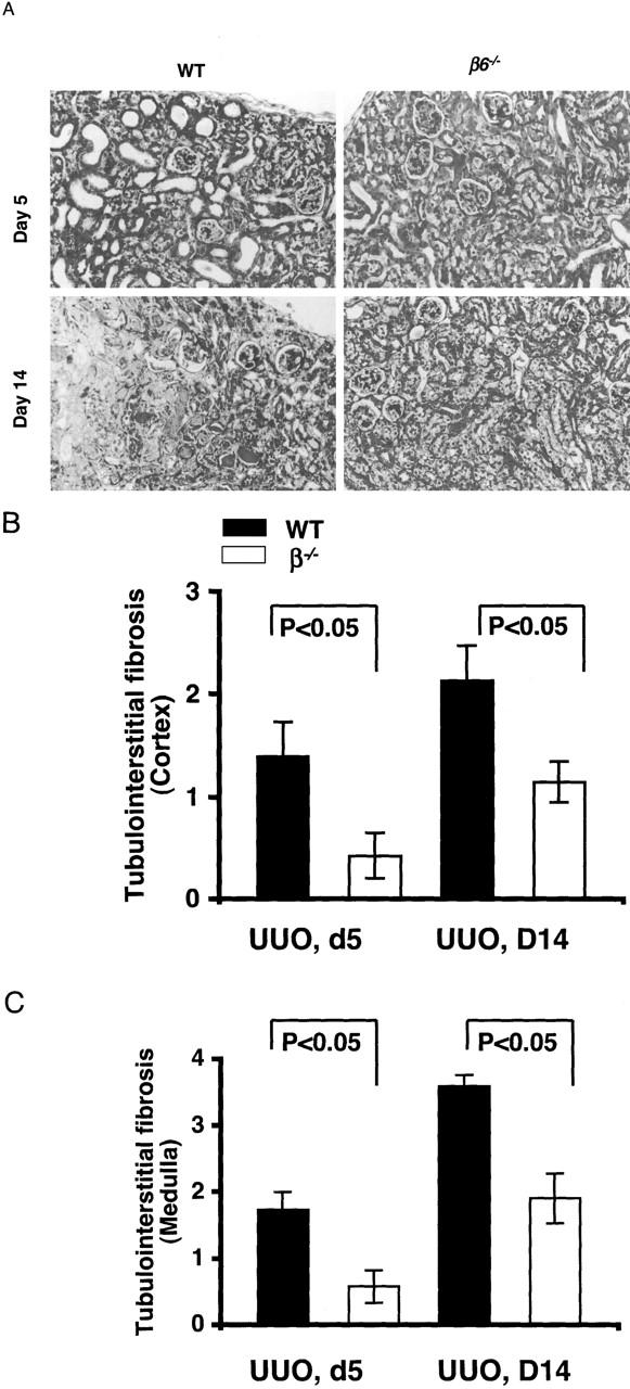
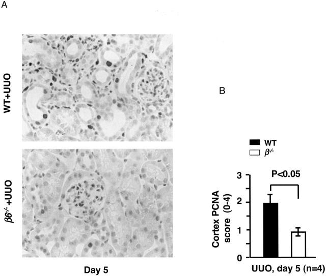
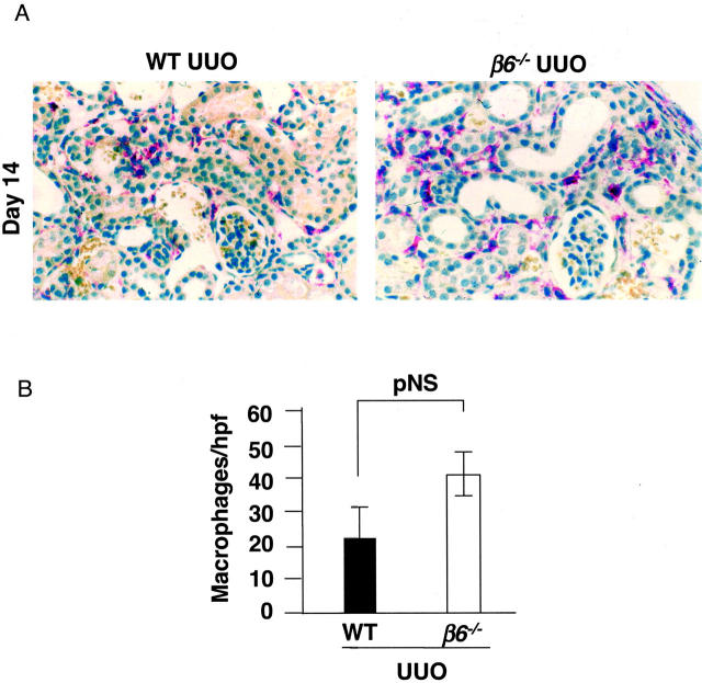
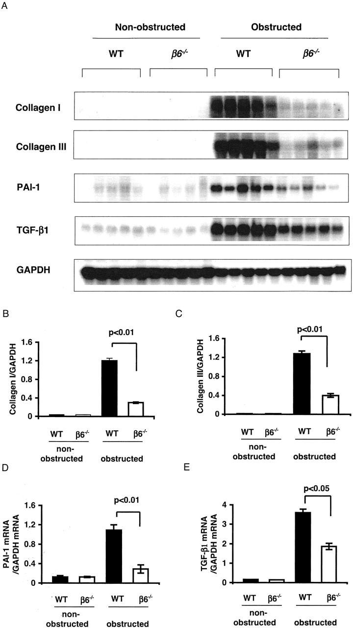
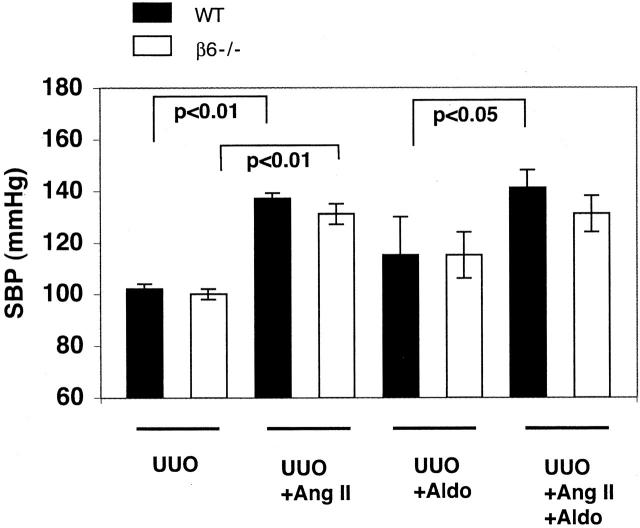
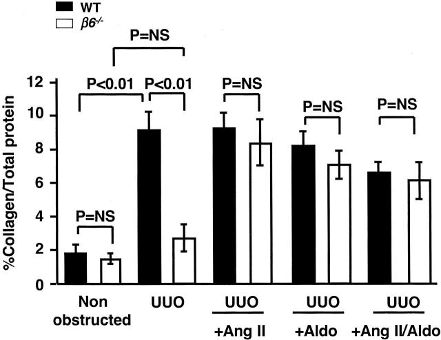
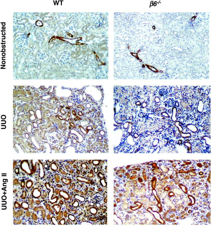
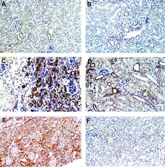
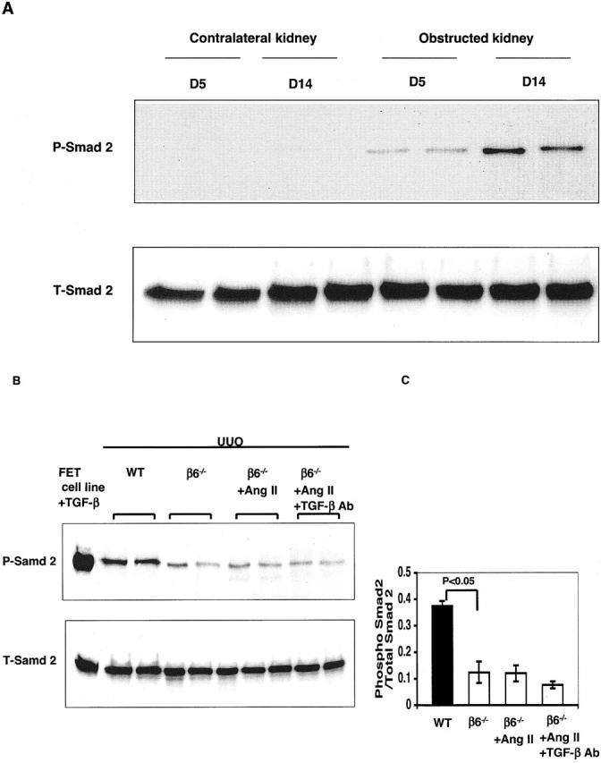
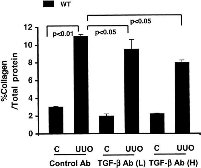
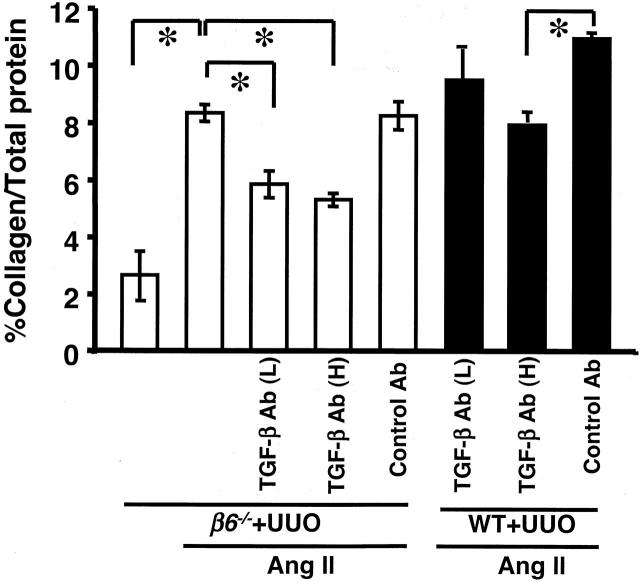
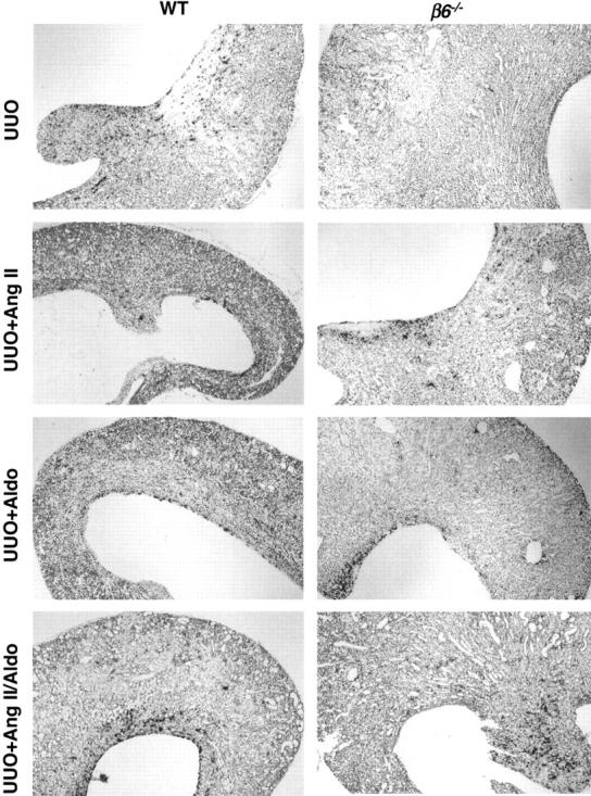
Transforming growth factor-beta1 (TGF-beta1) and the renin-angiotensin-aldosterone system are key mediators in kidney fibrosis. Integrin alphavbeta6, a heterodimeric matrix receptor expressed in epithelia, binds and activates latent TGF-beta1. We used beta6 integrin-null mice (beta6(-/-)) to determine the role of local TGF-beta1 activation in renal fibrosis in the unilateral ureteral obstruction (UUO) model. Obstructed kidneys from beta6(-/-) mice showed less injury than obstructed kidneys from wild-type (WT) mice, associated with lower collagen I, collagen III, plasminogen activator inhibitor (PAI-1), and TGF-beta1 mRNA levels and lower collagen content. Infusion with either angiotensin II (Ang II) or aldosterone (Aldo) or combination in beta6(-/-) UUO mice significantly increased collagen contents to levels comparable to those in identically treated WT. Active TGF-beta protein expression in beta6(-/-) mice was less in UUO kidneys with or without Ang II infusion compared to matched WT mice. Activated Smad 2 levels in beta6(-/-) obstructed kidneys were lower than in WT UUO mice, and did not increase when fibrosis was induced in beta6(-/-) UUO mice by Ang II infusion. Anti-TGF-beta antibody only partially decreased this Ang II-stimulated fibrosis in beta6(-/-) UUO kidneys. In situ hybridization and immunostaining showed low expression of PAI-1 mRNA and protein in tubular epithelium in beta6(-/-) UUO kidneys, with increased PAI-1 expression in response to Ang II, Aldo, or both. Our results indicate that interruption of alphavbeta6-mediated activation of TGF-beta1 can protect against tubulointerstitial fibrosis. Further, the robust induction of tubulointerstitial fibrosis without increase in activated Smad 2 levels in obstructed beta6(-/-) mice by Ang II suggests the existence of a TGF-beta1-independent pathway of induction of fibrosis through angiotensin.


Figures

Figure 1.
Figure 1.
Kidney morphological changes in obstructed kidneys. A: β6−/− mice had less fibrosis at days 5 and 14 after UUO than time-matched WT. B and C: Semiquantitative scoring of fibrosis (0 to 4 + scale) showed less injury in β6−/− UUO in cortex (B) and medulla (C) compared to WT UUO (n = 4 for each group at day 5, n = 8 for each group at day 14). Data are shown as mean ± SEM. Masson’s trichrome stain; original magnifications, ×200.
Figure 2.
Figure 2.
Tubular cell proliferation after UUO. A: PCNA immunostaining in tubules and interstitium in WT and β6−/− UUO kidneys at day 5. B: Semiquantitative scoring of PCNA-positive tubulointerstitial cells in cortex at day 5 (n = 4 for each group). Data are shown as mean ± SEM. Original magnifications, ×400.
Figure 3.
Figure 3.
F4/80 + interstitial macrophages in obstructed kidneys at day 14. A: Macrophages (in red) in WT and β6−/− UUO kidneys. B: Quantitation of average macrophage number per high-power field (n = 8 for each group). Data are shown as mean ± SEM. Original magnification, ×400.
Figure 4.
Figure 4.
Northern blot analysis of kidney mRNA expression. Collagen I (A, B), collagen III (A, C), PAI-1 (A, D), and TGF-β1 (A, E) mRNA expressions in kidneys were significantly decreased in β6−/− UUO versus WT UUO at day 14 (n = 5 in each group). Data are shown as mean ± SEM.
Figure 5.
Figure 5.
Systolic blood pressure (SBP). Ang II infusion alone or in combination with Aldo for 14 days increased systolic blood pressure significantly in both UUO WT and UUO β6−/− mice. Infusion with Aldo alone slightly increased systolic blood pressure (n = 3 to 8 in each group). Data are shown as mean ± SEM.
Figure 6.
Figure 6.
Kidney collagen contents. Total collagen content (expressed as percent total protein) was increased in WT UUO at day 14 compared to the nonobstructed, contralateral kidney, in contrast to no significant change in β6−/− in response to UUO. Infusion of Ang II or Aldo or combination restored full fibrotic response in β6−/− UUO (n = 8 each for WT and β6−/− with UUO only; n = 3 to 8 each for WT and β6−/− with UUO plus infusion). Data are shown as mean ± SEM.
Figure 7.
Figure 7.
TSP-1 protein expression. TSP-1 protein was identified in nonobstructed kidneys in smooth muscle cells of arterioles and arteries in both WT and β6−/− mice. TSP-1 was markedly increased in obstructed WT kidneys in cortical tubular epithelial cells, with a diffuse and strong granular pattern at day 14. In contrast, less TSP-1 was present in injured tubules in obstructed β6−/−. Infusion with Ang II in obstructed β6−/− lead to a stronger, more diffuse granular pattern of TSP-1 expression, similar to that seen in Ang II-infused WT UUO (n = 3 to 6 for each group). Data are shown as mean ± SEM. Original magnifications, ×200.
Figure 8.
Figure 8.
Active TGF-β expression. Active TGF-β was expressed in low levels in nonobstructed kidneys from both WT (A) and β6−/− mice (B). At day 14, active TGF-β, mostly expressed in tubular epithelial cells, as well as interstitial cells and glomerular parietal cells, was markedly increased in WT UUO kidney (C). In contrast, much less active TGF-β was detected in tubules in β6−/− UUO kidney (D). Ang II infusion resulted in fully activated TGF-β expression in WT UUO kidney (E), but only mild increase of active TGF-β in β6−/− UUO mice at day 14 (F). n = 3 to 6 for each group. Original magnifications, ×200.
Figure 9.
Figure 9.
Representative Western blot analysis of Smad 2 in UUO kidneys. A: Phosphorylated Smad 2 (P-Smad 2) and total Smad 2 (T-Smad 2) in nonobstructed contralateral and obstructed kidneys at day 5 and day 14 after UUO in WT mice. P-Smad 2 in obstructed WT kidneys was dramatically increased from day 5 to day 14, contrasting with lower P-Smad 2 in obstructed β6−/− kidneys (B, C). Ang II infusion in β6−/− UUO mice did not increase P-Smad 2 levels (B, C). Anti-TGF-β antibody treatment in β6−/− UUO mice infused with Ang II only numerically decreased P-Smad 2 (B, C). PET cell lysate stimulated with TGF-β1 served as a positive control (B) (n = 5 for each animal group). Data are shown as mean ± SEM.
Figure 10.
Figure 10.
. Effect of TGF-β antibody on kidney collagen content in WT UUO mice. Control antibody had no effect on total collagen content (expressed as percent total protein) increased in obstructed (UUO) WT kidney versus nonobstructed contralateral kidney (C). Total collagen content was partially but significantly inhibited after either low (L) or high (H) doses of TGF-β antibody (Ab) treatment (n = 4 to 5 for each group). Data are shown as mean ± SEM.
Figure 11.
Figure 11.
Effect of TGF-β antibody on kidney collagen content in Ang II-infused UUO mice. Total collagen content (expressed as percent total protein) in β6−/− UUO kidney was increased after Ang II infusion, and was only partially inhibited after either high (H) or low (L) doses of TGF-β antibody (Ab) treatment. High dose of TGF-β antibody had similar effects on kidney collagen content in Ang II-infused WT UUO kidney (n = 3 to 8 for each group). *, P < 0.01. Data are shown as mean ± SEM.
Figure 12.
Figure 12.
PAI-1 mRNA expression by in situ hybridization. PAI-1 mRNA expression was marked in WT UUO, contrasting very little expression in β6−/− UUO. After Ang II or Aldo or combination infusion, PAI-1 mRNA expression was similar in β6−/− UUO and WT UUO, correlating with similar degree of collagen accumulation (n = 3 to 6 for each group). Original magnifications, ×40.
Figure 13.
Figure 13.
PAI-1 expression in UUO kidney. PAI-1 immunoreactivity in WT nonobstructed kidneys (A); WT UUO kidneys (B); WT UUO kidneys after infusion with Ang II (C) or Aldo (D); and in β6−/− UUO kidney without (E) or with (F) Ang II infusion (n = 3 to 6 for each group). Original magnifications: ×400 (A–C, E, F); ×200 (D).

References

    1. Sharma K, Ziyadeh FN: The emerging role of transforming growth factor-beta in kidney diseases. Am J Physiol 1994, 266:F829-F842 - PubMed
    1. Munger JS, Harpel JG, Giancotti FG, Rifkin DB: Interactions between growth factors and integrins: latent forms of transforming growth factor-beta are ligands for the integrin alphavbeta1. Mol Biol Cell 1998, 9:2627-2638 - PMC - PubMed
    1. Breuss JM, Gallo J, DeLisser HM, Klimanskaya IV, Folkesson HG, Pittet JF, Nishimura SL, Aldape K, Landers DV, Carpenter W, Gillett N, Sheppard D, Matthay MA, Albelda SM, Kramer RH, Pytela R: Expression of the beta 6 integrin subunit in development, neoplasia and tissue repair suggests a role in epithelial remodeling. J Cell Sci 1995, 108:2241-2251 - PubMed
    1. Arend LJ, Smart AM, Briggs JP: Mouse beta(6) integrin sequence, pattern of expression, and role in kidney development. J Am Soc Nephrol 2000, 11:2297-2305 - PubMed
    1. Huang X-Z, Wu JF, Cass D, Erle DJ, Corry D, Young SG, Farese RVJ, Sheppard D: Inactivation of the integrin b6 subunit gene reveals a role of epithelial integrins in regulating inflammation in the lungs and skin. J Cell Biol 1996, 133:921-928 - PMC - PubMed

Publication types

MeSH terms