. 2003 Oct;163(4):1301-11.
doi: 10.1016/S0002-9440(10)63489-X.

Oxidative stress and oval cell accumulation in mice and humans with alcoholic and nonalcoholic fatty liver disease

Affiliations

Oxidative stress and oval cell accumulation in mice and humans with alcoholic and nonalcoholic fatty liver disease

Tania Roskams et al. Am J Pathol. 2003 Oct.

Abstract

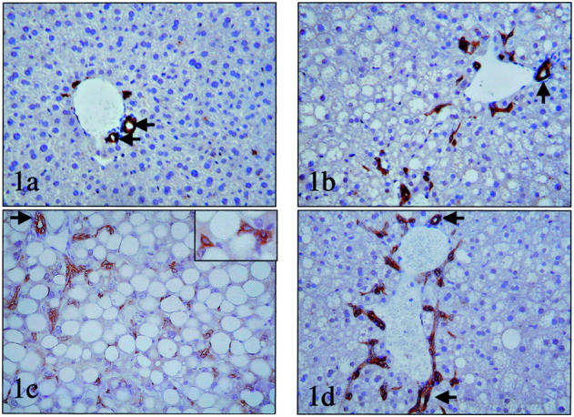
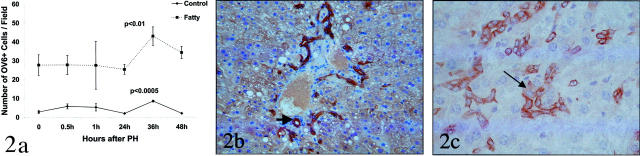
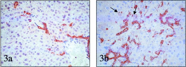
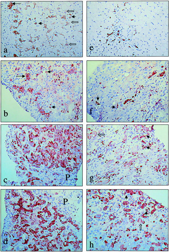
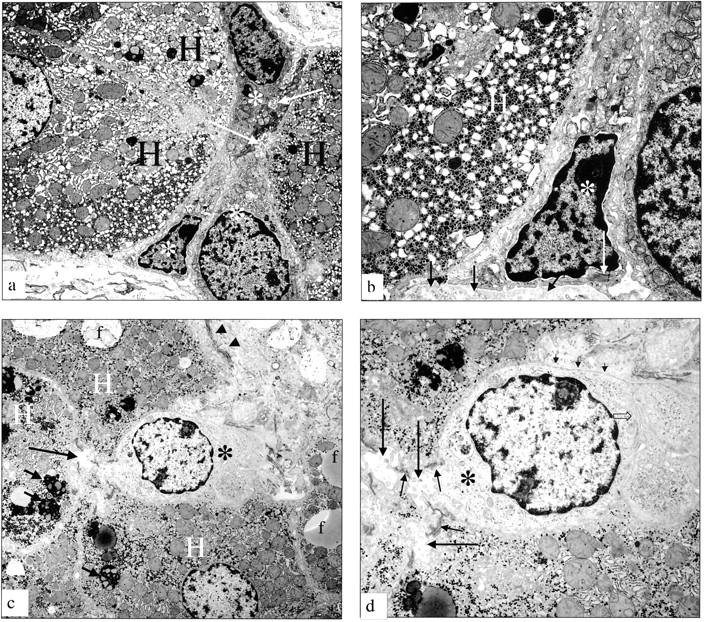
In animals, the combination of oxidative liver damage and inhibited hepatocyte proliferation increases the numbers of hepatic progenitors (oval cells). We studied different murine models of fatty liver disease and patients with nonalcoholic fatty liver disease or alcoholic liver disease to determine whether oval cells increase in fatty livers and to clarify the mechanisms for this response. To varying degrees, all mouse models exhibit excessive hepatic mitochondrial production of H(2)O(2), a known inducer of cell-cycle inhibitors. In mice with the greatest H(2)O(2) production, mature hepatocyte proliferation is inhibited most, and the greatest number of oval cells accumulates. These cells differentiate into intermediate hepatocyte-like cells after a regenerative challenge. Hepatic oval cells are also increased significantly in patients with nonalcoholic fatty liver disease and alcoholic liver disease. In humans, fibrosis stage and oval cell numbers, as well as the number of intermediate hepatocyte-like cells, are strongly correlated. However, cirrhosis is not required for oval cell accumulation in either species. Rather, as in mice, progenitor cell activation in human fatty liver diseases is associated with inhibited replication of mature hepatocytes. The activation of progenitor cells during fatty liver disease may increase the risk for hepatocellular cancer, similar to that observed in the Solt-Farber model of hepatocarcinogenesis in rats.


Figures

Figure 1.
Figure 1.
Oval cell accumulation in mice with fatty livers. Immunohistochemistry for bile duct-type cytokeratins was used to demonstrate oval cells in liver sections from healthy control mice (OV6 immunostain) (a) and mice with fatty livers caused by chronic EtOH consumption (OV6 immunostain) (b), feeding MCD diets (polyclonal cytokeratin stain). Inset shows detail of oval cells (c) or genetic obesity (ob/ob)(polyclonal cytokeratin stain) (d). Interlobular bile ducts (small arrow) were excluded from the countings, while the other immunoreactive cells were considered ductules/putative liver progenitor cells. Original magnifications: ×200; ×400 (inset in c).
Figure 2.
Figure 2.
Accumulation of putative liver progenitor cells in liver remnants after PH. a: Immunohistochemistry was used to demonstrate OV6 (+) cells in liver sections from two groups of healthy control mice (lean littermates of ob/ob mice, n = 12; wild-type mice pair-fed so that caloric intake matched EtOH-fed mice, n = 12) and two groups of mice with fatty livers (ob/ob mice, n = 12; EtOH-fed mice, n = 12) at various time points after PH. Because results in both control groups were similar, data are pooled and shown as the control line on this graph (n = 4 mice/time point). Similarly, there were no significant differences between ob/ob mice and EtOH-fed mice, so these results are pooled and results are shown as the fatty line on this graph (n = 4 mice/time point). *, P < 0.0005 versus control at time 0; #, P < 0.01 for fatty at time 0. b: Photomicrograph of Ov-6 (+) cells in a representative mouse with fatty liver at 36 hours after PH. In addition to cords of small oval cells, numerous intermediate hepatocyte-like cells showing a submembranous staining pattern for Ov-6 can be seen. c: High-magnification photomicrograph of CK7 (+) cells in another representative mouse with fatty liver at 48 hours after PH. Note the appearance of a small hepatocyte-like cell (arrow). This cell is immediately adjacent to cords of small oval cells. Cytokeratin 7 stain; original magnification, ×400 (c).
Figure 3.
Figure 3.
Accumulation of putative liver progenitor cells after treatment with the tumor-promoting drug, ethionine. a: Oval cells in the liver of a representative lean mouse fed one-half MCD diet plus ethionine. b: Oval cells in the liver of a representative ob/ob mouse fed control chow plus ethionine. Note intermediate hepatocyte-like cells (arrows). mOV-6 staining; original magnifications, ×200.
Figure 4.
Figure 4.
Accumulation of putative liver progenitor cells in patients with NAFLD and AFLD. Representative photomicrographs from human samples with ALD (a–d) and NAFLD (e–f). a: Paraffin section of a cirrhotic stage of ALD (Brunt stage 4) immunostained for cytokeratin 7, showing single small oval cells (small arrows), extending far into the parenchyma. Intermediate hepatocyte-like cells (open arrows) are present, often in continuity with oval cells. These cells are numerous in the cirrhotic stage of ALD, suggesting more differentiation toward hepatocytes in this stage of the disease. b: Frozen section of ALD Brunt stage 4 (cirrhotic liver) immunostained for cytokeratin 19, showing reactive ductules (large arrow) and oval cells (small arrows). Intermediate hepatocyte-like cells are not reactive for cytokeratin 19. c and d: Frozen section of ALD Brunt stage 4 (cirrhotic liver) immunostained for Ov-6 (c) and chromogranin-A (d), showing a strikingly high number of intermediate hepatocyte-like cells, suggesting a high degree of differentiation of oval cells toward hepatocytes in the cirrhotic stage of the disease. Cytokeratin 7, OV6, and chromogranin-A are immunoreactive in intermediate cells, while cytokeratin 19 is not or hardly; such as we have previously shown, cytokeratin 19 is a marker that is lost early during differentiation from oval cells toward hepatocytes. P, portal tract. e: Paraffin section of NAFLD Brunt stage 2, immunostained for cytokeratin 7, showing the putative progenitor cells, extending into the steatotic parenchyma. In this stage, no intermediate hepatocyte-like cells are seen. f: Frozen section of NAFLD Brunt stage 3, immunostained for cytokeratin 19, showing single progenitor cells (small arrows) and reactive ductules. g: Frozen section of NAFLD Brunt stage 3, immunostained for Ov-6. Oval cells (small arrows) and intermediate hepatocyte-like cells (open arrow) are present. Note that intermediate hepatocytes are less numerous compared to the cirrhotic stage of ALD (shown in c). h: Frozen section of NAFLD Brunt stage 3, immunostained for chromogranin-A, showing putative progenitor cells (small arrows) and intermediate hepatocyte-like cells (open arrow), which are less numerous compared to the cirrhotic stage of ALD (shown in d). Original magnifications, ×160.
Figure 5.
Figure 5.
Electron micrographs of putative progenitor cells in patients with FLD. a: Electron micrograph of a liver biopsy from a patient with ALD (active cirrhosis). A group of small epithelial hepatic progenitor cells (white asterisk) are arranged between hepatocytes (H). These cells range from very immature (a, bottom) to somewhat differentiating toward the hepatocyte lineage. The small cells form a hemicanaliculus (arrow) with the neighboring hepatocytes, showing that the differentiation direction is toward hepatocytes. b: Electron micrograph of a liver biopsy from a patient with ALD (active cirrhosis). Higher magnification of the most immature hepatic progenitor cell (asterisk) shown in a. H, adjacent hepatocyte. Bundles of tonofilaments (long white arrow) are seen in the cytoplasm; these tonofilaments distinguish these cells from nonepithelial cells such as endothelial cells or Kupffer cells; the cell rests on a basement membrane (short arrows). c: Electron micrograph of a liver from a patient with NAFLD. A small epithelial hepatic progenitor cell (asterisk) is wedged in between hepatocytes (H). Note fat droplets (f) and lipofuscin granules (small arrows). Collagen bundles are present in Disse’s space (arrowheads). d: Electron micrograph of a liver from a patient with NAFLD showing a higher magnification of the same progenitor cell (asterisk) from c. A tortuous bile canaliculus (large arrow) sealed by junctional complexes of the desmosomal type, typical for epithelial cells (small arrows) is seen between two hepatocytes (H) and the progenitor cell (asterisk). Note the basement membrane (short arrows) and villous interdigitations (open arrow) with neighboring progenitor cell, which are features of the biliary lineage. Original magnifications: ×7150 (a, c); ×18,400 (b); ×14,500 (d).

References

    1. Thorgeirsson SS, Factor VM, Snyderwine EG: Transgenic mouse models in carcinogenesis research and testing. Toxicol Lett 2000, 112–113:553-555 - PubMed
    1. Evarts RP, Nagy P, Marsden E, Thorgeirsson SS: A precursor-product relationship exists between oval cells and hepatocytes in rat liver. Carcinogenesis 1987, 8:1737-1740 - PubMed
    1. Sell S, Dunsford HA: Evidence for the stem cell origin of hepatocellular carcinoma and cholangiocarcinoma. Am J Pathol 1989, 134:1347-1363 - PMC - PubMed
    1. Libbrecht L, Meerman L, Kuipers F, Roskams T, Desmet V, Jansen P: Liver pathology and hepatocarcinogenesis in a long-term mouse model of erythropoietic protoporphyria. J Pathol 2003, 199:191-200 - PubMed
    1. Hsia CC, Evarts RP, Nakatsukasa H, Marsden ER, Thorgeirsson SS: Occurrence of oval-type cells in hepatitis B virus-associated human hepatocarcinogenesis. Hepatology 1992, 16:1327-1333 - PubMed

Publication types