. 2003 Oct;163(4):1457-65.
doi: 10.1016/S0002-9440(10)63503-1.

Nontransgenic hyperexpression of a complement regulator in donor kidney modulates transplant ischemia/reperfusion damage, acute rejection, and chronic nephropathy

Affiliations

Nontransgenic hyperexpression of a complement regulator in donor kidney modulates transplant ischemia/reperfusion damage, acute rejection, and chronic nephropathy

Julian R Pratt et al. Am J Pathol. 2003 Oct.

Abstract

Complement activation during ischemia and reperfusion contributes to the development of tissue injury with severe negative impact on outcomes in transplantation. To counter the effect of complement, we present a strategy to deliver a novel complement regulator stabilized on cell surfaces within donor organs. The membrane-bound complement regulator is able to inhibit complement activation when the donor organ is revascularized and exposed to host-circulating complement. Application of this construct to donor kidneys protected transplanted tissues from ischemia/reperfusion injury and reduced the deposition of activated complement and histological signs of damage under conditions in which a nontargeted control construct was ineffective. Treatment of donor organs in this way improved graft performance in the short and long term. An analysis of the immune response in allograft recipients showed that reducing graft damage at the time of transplantation through complement regulation also modulated the alloresponse. Additionally, the results of perfusion studies with human kidneys demonstrated the feasibility of targeting endothelial and epithelial surfaces with this construct, to allow investigation in clinical transplantation.


Figures

Figure 1.
Figure 1.
Comparative structure of human CR1 and m-pSCR1-3. a: Human CR1 exists on the cell surface as a string of short consensus repeat (SCR) domains attached via a transmembrane portion to a short cytoplasmic domain, each SCR being of 60 to 70 amino acids. Several allotypes exist, differing in the number of SCRs. The most common form, allotype A, consists of 30 SCRs and exhibits a further level of internal homology in the form of long homologous repeats (LHRs) of every seven SCRs totaling 235 kd. b: m-pSCR1-3 consists of recombinant SCR(1-3) of LHR-A of CR1 attached to the membrane-binding tag. The tag consists of a myristoyl tail chemically linked to a synthetic positively charged peptide whose terminal cysteine enables attachment of the SCR(1-3) of CR1. This water-soluble molecule is ∼23 kd in size and retains the known complement regulatory mechanisms of intact CR1 as well as the ability to bind cells. The construct is designed to insert itself into the hydrophobic core of the lipid bilayer of cell membranes and binding is stabilized by the basic peptide region allowing presentation of the functional moiety, in this instance human SCR(1-3) of CR1.
Figure 2.
Figure 2.
Membrane binding of m-pSCR(1-3). a: m-pSCR1-3 was detected using Cy3-labeled anti-SCR(1-3) mAb (3E10) on cultured transformed rat renal endothelial cells exposed to 2.5 μg/ml of m-pSCR1-3. The dome of the cell is outside the focal plain of the photomicrograph giving the appearance of a central dark area. b: Staining was not present on endothelial cells incubated with SCR1-3 lacking the m-p tail. c: Dose-dependent cell-surface binding to rat endothelial cells in vitro detected by FACS in the presence of the membrane-binding tag. d: No binding occurred at any concentration of SCR(1-3). Original magnification, ×1000 (a).
Figure 3.
Figure 3.
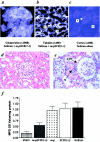
Immunohistopathology of perfused donor kidneys Isolated rat kidneys were flushed with 5 ml of kidney perfusion solution containing 200 μg of m-pSCR(1-3). The tissue distribution of m-pSCR1-3 is shown after immunofluorescent staining with Cy3-labeled 3E10. a: Glomerulus with capillary loops stained for m-pSCR(1-3). b: Basolateral surfaces of tubules (T) and peritubular capillary wall (C) outlined by the staining for m-pSCR(1-3). c: The cortex of a control kidney perfused with carrier solution alone and stained with Cy3-labeled 3E10. Glomerulus (G) and tubules (T) are negative. H&E staining of illustrative donor kidney sections 24 hours after transplantation showed well-preserved architecture if perfused with m-pSCR1-3 (d) whereas carrier-only perfused kidneys (e) showed marked tubular flattening (ATN) and some infiltration of neutrophils (N). Tissue myeloperoxidase activity 24 hours after transplantation (f) showed significantly reduced MPO activity in m-pSCR1-3-treated grafts ▒ compared with control-treated grafts perfused only with carrier solution ▪ (P = 0.03). MPO levels in SCR(1-3) □- and m-p ░⃞-treated grafts were not significantly different from controls. Sham-treated animals ▩ had a laparotomy 24 hours before MPO measurement (n = 4/group).
Figure 4.
Figure 4.
Membrane attack complex formation at day 1 after transplantation. Immunohistochemistry for C5b-9 membrane attack complex showing a normal, sham-operated kidney (a), a graft treated with perfusion solution alone (b), effect of treatment with perfusion solution containing untagged SCR(1-3) (c), effect of treatment with the m-p tag alone (d), and effect of treatment with m-pSCR(1-3) (e). Control-treated grafts undergoing ischemia and reperfusion exhibited widespread tubular deposition of C5b-9. Grafts protected by m-pSCR1-3 showed markedly reduced deposition of C5b-9. Original magnifications, ×1000.
Figure 5.
Figure 5.
Functional and histological analysis of renal isografts. a: Blood urea nitrogen up to 7 days after transplantation in recipients of grafts treated with m-pSCR1-3 —▪— , perfusion solution only ◆̶, SCR(1-3) lacking the m-p tail —▴— , or the m-p tag alone –▾– or in sham-operated rats undergoing nephrectomy without transplantation …•…. Data are shown from day 3 because this was the point from which renal function was dependent on the transplanted kidney alone. Treatment with m-pSCR1-3 led to significantly improved renal function compared to any one of the control groups (P < 0.001; n = 6 animals/group). b: Blood urea nitrogen throughout 20 weeks after transplantation after perfusion of donor graft with perfusion solution alone -▴-, or m-pSCR1-3 –•– . Protection conferred by m-pSCR1-3 in the first week led to improved function throughout 20 weeks (P < 0.001; n = 6 m-pSCR1-3-treated grafts and n = 5 perfusion solution-treated controls). H&E staining of illustrative donor kidney sections showed well-preserved tubular structure in m-pSCR1-3-treated grafts at 20 weeks (c) compared to carrier-treated controls (d). Vessels also had a near normal appearance in the m-pSCR(1-3)-treated grafts (e), compared to carrier-treated grafts at 20 weeks in which arteriolar intimal proliferation was present (f). No elastic duplication was seen in untreated (g) or treated grafts (h). Original magnifications: ×400 (c, d); ×1000 (e, f).
Figure 6.
Figure 6.
Effects of m-pSCR1-3 on renal allografts. Histological analysis of F344 to Lewis renal allografts at day 14 after transplant showed signs of cellular infiltration in all transplants. However, the degree of interstitial infiltration of mononuclear cells and evidence for tubulitis was less in m-pSCR1-3-treated grafts (a and b) compared to control transplants treated with perfusion solution only (c and d; n = 8/group). The m-pSCR1-3-treated grafts also showed significantly improved renal function compared to perfusion solution-treated controls (e) between days 4 and 7 (P < 0.0001), which was 24 hours after removal of the remaining native kidney. BUN levels <10 mg/dL were taken to be within normal range (for animals with two functioning kidneys). Morphometric analysis of antibody-stained frozen sections (f) showed reduced CD45 and CD3 staining in m-pSCR1-3-treated kidneys at day 14 after transplantation, compared to controls treated with perfusion solution only. Data are expressed in arbitrary units (pixels) and derived from 15 representative cortical fields from three transplanted kidneys in each group. Proliferation of spleen T cells isolated from transplant recipients at day 14 after transplantation (g) showed significantly reduced alloresponse in the recipients of m-pSCR1-3-treated kidneys compared to recipients of control-treated kidneys, analyzed throughout 7 days by MTS assay (n = 4 animals/group assayed in triplicate, in comparing the treatment group with either control group, P < 0.01). Original magnifications, ×250 (a–d).
Figure 7.
Figure 7.
Binding of m-pSCR1-3 to human kidney. Immunohistochemistry of human kidneys perfused with preservation solution containing m-pSCR1-3 showed (a and b) that the agent bound to glomeruli (G) and to tubules (Tu). c: The high-power view reveals a pattern of m-pSCR1-3 staining consistent with binding to the basolateral tubule surface (B). d: Human kidney perfused without m-pSCR1-3 showed no staining. Original magnifications: ×1000 (a, b, d); ×2000 (c).

References

    1. Baldwin WM, Pruitt SK, Brauer RB, Daha MR, Sanfillipo F: Complement in organ transplantation: contributions to inflammation, injury and rejection. Transplantation 1995, 59:797-802 - PubMed
    1. Weisman HF, Bartow T, Leppo MK, Marsh HC, Jr, Carson GR, Concino MF, Boyle MP, Roux KH, Weisfeldt ML, Fearon DT: Soluble human complement receptor type 1: in vivo inhibitor of complement suppressing post-ischemic myocardial inflammation and necrosis. Science 1990, 249:146-151 - PubMed
    1. Hill J, Lindsay TF, Ortiz F, Yeh CG, Hechtman HB, Moore FD, Jr: Soluble complement receptor type 1 ameliorates the local and remote organ injury after intestinal ischemia-reperfusion in the rat. J Immunol 1992, 149:1723-1728 - PubMed
    1. Mulligan MS, Yeh CG, Rudolph AR, Ward PA: Protective effects of soluble CR1 in complement and neutrophil-mediated tissue injury. J Immunol 1992, 148:1479-1486 - PubMed
    1. Passwell J, Schreiner GF, Nonaka M, Beuscher HU, Colten HR: Local extrahepatic expression of complement genes C3, factor B, C2, and C4 is increased in murine lupus nephritis. J Clin Invest 1988, 82:1676-1684 - PMC - PubMed

Publication types

MeSH terms