. 2003 Oct;163(4):1587-98.
doi: 10.1016/S0002-9440(10)63515-8.

Carbon monoxide inhalation protects rat intestinal grafts from ischemia/reperfusion injury

Affiliations

Carbon monoxide inhalation protects rat intestinal grafts from ischemia/reperfusion injury

Atsunori Nakao et al. Am J Pathol. 2003 Oct.

Abstract

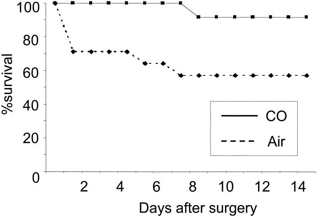
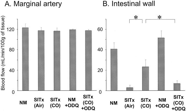
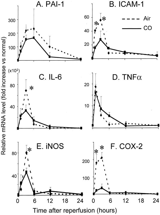
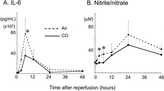
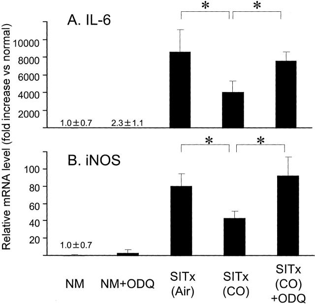
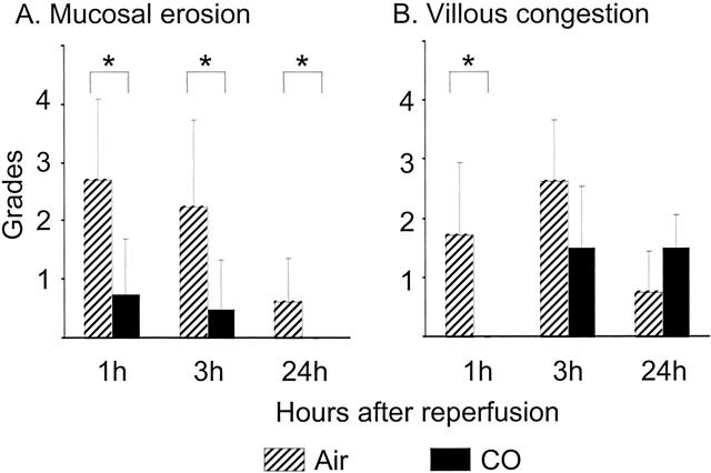
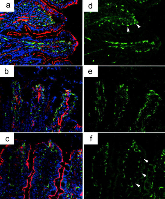
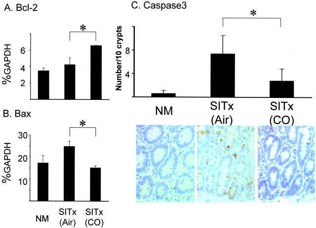
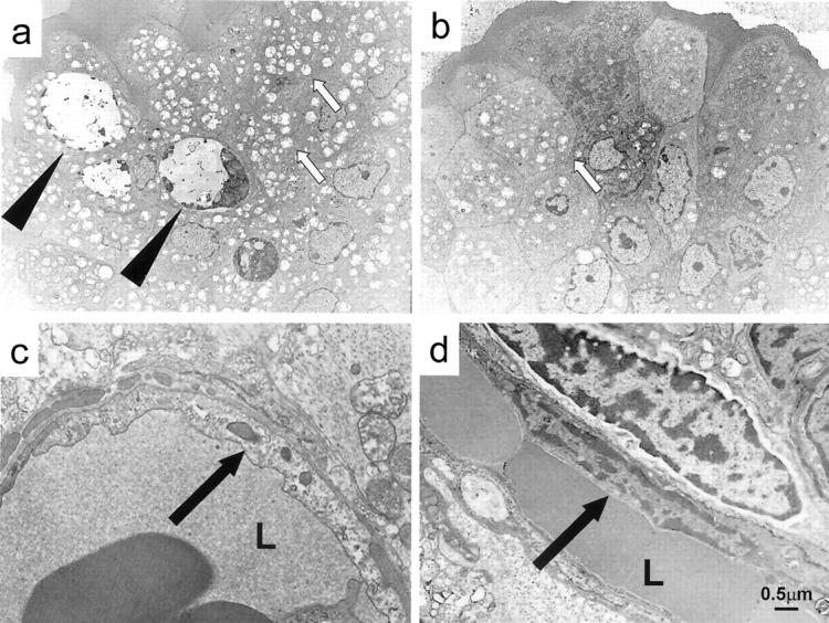
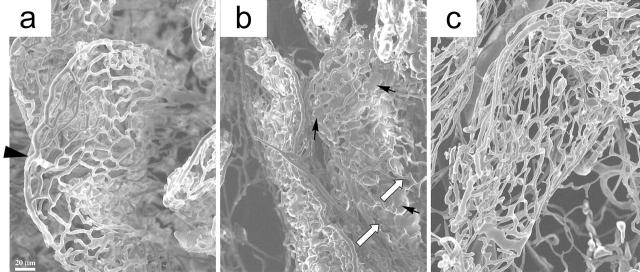
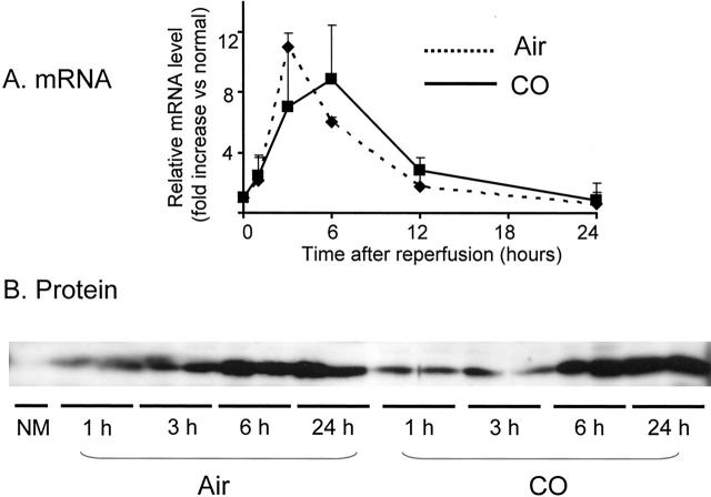
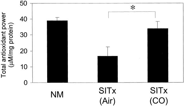
Carbon monoxide (CO), a byproduct of heme catalysis by heme oxygenases, has been shown to exert anti-inflammatory effects. This study examines the cytoprotective efficacy of inhaled CO during intestinal cold ischemia/reperfusion injury associated with small intestinal transplantation. Orthotopic syngenic intestinal transplantation was performed in Lewis rats after 6 hours of cold preservation in University of Wisconsin solution. Three groups were examined: normal untreated controls, control intestinal transplant recipients kept in room air, and recipients exposed to CO (250 ppm) for 1 hour before and 24 hours after surgery. In air grafts, mRNA levels for interleukin-6, cyclooxygenase-2, intracellular adhesion molecule (ICAM-1), and inducible nitric oxide synthase rapidly increased after intestinal transplant. Histopathological analysis revealed severe mucosal erosion, villous congestion, and inflammatory infiltrates. CO effectively blocked an early up-regulation of these mediators, showed less severe histopathological changes, and resulted in significantly improved animal survival of 92% from 58% in air-treated controls. CO also significantly reduced mRNA for proapoptotic Bax, while it up-regulated anti-apoptotic Bcl-2. These changes in CO-treated grafts correlated with well-preserved CD31(+) vascular endothelial cells, less frequent apoptosis/necrosis in intestinal epithelial and capillary endothelial cells, and improved graft tissue blood circulation. Protective effects of CO in this study were mediated via soluble guanylyl cyclase, because 1H-(1,2,4)oxadiazole (4,3-alpha) quinoxaline-1-one (soluble guanylyl cyclase inhibitor) completely reversed the beneficial effect conferred by CO. Perioperative CO inhalation at a low concentration resulted in protection against ischemia/reperfusion injury to intestinal grafts with prolonged cold preservation.


Figures

Figure 1.
Figure 1.
Effect of CO on animal survival after 6 hours of cold preservation of intestinal grafts. Fourteen-day survival of air-treated recipients (58.3%, 7 of 12) significantly (P < 0.05) improved with CO to 91.7% (11 of 12).
Figure 2.
Figure 2.
Laser Doppler flowmeter measurements of graft blood flows of marginal artery (A) and intestinal wall (B). A: Blood flows of intestinal marginal artery were similar in all groups. B: CO inhalation significantly increased intestinal graft blood flow to 23.2 ± 7.0 ml/minute/100 g compared to <4 ml/minute/100 g in air-treated grafts. Intestinal microcirculation of normal and ODQ-treated unoperated animals was ∼40 ml/minute/100 g. ODQ (20 m/kg) administration to recipient 30 minutes before CO treatment completely abrogated CO effect and intestinal blood flow was 6 ml/minute/100 g. n = 3 to 4 for each group. *, P < 0.05. NM, normal.
Figure 3.
Figure 3.
mRNA expression in intestinal grafts. A: mRNA for PAI-1 was up-regulated during I/R injury in air-treated controls with a peak at 6 hours. CO treatment tended toward a reduction in the expression of PAI-1 but statistical significance was not achieved. Cytokine and inflammatory mediator mRNA expressions of ICAM-1 (B), IL-6 (C), TNF-α (D), iNOS (E), and COX-2 (F) were evaluated in CO versus air-treated grafts. These mediators, except for TNF-α, were significantly reduced with CO inhalation. Of note, COX-2 was reduced with CO to <30% of control. n = 4 to 6 for each group. *, P < 0.05.
Figure 4.
Figure 4.
Analysis of serum IL-6 (A) and nitrate/nitrite (B). A: Serum IL-6 rapidly elevated and peaked at 6 hours in air-treated controls. Similarly to mRNA expression, CO treatment significantly decreased serum IL-6. B: Serum nitrate/nitrite gradually increased for 24 hours during I/R injury in air-treated animals, which was reduced by CO with statistical significance (P < 0.05) at 3 and 6 hours.
Figure 5.
Figure 5.
ODQ reverses CO-induced anti-inflammatory effects. Reduction of mRNA for IL-6 and iNOS by CO treatment was abrogated in animals treated with ODQ. ODQ administration to CO-treated animals restored IL-6 and iNOS mRNA expression comparable to air control grafts 3 hours after reperfusion. ODQ did not alter mRNA expression of these mediators in unoperated normal animals. n = 3 for each group. *, P < 0.05. NM, normal.
Figure 6.
Figure 6.
Severity of mucosal injury assessed by routine histopathology. Intestinal cold I/R injury associated with intestinal epithelial cell loss and mucosal erosion (A), and villous congestion observed in air-treated control grafts (B). CO-treated grafts showed significantly less severe injury. n = 6 for each group. *, P < 0.05.
Figure 7.
Figure 7.
CD31 expression of vascular endothelial cells. a and d: Immunofluorescent stain of CD31 revealed an abundant expression of CD31 on the vascular endothelial cells in normal intestinal lamina propria. F-actin, which indicates intestinal epithelial microvilli, is also well visualized on the entire villi of normal intestine. b and e: CD31 expression was faint and interrupted in air-treated intestinal grafts after cold I/R injury, and F-actin stain was either disrupted in the upper half of the villi or weak in the remaining villi (air-treated grafts, 1 hour after reperfusion). c and f: CD31-positive vascular endothelial cells were preserved in the lamina propria of CO-treated grafts with F-action stain revealing normal intestinal microvilli preservation (CO-treated graft, 1 hour after SITx). Green, CD31; red, F-actin; blue, nuclei. Original magnifications, ×400.
Figure 8.
Figure 8.
Apoptosis-related molecules in intestinal grafts. A: There is a twofold increase of anti-apoptotic Bcl-2 in CO-treated grafts compared to air-treated controls at 1 hour after reperfusion. B: Bax, a proapoptotic gene, was significantly up-regulated 1 hour in air control (P < 0.05), which was not observed in CO-treated grafts. C: Numbers of activated caspase 3-positive cells increased in crypt epithelial cells of air control. CO significantly (P < 0.05) reduced caspase 3-stained apoptotic crypt epithelial cells. NM, normal intestine.
Figure 9.
Figure 9.
Transmission electron microscopy of intestinal grafts. a: Numerous vacuolization (white arrows) and apoptotic bodies (black arrows) are present in epithelial cells of air exposed control grafts. b: CO-treated grafts contain minimum apoptotic bodies and considerably less vacuolization. c: In the air-exposed controls, vascular endothelial cells in the lamina propria showed karyolitic nuclei with disorganized internal architecture (arrow). d: Endothelial cells in the CO-treated grafts were well maintained and demonstrated a normal intracellular architecture (arrow). a–d: 1 hour after reperfusion. L, lumen of blood vessels. Scale bar, 0.5 μm. Original magnifications: ×1500 (a, b); ×5000 (c, d).
Figure 10.
Figure 10.
Scanning electron microscopy of graft vascular cast. a: Normal intestine showed typical hollow arrangement of vessels that comprise the villi vasculature (arrowhead, afferent artery reaching the apex of the villous and giving rise to the villous capillary). b: Severe leakage (collection of methyl methacrylate, black arrow) with interruption of vascular cast (white arrow) was evident in the internal sections of the villi (air-treated, 1 hour). c: Nearly normal villous vasculature was obtained without leakage (CO-treated, 1 hour). Scale bar, 20 μm.
Figure 11.
Figure 11.
HO-1 mRNA (A) and protein (B) levels during intestinal I/R injury with and without CO inhalation. A: The real-time PCR for HO-1 showed an up-regulation of HO-1 mRNA expression in air- and CO-treated grafts with peaks at 3 and 6 hours after transplantation. B: Western blot showed a gradual increase of HO-1 protein expression 1 hour after reperfusion. CO treatment did not alter HO-1 protein expression. NM, normal.
Figure 12.
Figure 12.
Antioxidant power in intestinal grafts. Intestinal I/R injury resulted in a significant reduction of antioxidant power in the air-treated control. In contrast, the CO-treated group showed preserved antioxidant power. *, p < 0.05.

References

    1. Abu-Elmagd K, Reyes J, Bond G, Mazariegos G, Wu T, Murase N, Sindhi R, Martin D, Colangelo J, Zak M, Janson D, Ezzelarab M, Dvorchik I, Parizhskaya M, Deutsch M, Demetris A, Fung J, Starzl TE: Clinical intestinal transplantation: a decade of experience at a single center. Ann Surg 2001, 234:404-416 - PMC - PubMed
    1. Gores GJ, Nieminen AL, Fleishman KE, Dawson TL, Herman B, Lemasters JJ: Extracellular acidosis delays onset of cell death in ATP-depleted hepatocytes. Am J Physiol 1988, 255:C315-C322 - PubMed
    1. Buderus S, Siegmund B, Spahr R, Krutzfeldt A, Piper HM: Resistance of endothelial cells to anoxia-reoxygenation in isolated guinea pig hearts. Am J Physiol 1989, 257:H488-H493 - PubMed
    1. Freeman BA, Crapo JD: Biology of disease: free radicals and tissue injury. Lab Invest 1982, 47:412-426 - PubMed
    1. Clavien PA, Harvey PR, Strasberg SM: Preservation and reperfusion injuries in liver allografts. An overview and synthesis of current studies. Transplantation 1992, 53:957-978 - PubMed

Publication types

MeSH terms