. 2003 Oct;163(4):1663-75.
doi: 10.1016/S0002-9440(10)63523-7.

Proteasome inhibitor (MG-132) treatment of mdx mice rescues the expression and membrane localization of dystrophin and dystrophin-associated proteins

Affiliations

Proteasome inhibitor (MG-132) treatment of mdx mice rescues the expression and membrane localization of dystrophin and dystrophin-associated proteins

Gloria Bonuccelli et al. Am J Pathol. 2003 Oct.

Abstract

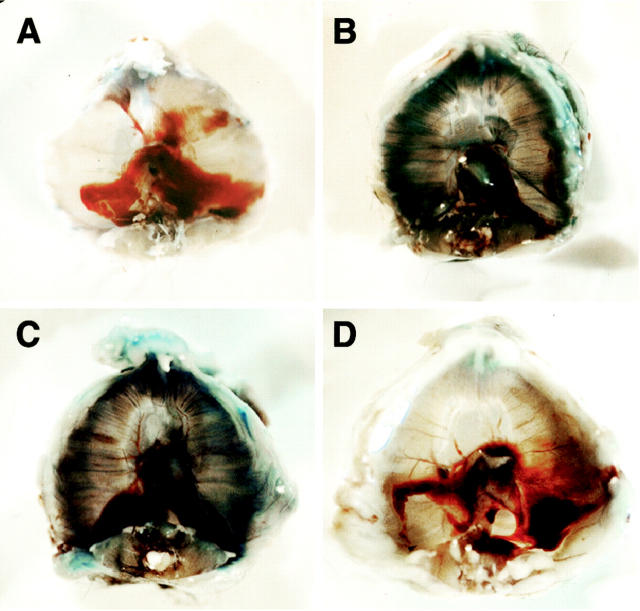
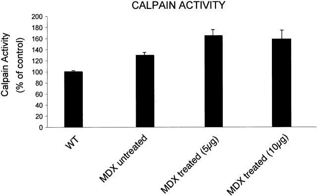
Dystrophin, the protein product of the Duchenne muscular dystrophy (DMD) gene, is absent in the skeletal muscle of DMD patients and mdx mice. At the plasma membrane of skeletal muscle fibers, dystrophin associates with a multimeric protein complex, termed the dystrophin-glycoprotein complex (DGC). Protein members of this complex are normally absent or greatly reduced in dystrophin-deficient skeletal muscle fibers, and are thought to undergo degradation through an unknown pathway. As such, we reasoned that inhibition of the proteasomal degradation pathway might rescue the expression and subcellular localization of dystrophin-associated proteins. To test this hypothesis, we treated mdx mice with the well-characterized proteasomal inhibitor MG-132. First, we locally injected MG-132 into the gastrocnemius muscle, and observed the outcome after 24 hours. Next, we performed systemic treatment using an osmotic pump that allowed us to deliver different concentrations of the proteasomal inhibitor, over an 8-day period. By immunofluorescence and Western blot analysis, we show that administration of the proteasomal inhibitor MG-132 effectively rescues the expression levels and plasma membrane localization of dystrophin, beta-dystroglycan, alpha-dystroglycan, and alpha-sarcoglycan in skeletal muscle fibers from mdx mice. Furthermore, we show that systemic treatment with the proteasomal inhibitor 1) reduces muscle membrane damage, as revealed by vital staining (with Evans blue dye) of the diaphragm and gastrocnemius muscle isolated from treated mdx mice, and 2) ameliorates the histopathological signs of muscular dystrophy, as judged by hematoxylin and eosin staining of muscle biopsies taken from treated mdx mice. Thus, the current study opens new and important avenues in our understanding of the pathogenesis of DMD. Most importantly, these new findings may have clinical implications for the pharmacological treatment of patients with DMD.


Figures

Figure 1.
Figure 1.
Localized treatment of mdx mice with MG-132: Immunohistochemistry. The right hindlimbs of mdx mice were locally injected with MG-132 (at a final concentration of 20 μmol/L). After 24 hours, the mice were sacrificed, and the gastrocnemius muscle was isolated. The left hindlimb of each mouse served as internal control. Frozen sections prepared from untreated and MG-132-treated mdx gastrocnemius muscles were immunostained with antibodies directed against β-dystroglycan (A), α-dystroglycan (B), α-sarcoglycan (C), dystrophin (D), and nNOS (E). Then, samples were observed under an Olympus IX 70 inverted microscope. β-dystroglycan, α-dystroglycan, α-sarcoglycan, dystrophin, and nNOS were absent or extremely reduced at the sarcolemma of skeletal muscle fibers from untreated mdx mice (upper panels). On the contrary, β-dystroglycan, α-dystroglycan, α-sarcoglycan, and dystrophin appear highly expressed at the plasma membranes of the muscle fibers from MG-132-treated mdx mice. In certain fibers, it is possible to appreciate some cytoplasmic staining. However, nNOS expression levels are not increased in MG-132-treated muscles, as compared to untreated controls. Importantly, the pairs of images were acquired at the same exposure time.
Figure 2.
Figure 2.
Localized treatment of mdx mice with MG-132: Western blot analysis. The right hindlimbs of mdx mice were locally injected with MG-132 (at a final concentration 20 μmol/L). After 24 hours, the mice were sacrificed, and gastrocnemius muscle was isolated. The left hindlimb of each mouse served as internal control. Protein lysates were prepared from untreated and MG-132-treated mdx skeletal muscles. Equal amounts of total protein were separated by SDS-PAGE and transferred to nitrocellulose. Then, the blots were incubated with specific antibodies directed against β-dystroglycan, α-dystroglycan, α-sarcoglycan, dystrophin, and nNOS. Note that proteasomal inhibitor treatment induces an increase of the expression levels of β-dystroglycan, α-dystroglycan, α-sarcoglycan, and dystrophin in skeletal muscles from mdx mice. Interestingly, MG-132 treatment causes the accumulation of the precursor form of α-dystroglycan in skeletal muscle. Consistent with the results obtained from immunofluorescence analysis, nNOS expression levels are only moderately increased in MG-132-treated skeletal muscles, as compared with untreated controls.
Figure 3.
Figure 3.
EBD staining of the diaphragm in mdx mice after systemic treatment with MG-132. To systemically administrate MG-132, we subcutaneously implanted osmotic pumps in 6-month-old mdx mice. Using this approach, we were able to deliver either MG-132 at a constant rate (either 1 μg, 5 μg, or 10 μg/kg/24 hours), over an 8-day period. As a negative control, 6-month-old mdx mice were implanted with osmotic pumps containing inhibitor-diluent (PBS). To evaluate muscle fiber damage, 20 hours before the end of treatment we injected EBD intraperitoneally into wild-type mice, untreated (PBS only) mdx mice and MG-132-treated mdx mice. EBD is used as marker of damaged fibers. After isolation, the diaphragms were rinsed in PBS, fixed in 10% formalin, and evaluated macroscopically. No evident uptake of the blue tracer was detected in fibers of the diaphragm from wild-type mice (A), indicating that the muscle fibers are not damaged, as expected. In contrast, many blue fibers were detected in the diaphragms of the untreated mdx mice (B), and from MG-132-treated mdx mice (delivered at a rate of 5 μg/kg/24 hours) (C). However, the diaphragms from MG-132-treated mdx mice (delivered at a rate of 10 μg/kg/24 hours) exhibit little or no blue fibers, suggesting that systemic administration of the proteasomal inhibitor can effectively improve the integrity of the skeletal muscle fiber plasma membrane in mdx mice (D).
Figure 4.
Figure 4.
EBD staining of mdx gastrocnemius muscle biopsies after systemic treatment with MG-132. We systemically administered the proteasomal inhibitor in 6-month-old mdx mice, by subcutaneous implantation of osmotic pumps. Using this approach, we were able to deliver MG-132 at constant rate (either 1 μg, or 5 μg or 10 μg/kg/24 hours) or inhibitor diluent (PBS) for a period of 8 days. To evaluate muscle fiber damage, 20 hours before the end of the treatment, we intraperitoneally injected EBD into wild-type mice, untreated (PBS only) mdx mice and MG-132-treated mdx mice. EBD staining is a marker of damaged muscle fibers. As EBD emits red autofluorescence, we examined frozen sections of gastrocnemius muscle biopsies taken from wild-type mice, untreated (PBS only) mdx mice and MG-132-treated mdx mice by fluorescence microscopy. A: Strong red autofluorescent signal is present in myofibers from untreated mice. B: In contrast, myofibers from MG-132-treated mice (delivered at rate of 10 μg/kg/24 hours) did not show any red fluorescent signal; similar results were obtained with wild-type mice (not shown). These results suggest that MG-132 systemic treatment effectively ameliorates the skeletal muscle fiber damage.
Figure 5.
Figure 5.
Histological analysis of H&E-stained skeletal muscle fibers after systemic treatment with MG-132. Mdx mice were systemically treated with MG-132 (delivered at rate of 10 μg/kg/24) for 8 days. H&E staining was performed on frozen skeletal muscle tissue sections from untreated mdx mice and MG-132-treated mdx mice. Skeletal muscle biopsies from untreated mdx mice (PBS) show signs of myopathic changes, including variability in fiber diameter, lymphocytic infiltration, lipidic droplets, and muscle fiber regeneration with central nuclei. In contrast, analysis of skeletal muscle biopsies from MG-132-treated mdx mice revealed few centrally nucleated muscle fibers and no signs of lymphocytic invasion, suggesting that the inflammatory process was reduced, and that the histopathological appearance was greatly improved. A: Original magnification, ×20. B: Original magnification, ×40.
Figure 6.
Figure 6.
Systemic treatment of mdx mice with proteasomal inhibitor: immunohistochemistry. To systemically administer the proteasomal inhibitor, we subcutaneously implanted osmotic pumps in 6-month-old mdx mice. Using this approach, we were able to deliver MG-132 at a constant rate (either 1 μg, 5 μg, or 10 μg/kg/24 hours) or inhibitor-diluent (PBS) for a period of 8 days. Frozen sections of gastrocnemius muscles from untreated (PBS only) mdx mice and MG-132-treated mdx mice were subjected to immunofluorescence analysis with antibodies directed against β-dystroglycan (A), α-dystroglycan (B), α-sarcoglycan (C), dystrophin (D), nNOS (E), and Cav-3 (F). As expected, β-dystroglycan, α-dystroglycan, α-sarcoglycan, dystrophin, and nNOS were absent or greatly reduced in skeletal muscle from untreated mdx mice (PBS only), whereas Cav-3 was augmented in skeletal muscles from the same mice (upper panels). Surprisingly, the expression levels of β-dystroglycan, α-dystroglycan, α-sarcoglycan, and dystrophin were rescued in skeletal muscle from MG-132-treated mdx mice. However, after MG-132 systemic treatment, we did not detect any significant increases in the expression levels of nNOS and Cav-3 in skeletal muscles fibers. Interestingly, we achieved rescue of the expression levels of α-dystroglycan, α-sarcoglycan, and dystrophin by delivering MG-132 at the rate of 10 μg/kg/24 hours, whereas the rescue of β-dystroglycan was accomplished by delivering MG-132 at the rate of 1 μg/kg/24 hours. Importantly, pairs of images (treated versus untreated) were acquired with the same exposure time.
Figure 7.
Figure 7.
Systemic treatment of mdx mice with proteasomal inhibitor: Western blot analysis. To systemically administer the proteasomal inhibitor, we subcutaneously implanted osmotic pumps in 6-month-old mdx mice. Using this approach, we were able to deliver MG-132 at a constant rate (either 1 μg, 5 μg, or 10 μg/kg/24 hours) or inhibitor-diluent (PBS only) for a period of 8 days. Lysates of gastrocnemius muscle biopsies were prepared from untreated mdx mice and MG-132-treated mdx mice. Equal amounts of total protein were separated by SDS-PAGE and transferred onto nitrocellulose. The expression levels of β-dystroglycan, α-dystroglycan, α-sarcoglycan, dystrophin, nNOS, and Cav-3 were assessed by Western blot analysis, using specific antibodies. Interestingly, the expression levels of β-dystroglycan, α-dystroglycan, α-sarcoglycan, and dystrophin are increased in skeletal muscles from MG-132-treated mdx mice, as compared to their untreated (PBS only) counterparts. The rescue of β-dystroglycan was achieved by delivering MG-132 at the rate of 1 μg/kg/24 hours of MG-132, while the rescue of the expression levels of α-dystroglycan, α-sarcoglycan, and dystrophin was accomplished by delivering MG-132 at a rate of 10 μg/kg/24 hours. Note that the systemic treatment rescues the precursor form of the α-dystroglycan, consistent with the results we obtained with localized treatment using MG-132 (see Figure 2 ▶ ). Consistent with the results we obtained by immunofluorescence analysis, nNOS expression levels are not increased in skeletal muscles from MG-132-treated mdx mice, as compared with their untreated counterparts. Interestingly, treatment of mdx mice with MG-132 actually reduces the expression levels of Cav-3.
Figure 8.
Figure 8.
Treatment with MG-132 does not significantly affect calpain activity in muscle samples derived from mdx mice. Calpain activity in skeletal muscles samples was measured using a fluorimetric assay (Oncogene Research Products). Calpain activity is expressed as % of control (wild-type untreated mice). The data shown are means ± SD of four independent experiments, each performed in duplicate. Note that calpain activity levels are increased in mdx mice versus wild-type mice. Importantly, there is no significant change in the levels of calpain activity in MG-132-treated mdx mice (MG-132 delivered at rate of 5 μg and 10 μg/kg/24 hours), as compared with untreated mdx mice.

References

    1. Hoffman EP, Brown RH, Kunkel LM: Dystrophin: the protein product of the Duchenne muscular dystrophy locus. Cell 1987, 51:919-928 - PubMed
    1. Ervasti JM, Campbell KP: Membrane organization of the dystrophin-glycoprotein complex. Cell 1991, 66:1121-1131 - PubMed
    1. Brenman JE, Chao DS, Xia H, Aldape K, Bredt DS: Nitric oxide synthase complexed with dystrophin and absent from skeletal muscle sarcolemma in Duchenne muscular dystrophy. Cell 1995, 82:743-752 - PubMed
    1. Crosbie RH, Yamada H, Venzke DP, Lisanti MP, Campbell KP: Caveolin-3 is not an essential component of the dystrophin glycoprotein complex. FEBS Lett 1998, 427:279-282 - PubMed
    1. Song KS, Scherer PE, Tang Z-L, Okamoto T, Li S, Chafel M, Chu C, Kohtz DS, Lisanti MP: Expression of caveolin-3 in skeletal, cardiac, and smooth muscle cells: caveolin-3 is a component of the sarcolemma and co-fractionates with dystrophin and dystrophin-associated glycoproteins. J Biol Chem 1996, 271:15160-15165 - PubMed

Publication types

MeSH terms