. 2003 Oct;85(4):2519-24.
doi: 10.1016/S0006-3495(03)74674-8.

Packaging of single DNA molecules by the yeast mitochondrial protein Abf2p

Affiliations

Packaging of single DNA molecules by the yeast mitochondrial protein Abf2p

Laurence R Brewer et al. Biophys J. 2003 Oct.

Abstract

Mitochondrial and nuclear DNA are packaged by proteins in a very different manner. Although protein-DNA complexes called "nucleoids" have been identified as the genetic units of mitochondrial inheritance in yeast and man, little is known about their physical structure. The yeast mitochondrial protein Abf2p was shown to be sufficient to compact linear dsDNA, without the benefit of supercoiling, using optical and atomic force microscopy single molecule techniques. The packaging of DNA by Abf2p was observed to be very weak as evidenced by a fast Abf2p off-rate (k(off) = 0.014 +/- 0.001 s(-1)) and the extremely small forces (<0.6 pN) stabilizing the condensed protein-DNA complex. Atomic force microscopy images of individual complexes showed the 190-nm structures are loosely packaged relative to nuclear chromatin. This organization may leave mtDNA accessible for transcription and replication, while making it more vulnerable to damage.


Figures
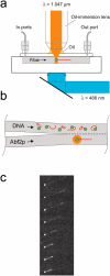
FIGURE 1
FIGURE 1
(a) Side view of the flow cell showing the trapping and excitation laser beams. (b) Top view of the flow cell. An individual DNA molecule held by an optical trap (orange) via its attached bead, and extended by flowing buffer, is moved into protein solution by translating the stage holding the flow cell perpendicular to the direction of flow. DNA was stained with YOYO-1 dye, allowing the compaction to be observed using fluorescence microscopy. The molecule was then moved back to the DNA side of the flow cell (which was protein-free), and the decompaction of the molecule was observed as protein left it, ultimately returning to its original length. (c) Time-lapse images of a lambda-phage DNA dimer (35 μm contour length) undergoing compaction by Abf2p. The Abf2p concentration is 2 μM. The time interval between successive frames is 0.5 s. The buffer flow speed is 63 μm/s.
FIGURE 2
FIGURE 2
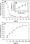
(a) Compaction of a single DNA molecule by Abf2p in 1.43 μM Abf2p (kon = 0.45 ± 0.1 μM−1 s−1). (b) Linear variation of Abf2p binding rate (kon[Abf2p]) with Abf2p concentration (kon = 0.36 ± 0.1 μM−1 s−1). (c) Decompaction of the same DNA molecule as in a (koff = 7.0 ± 1.3 × 10⁻³ s−1). The data in a and c are given by black points, and the exponential fit is represented by a red line.
FIGURE 3
FIGURE 3
DNA compaction versus initial extension for 67 molecules in 2 μM Abf2p, taken at buffer flow speeds between 50 and 110 μm/s. The compaction is defined as the difference between the initial (no protein) and final (in protein) DNA extensions divided by the initial DNA extension. For the range of DNA contour lengths and buffer flow velocities used in our experiment, the fractional extension of the DNA was fairly constant: 76 ± 5% (Perkins et al., 1995; Stigter and Bustamante, 1998). The incomplete compaction is thought to be due to the extension of the Abf2p-DNA complex by the flowing buffer. The longer the initial DNA contour length, the greater the hydrodynamic drag of the compacted Abf2p-DNA complex, and the less compaction is observed. The linear least squares fit is represented by a green line.
FIGURE 4
FIGURE 4
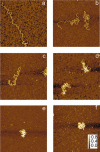
AFM images (image scale is 1 μm along each axis, height scale (f) is in nm) of Abf2p protein bound to single linear dsDNA molecules (pBR322, 1.5 μm contour length, 4361 bp). The DNA is bent and compacted as the concentration of Abf2p increases, ultimately forming a compact, 190 ± 90 nm diameter, round object. The ratio of Abf2p molecules to DNA bp is as follows with Abf2p concentrations indicated in parenthesis: (a) 0.0 Abf2p/bp (0 μM), (b) 0.04 Abf2p/bp (0.075 μM), (c) 0.1 Abf2p/bp (0.175 μM), (d) 0.2 Abf2p/bp (0.35 μM), (e) 0.45 Abf2p/bp (0.75 μM), and (f) 0.75 Abf2p/bp (1.25 μM). For an Abf2p footprint of 27 bp, the DNA coverage is complete for an Abf2p-to-bp ratio of 0.037.

References

    1. Alam, T. I., T. Kanki, T. Muta, K. Ukaji, Y. Abe, H. Nakayama, K. Takio, N. Hamasaki, and D. Kang. 2003. Human mitochondrial DNA is packaged with TFAM. Nucleic Acids Res. 31:1640–1645. - PMC - PubMed
    1. Baumann, C. G., V. A. Bloomfield, S. B. Smith, C. Bustamante, M. D. Wang, and S. M. Block. 2000. Stretching of single collapsed DNA molecules. Biophys. J. 78:1965–1978. - PMC - PubMed
    1. Brewer, L. R., M. Corzett, and R. Balhorn. 1999. Protamine-induced condensation and decondensation of the same DNA molecule. Science. 286:120–123. - PubMed
    1. Brower-Toland, B. D., C. L. Smith, R. C. Yeh, J. T. Lis, C. L. Peterson, and M. D. Wang. 2002. Mechanical disruption of individual nucleosomes reveals a reversible multistage release of DNA. Proc. Natl. Acad. Sci. USA. 99:1960–1965. - PMC - PubMed
    1. Caron, F., C. Jacq, and J. Rouviere-Yaniv. 1979. Characterization of a histone-like protein extracted from yeast mitochondria. Proc. Natl. Acad. Sci. USA. 76:4265–4269. - PMC - PubMed

Publication types

MeSH terms