. 2003 Oct;85(4):2589-98.
doi: 10.1016/S0006-3495(03)74681-5.

NMR solution structure and topological orientation of monomeric phospholamban in dodecylphosphocholine micelles

Affiliations

NMR solution structure and topological orientation of monomeric phospholamban in dodecylphosphocholine micelles

Jamillah Zamoon et al. Biophys J. 2003 Oct.

Abstract

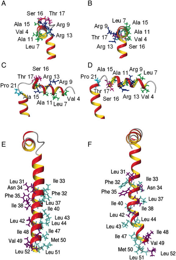
Phospholamban is an integral membrane protein that regulates the contractility of cardiac muscle by maintaining cardiomyocyte calcium homeostasis. Abnormalities in association of protein kinase A with PLB have recently been linked to human heart failure, where a single mutation is responsible for dilated cardiomyopathy. To date, a high-resolution structure of phospholamban in a lipid environment has been elusive. Here, we describe the first structure of recombinant, monomeric, biologically active phospholamban in lipid-mimicking dodecylphosphocholine micelles as determined by multidimensional NMR experiments. The overall structure of phospholamban is "L-shaped" with the hydrophobic domain approximately perpendicular to the cytoplasmic portion. This is in agreement with our previously published solid-state NMR data. In addition, there are two striking discrepancies between our structure and those reported previously for synthetic phospholamban in organic solvents: a), in our structure, the orientation of the cytoplasmic helix is consistent with the amphipathic nature of these residues; and b), within the hydrophobic helix, residues are positioned on two discrete faces of the helix as consistent with their functional roles ascribed by mutagenesis. This topology renders the two phosphorylation sites, Ser-16 and Thr-17, more accessible to kinases.


Figures

FIGURE 1
FIGURE 1
Fully assigned 1H/15N HSQC spectrum of AFA-PLB in 600 mM DPC micelles. The spectrum was recorded at 600 MHz at 50°C.
FIGURE 2
FIGURE 2
Two-dimensional strips from three-dimensional 1H/15N/13C HNCACB spectrum of AFA-PLB in DPC micelles. The data were recorded at 800 MHz at 50°C. Residue numbers denote the sequential connectivities.
FIGURE 3
FIGURE 3
Chemical shift index histograms of AFA-PLB obtained from triple resonance experiments (see Methods section).
FIGURE 4
FIGURE 4
Summary of the backbone NOEs obtained from 1H/15N NOESY-HSQC experiments (top). Deuterium/proton exchange factors obtained for AFA-PLB in DPC micelles at 37°C and 50°C, respectively (bottom).
FIGURE 5
FIGURE 5
Histogram illustrating the number of intra- and interresidue NOEs as a function of the residue. The asterisk denotes the presence of ambiguous NOEs that were not included in the structural calculations. However, their assignments were verified a posteriori.
FIGURE 6
FIGURE 6
Stereo view of the superposition of the 20 lowest energy structures of AFA-PLB: (A) Backbone representation of the 20 conformers (superposition of residues 2–50). (B) Superposition of residues 2–16 of domain IA of AFA-PLB. (C) Superposition of residues 17–21 of the flexible loop domain IB. (D) Superposition of residues 22–50 of domain II of AFA-PLB.
FIGURE 7
FIGURE 7
Network of NOEs found in the loop region imposing the “L-shape” topology on AFA-PLB in micelles and the corresponding distribution of φ and ψ angles defining the turn.
FIGURE 8
FIGURE 8
Paramagnetic quenching obtained for AFA-PLB in DPC micelles using Mn2+ and 16-doxyl stearic acid. (A) Intensity retention plot for AFA-PLB in the presence of 0.8 mM of MnCl2 (1:1 Mn2+/protein molar ratio). (B) Intensity retention plot for AFA-PLB in the presence of 2.4 mM of MnCl2 (3:1 Mn2+/protein molar ratio). (C) Intensity retention plot for AFA-PLB in the presence of 3.2 mM of 16-doxyl stearic acid (4:1 16-doxyl stearic acid/protein molar ratio). (D) Intensity retention plot for AFA-PLB in the presence of 6.4 mM of 16-doxyl stearic acid (1:8 16-doxyl stearic acid/protein molar ratio). (E) Surface plot of AFA-PLB illustrating solvent-exposed (coral) and buried (green) residues for histograms in A and C. (F) Electrostatic potential plot of AFA-PLB showing the hydrophobic patch centered at Leu-7.
FIGURE 9
FIGURE 9
(A, C, and E) Ribbon representation of the minimized average structure of AFA-PLB in DPC micelles (B, D, and F) compared to that in organic solvent. The structure in DPC (A and C) exhibits all the hydrophobic residues of the cytoplasmic domain (green) facing the micellar interior and the hydrophilic residues (blue), including the phosphorylation sites Ser-16 and Thr-17 (magenta), pointing toward the bulk solvent. This is not the case for the structure in organic solvent (B and D). Concomitantly, the residues of domain II, defined by mutagenesis studies to enhance (aqua) or reduce (violet) monomer formation and inhibitory activity, are aligned on two discrete faces for the AFA-PLB structure in DPC micelles (E) but interspersed for the structure in organic solvent (F).

References

    1. Arkin, I. T., P. D. Adams, A. T. Brunger, S. O. Smith, and D. M. Engelman. 1997. Structural perspectives of phospholamban, a helical transmembrane pentamer. Annu. Rev. Biophys. Biomol. Struct. 26:157–179. - PubMed
    1. Arkin, I. T., M. Rothman, C. F. Ludlam, S. Aimoto, D. M. Engelman, K. J. Rothschild, and S. O. Smith. 1995. Structural model of the phospholamban ion channel complex in phospholipid membranes. J. Mol. Biol. 248:824–834. - PubMed
    1. Buck, B., J. Zamoon, T. L. Kirby, T. M. DeSilva, C. Karim, D. Thomas, and G. Veglia. 2003. Overexpression, purification and characterization of recombinant Ca-ATPase regulators for high-resolution solution and solid-state NMR studies. Prot. Exp. Pur. 30:253–261. - PubMed
    1. Brunger, A. T., P. D. Adams, G. M. Clore, W. L. DeLano, P. Gros, R. W. Grosse-Kunstleve, J. S. Jiang, J. Kuszewski, M. Nilges, N. S. Pannu, R. J. Read, L. M. Rice, T. Simonson, and G. L. Warren. 1998. Crystallography & NMR system: a new software suite for macromolecular structure determination. Acta Crystallogr. D. Biol. Crystallogr. 54:905–21. - PubMed
    1. Cornilescu, G., F. Delaglio, and A. Bax. 1999. Protein backbone angle restraints from searching a database for chemical shift and sequence homology. J. Biomol. NMR. 13:289–302. - PubMed

Publication types

LinkOut - more resources