. 2003 Sep 24;23(25):8664-72.
doi: 10.1523/JNEUROSCI.23-25-08664.2003.

Postsynaptic protein kinase A reduces neuronal excitability in response to increased synaptic excitation in the Drosophila CNS

Affiliations

Postsynaptic protein kinase A reduces neuronal excitability in response to increased synaptic excitation in the Drosophila CNS

Richard A Baines. J Neurosci. .

Abstract

Previous work has identified a role for synaptic activity in the development of excitable properties of motoneurons in the Drosophila embryo. In this study the underlying mechanism that enables two such neurons, termed aCC and RP2, to respond to increased exposure to synaptic excitation is characterized. Synaptic excitation is increased in genetic backgrounds that lack either a cAMP-specific phosphodiesterase (EC:3.1.4, dunce) or acetylcholinesterase (EC:3.1.1.7, ace), the enzyme that terminates the endogenous cholinergic excitation of these motoneurons. Analysis of membrane excitability in aCC/RP2, in either background, shows that these neurons have a significantly reduced capability to fire action potentials (APs) in response to injection of depolarizing current. Analysis of underlying voltage-gated currents show that this effect is associated with a marked reduction in magnitude of the voltage-dependent inward Na+ current (INa). Partially blocking INa in these motoneurons, using low concentrations of TTX, demonstrates that a reduction of INa is, by itself, sufficient to reduce membrane excitability. An analysis of firing implicates an increased AP threshold to underlie the reduction in membrane excitability observed because of heightened exposure to synaptic excitation. Genetic or pharmacological manipulations that either elevate cAMP or increase protein kinase A (PKA) activity in wild-type aCC/RP2 mimic both the reductions in membrane excitability and INa. In comparison, increasing cAMP catabolism or inhibition of PKA activity is sufficient to block the induction of these activity-dependent changes. The induced changes in excitability can be rapid, occurring within 5 min of exposure to a membrane-permeable cAMP analog, indicative that threshold can be regulated in these neurons by a post-translational mechanism that is dependent on phosphorylation.


Figures
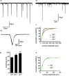
Figure 1.
Figure 1.
Synaptic excitation of aCC/RP2 is increased in dnc. Ai, Whole-cell voltage-clamp recordings (Vh -60 mV) from aCC/RP2 (WT aCC shown) reveal large inward currents that are the result of the evoked release of presynaptic ACh (see Baines et al.,1999,2001). These currents are relatively long-lived, lasting for up to 400 msec (see Aii). B, The average amplitude of excitatory currents recorded in aCC/RP2 is significantly greater in both dnc1 and dnc2 relative to WT. *p ≤ 0.05; **p ≤ 0.01. C, Cumulative probability plots of individual excitatory current amplitudes show that there is an increase in larger amplitude currents in both alleles of dnc as compared with WT. D, In the presence of 0.1 μm TTX the evoked excitatory currents are abolished, leaving only those currents that are elicited from the spontaneous release of vesicles in the presynaptic interneurons. E, Spontaneous current amplitudes in aCC/RP2 between WT and dnc2 are not significantly different.
Figure 2.
Figure 2.
Membrane excitability of aCC/RP2 is reduced in both dnc and ace. Ai, Injection of depolarizing current (10 pA/500 msec) is sufficient to fire action potentials in aCC/RP2. Aii, Averaged APs fired in response to depolarizing current (10 pA/500 msec) show a significant reduction in both dnc1 and dnc2 as compared with WT (p ≤ 0.01). Bi, The duration between successive APs, measured from the peak of the preceding afterhyperpolarization to the peak of depolarization (shown by arrows and dotted lines), is increased significantly in dnc2 as compared with WT. Bii, AP duration is consistently longer in dnc2 as compared with WT, regardless of which AP is measured. Shown is the average inter-AP duration for the 2nd, 5th, 10th, and 15th AP to be fired by the depolarizing current pulse. Ci, Injection of constant current (10 pA/500 msec) also results in fewer APs being fired in aCC/RP2 in ace late-stage 17 embryos (19-21 hr after egg laying) as compared with heterozygous controls. Cii, Similar to the effect seen in dnc, the duration between successive APs also is increased significantly in ace mutants. *p ≤ 0.05; **p ≤ 0.01.
Figure 3.
Figure 3.
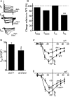
INa is reduced significantly in aCC/RP2 in both dnc and ace mutants. A, Whole-cell voltage-clamp analysis in aCC/RP2 reveals the presence of at least two outward K+ conductances (IKfast and IKslow), inward Ca2+, and inward Na+ currents (for a complete description of these currents, see Baines and Bate, 1998). B, The current density of INa is reduced significantly in dnc2 as compared with WT. Magnitude of the other currents is not significantly different from WT. Shown are WT currents that have been normalized to 100% (dashed line; actual values: WT, 77.4 ± 9.2, 69.8 ± 9.5, 21.0 ± 1.6, 28.0 ± 2.6 pA/pF; dnc2, 73.0 ± 4.5, 57.1 ± 3.9, 21.3 ± 3.3, 17.6 ± 1.4 pA/pF for IKfast, IKslow, ICa, and INa, respectively). **p ≤ 0.01. C, Current-voltage plots of INa in both WT and dnc2 show a reduction in current magnitude at all voltages above activation. Moreover, the membrane potential at which this current activates is not significantly different. D, The magnitude of INa in aCC/RP2 also is reduced significantly in ace late-stage 17 embryos as compared with heterozygous controls. *p ≤ 0.05. E, A plot of current-voltage relationships indicates no change in voltage dependence of INa in ace as compared with WT.
Figure 4.
Figure 4.
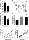
Reduction of INa is sufficient to suppress membrane excitability. A, The amplitude of INa recorded in aCC/RP2 is reduced significantly in the presence of a low concentration of 1 nm TTX (15.9 ± 3.8 vs 28.0 ± 2.6 pA/pF; n ≥ 5; *p ≤ 0.05). B, Current-voltage plots indicate no change in voltage dependence. The current-voltage plot for INa recorded in dnc2 is shown for comparison. C, In the presence of 1 nm TTX the number of APs fired by aCC/RP2 in response to depolarizing current (10 pA/500 msec) is reduced significantly as compared with WT lacking this neurotoxin (14.2 ± 1.8 vs 23 ± 0.8 APs; n ≥ 5; **p ≤ 0.01). D, The duration between successive APs elicited by current injection is increased significantly as compared with controls. The increase in duration observed in dnc2 is shown for comparison. Duration between the first and second APs to be fired is shown (see Fig. 2 Bi for an explanation of the measure that was used). **p ≤ 0.01. E, APs elicited by injection of constant current in aCC/RP2 in WT show a proportional increase with current strength (0-10 pA/500 msec). An increased amount of current is required to elicit a comparable number of APs in the presence of 1 nm TTX. This shift to the right is indicative of an increased threshold for AP firing. F, Similar current-firing plots for dnc2 and ace mutants show a similar, although reduced, shift to the right as compared with WT.
Figure 5.
Figure 5.
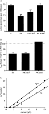
Postsynaptic cAMP/PKA activity is sufficient to reduce membrane excitability in aCC/RP2. A, Increasing cAMP in just aCC/RP2 by expression of UAS-rut (using RN2-O GAL4) is sufficient to reduce membrane excitability in these neurons to an extent similar to that observed in the alleles of dnc and ace (see Fig. 2). Expression of a constitutively active form of PKA (PKAact1) in just these two motoneurons is also sufficient to reduce membrane excitability. Inhibition of PKA (PKAinh1), however, is without effect as compared with controls (c, parental stocks). B, Expression of UAS-rut (rut) or UAS-PKAact1 in aCC/RP2, but not UAS-PKAinh1, is also sufficient to reduce INa in these neurons as compared with WT, which has been set to 100% (dashed line; actual values are 17.6 ± 3.1, 17.9 ± 3.4, 30.25 ± 2.6, 28.0 ± 2.6 pA/pF for rut, PKAact1, PKAinh1, and WT, respectively). Values are the mean ± SE; n ≥ 8 (**p ≤ 0.01). C, Analysis of the effect of rut expression on current-firing properties in aCC/RP2 shows that expression of this transgene shifts the relationship to the right, indicative of an increased AP threshold.
Figure 6.
Figure 6.
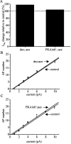
Blockade of cAMP/PKA activity is sufficient to prevent activity-dependent changes in excitability. A, Expression of either UAS-dnc or UAS-PKAinh1 in aCC/RP2 in an ace background prevents the activity-dependent reduction in INa normally seen in ace alone (compare with Fig. 3D). Actual values are 27 ± 1.9 versus 32 ± 4.2 pA/pF for control versus dnc;ace and 27 ± 2.7 versus 28 ± 2.7 pA/pF for control versus PKAinh1; ace (n = 8; mean ± SE; p > 0.05). B, C, Expression of either dnc or PKAinh1 in aCC/RP2 in an ace background is also sufficient to prevent the reduction in excitability observed in ace alone (compare with Fig. 4 F). In all cases the controls consist of parental stocks.
Figure 7.
Figure 7.
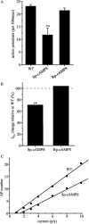
Regulation of membrane excitability is rapid. A, Exposure of aCC/RP2 to a membrane-permeant activator of PKA (Sp-cAMPS; 0.5 mm/5 min) is sufficient to induce a significant reduction in the number of APs fired in response to an injection of depolarizing current (10 pA/500 msec). Inhibition of PKA by using membrane-permeant Rp-cAMPS (0.5 mm/5 min) was without effect. B, Exposure to the PKA activator Sp-cAMPS (0.5 mm/5 min) is also sufficient to reduce INa significantly as compared with control (dashed line; normalized to 100%; 20.0 ± 1.7 vs 28.0 ± 2.6 pA/pF, respectively). Exposure to Rp-cAMPS did not affect INa. **p ≤ 0.01. C, Exposure of aCC/RP2 to Sp-cAMPS (0.5 mm/5 min) also increased the current required to elicit a comparable number of APs relative to WT.

References

    1. Adelman JP, Shen K-Z, Kavanaugh MP, Warren RA, Wu Y-N, Lagrutta A, Bond CT, North RA ( 1992) Calcium-activated potassium channels expressed from cloned complementary DNAs. Neuron 9: 209-216. - PubMed
    1. Atkinson NS, Robertson GA, Ganetzky B ( 1991) A component of calcium-activated potassium channels encoded by the Drosophila slo locus. Science 253: 551-555. - PubMed
    1. Baines RA, Bate M ( 1998) Electrophysiological development of central neurons in the Drosophila embryo. J Neurosci 18: 4673-4683. - PMC - PubMed
    1. Baines RA, Robinson SG, Fujioka M, Jaynes JB, Bate M ( 1999) Postsynaptic vesicle release is essential for synaptogenesis in Drosophila Curr Biol 9: 1267-1270. - PMC - PubMed
    1. Baines RA, Uhler JP, Thompson A, Sweeney ST, Bate M ( 2001) Altered electrical properties in Drosophila neurons developing without synaptic transmission. J Neurosci 21: 1523-1531. - PMC - PubMed

Publication types

MeSH terms