. 2003 Oct 14;100(21):12009-14.
doi: 10.1073/pnas.2030930100. Epub 2003 Sep 24.

HdmX stimulates Hdm2-mediated ubiquitination and degradation of p53

Affiliations

HdmX stimulates Hdm2-mediated ubiquitination and degradation of p53

Laëtitia K Linares et al. Proc Natl Acad Sci U S A. .

Abstract

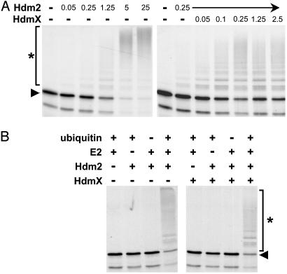
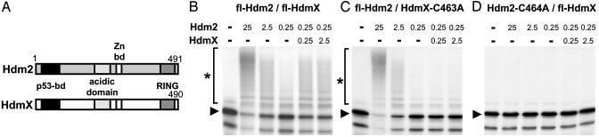
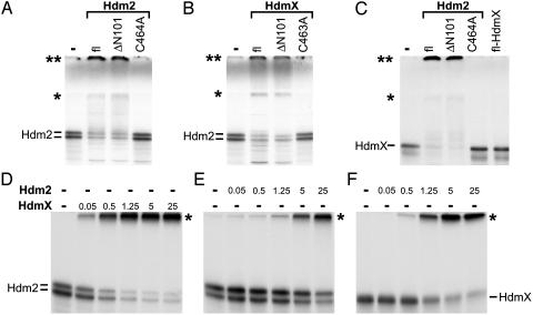
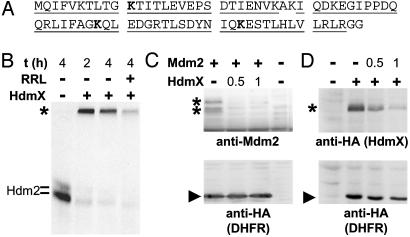
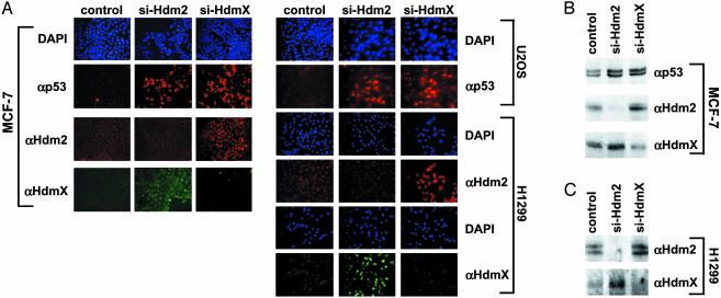
The RING finger proteins HdmX and Hdm2 share significant structural and functional similarity. Hdm2 is a member of the RING finger family of ubiquitin-protein ligases E3 and targets the tumor suppressor protein p53 for degradation. Although HdmX also binds to p53, HdmX does not induce p53 degradation. Moreover, HdmX has been reported to interfere with p53 degradation in overexpression experiments. To obtain insight into the mechanism by which HdmX interferes with p53 degradation, we studied the effect of HdmX on the E3 activity of Hdm2 in vitro. Surprisingly, this revealed that HdmX stimulates Hdm2-mediated ubiquitination of p53 and that HdmX facilitates ubiquitination of Hdm2 and vice versa. In addition, down-regulation of HdmX expression within cells results in the accumulation of both p53 and Hdm2. Because HdmX alone does not have appreciable E3 activity, these data indicate that HdmX acts as a stimulator, rather than as an inhibitor, of the E3 activity of Hdm2 and that, at least under certain conditions, HdmX is actively involved in the degradation of both p53 and Hdm2.


Figures

Fig. 1.
Fig. 1.
HdmX stimulates Hdm2-mediated ubiquitination of p53. (A) Increasing amounts of bacterially expressed GST fusion proteins of Hdm2 and HdmX, respectively, were incubated with in vitro translated radiolabeled p53 under standard ubiquitination conditions for 2 h. The reaction products were analyzed by SDS/PAGE followed by fluorography. Amounts of Hdm2 and HdmX used are indicated in 10–1 μg. (B) Determination of the requirements for ubiquitination of in vitro translated p53 by bacterially expressed Hdm2 (250 ng) and by Hdm2/HdmX (25 ng each), respectively. E2, UbcH5. The running positions of the nonmodified form and of the ubiquitinated forms of p53 are indicated by an arrowhead and an asterisk, respectively.
Fig. 2.
Fig. 2.
An intact RING finger motif of Hdm2 and HdmX is required for HdmX to stimulate p53 ubiquitination. (A) Schematic representation of the structure of Hdm2 and HdmX. bd, binding domain. (BD) In vitro ubiquitination of p53 in the presence of different amounts of the various Hdm2 and HdmX proteins as indicated. Amounts of Hdm2 and HdmX used are indicated in 10–1 μg. fl, full-length protein. C463A and C464A, the respective cysteine residues of HdmX and Hdm2, were replaced by alanine. The running positions of the nonmodified form of p53 and of the ubiquitinated forms of p53 are indicated by an arrowhead and an asterisk, respectively.
Fig. 3.
Fig. 3.
HdmX stimulates ubiquitination of Hdm2 and vice versa. In vitro-translated, radiolabeled Hdm2 (A and B) or HdmX (C) were incubated with various forms of bacterially expressed GST fusion proteins of Hdm2 (1 μg) and HdmX (1 μg) under standard in vitro ubiquitination conditions as indicated. fl, full-length. ΔN101 represents an Hdm2 or HdmX mutant, of which the N-terminal 101 aa of Hdm2 or HdmX were deleted; C464A and C463A, the respective cysteine residues of HdmX and Hdm2 were replaced by alanine. Note that, in this particular experiment, the ubiquitinated forms of both Hdm2 (A and B) and HdmX (C) run at two positions indicated by an asterisk and by a double asterisk. The position indicated by a single asterisk coincides with the top of the separating gel, and the position of the double asterisk coincides with the top of the stacking gel. The reason for the different migration behavior of the ubiquitinated forms (top of separating gel vs. top of stacking gel) is unknown, but note that analysis of the different forms by mass spectrometry revealed no differences with respect to the lysine residues of ubiquitin used for chain formation (see also legend to Fig. 4). (DF) Titration analysis of HdmX-mediated ubiquitination of Hdm2 (D) and of Hdm2-mediated ubiquitination of Hdm2 (E) and HdmX (F). Amounts of Hdm2 and HdmX used are indicated in 10–1 μg. The running positions of ubiquitinated forms of Hdm2 and HdmX are indicated by an asterisk.
Fig. 4.
Fig. 4.
HdmX stimulates Hdm2/Mdm2 degradation. (A) Amino acid sequence of ubiquitin. The amino acid residues identified by mass spectrometry in three independent experiments are underlined. The lysine residues used for chain formation are indicated in bold. Note that mass spectrometric analysis of samples taken from the top of the separating gel and from the top of the stacking gel (see Fig. 3) yielded identical results. (B) In vitro-translated radio-labeled Hdm2 was incubated with bacterially expressed HdmX (250 ng) at standard ubiquitination conditions. After 2 h, additional rabbit reticulocyte lysate (RRL) was added as a source of the 26S proteasome, where indicated, and the reactions were incubated for an additional 2 h. Note that the disappearance of the polyubiquitinated forms of Hdm2 depended on the presence of ATP, indicating that their disappearance was not due to the action of ubiquitin-specific isopeptidases that may be present in the reticulocyte lysate used (data not shown). The running position of ubiquitinated Hdm2 is indicated with an asterisk. (C and D) Expression constructs encoding HA-tagged HdmX, Mdm2, and HA-tagged DHFR were cotransfected into H1299 cells. Expression levels of the respective proteins were determined by Western blot analysis with the antibodies indicated. The running positions of HdmX and Mdm2, respectively, are indicated with an asterisk, and the running position of DHFR is indicated with an arrowhead. +, 0.5 μg of the respective expression plasmid was cotransfected with the indicated amount of the other construct.
Fig. 5.
Fig. 5.
Knockdown of HdmX expression results in increased p53 and Hdm2 levels. siRNAs specific for HdmX and Hdm2 were transfected into the cell lines indicated. As a control, siRNA against the E6 oncoprotein of human papillomavirus type 16, which is not expressed in the cell lines used, was used. (A) The levels of p53, Hdm2, and HdmX were determined 3 days after transfection by immunofluorescence. (B and C) The levels of p53, Hdm2, and HdmX were determined 2 days after transfection by Western blot analysis. MCF-7 cells and U2OS cells express endogenous wild-type p53, whereas p53 is not expressed in H1299 cells. Note that similar results were obtained with two different siRNAs against HdmX (si-HdmX-1, si-HdmX-2; see Materials and Methods) and Hdm2 (si-Hdm2-1, si-Hdm2-2).

References

    1. Shvarts, A., Steegenga, W. T., Riteco, N., van Laar, T., Dekker, P., Bazuine, M., van Ham, R. C., van der Houven van Oordt, W., Hateboer, G., van der Eb, A. J. & Jochemsen, A. G. (1996) EMBO J. 15, 5349–5357. - PMC - PubMed
    1. Sharp, D. A., Kratowicz, S. A., Sank, M. J. & George, D. L. (1999) J. Biol. Chem. 274, 38189–38196. - PubMed
    1. Michael, D. & Oren, M. (2002) Curr. Opin. Genet. Dev. 12, 53–59. - PubMed
    1. Montes de Oca Luna, R., Wagner, D. S. & Lozano, G. (1995) Nature 378, 203–206. - PubMed
    1. Parant, J., Chavez-Reyes, A., Little, N. A., Yan, W., Reinke, V., Jochemsen, A. G. & Lozano, G. (2001) Nat. Genet. 29, 92–95. - PubMed

Publication types

MeSH terms