. 2003 Nov;15(11):2612-25.
doi: 10.1105/tpc.015560. Epub 2003 Sep 24.

ALTERED RESPONSE TO GRAVITY is a peripheral membrane protein that modulates gravity-induced cytoplasmic alkalinization and lateral auxin transport in plant statocytes

Affiliations

ALTERED RESPONSE TO GRAVITY is a peripheral membrane protein that modulates gravity-induced cytoplasmic alkalinization and lateral auxin transport in plant statocytes

Kanokporn Boonsirichai et al. Plant Cell. 2003 Nov.

Abstract

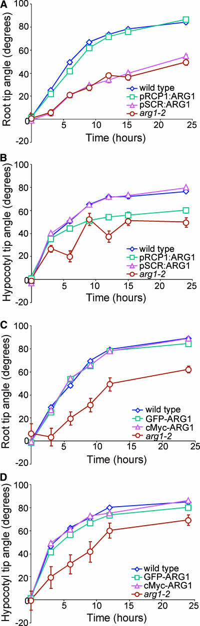
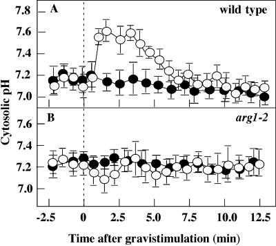
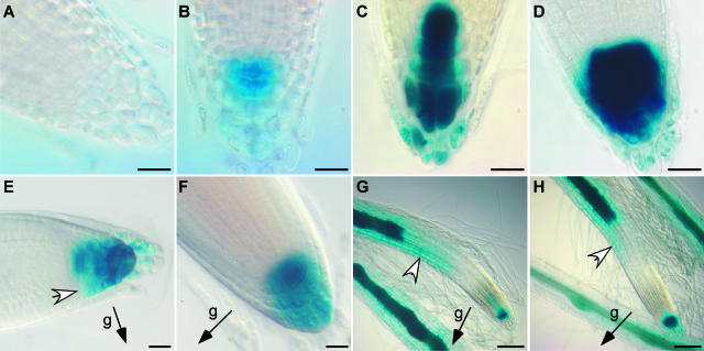
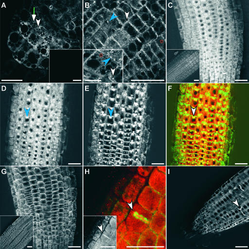
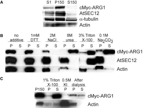
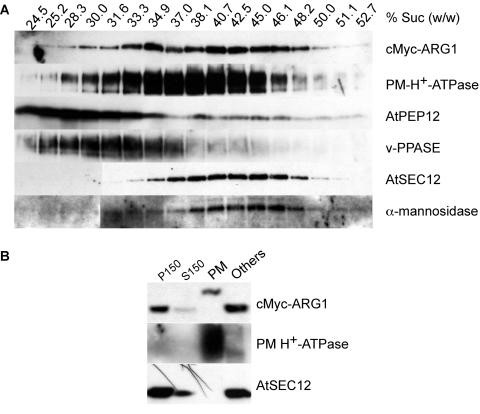
ARG1 (ALTERED RESPONSE TO GRAVITY) is required for normal root and hypocotyl gravitropism. Here, we show that targeting ARG1 to the gravity-perceiving cells of roots or hypocotyls is sufficient to rescue the gravitropic defects in the corresponding organs of arg1-2 null mutants. The cytosolic alkalinization of root cap columella cells that normally occurs very rapidly upon gravistimulation is lacking in arg1-2 mutants. Additionally, vertically grown arg1-2 roots appear to accumulate a greater amount of auxin in an expanded domain of the root cap compared with the wild type, and no detectable lateral auxin gradient develops across mutant root caps in response to gravistimulation. We also demonstrate that ARG1 is a peripheral membrane protein that may share some subcellular compartments in the vesicular trafficking pathway with PIN auxin efflux carriers. These data support our hypothesis that ARG1 is involved early in gravitropic signal transduction within the gravity-perceiving cells, where it influences pH changes and auxin distribution. We propose that ARG1 affects the localization and/or activity of PIN or other proteins involved in lateral auxin transport.


Figures

Figure 1.
Figure 1.
Gravitropic Reorientation Kinetics of Transgenic arg1-2. (A) and (B) Reorientation kinetics of root (A) and hypocotyl tips (B) of wild-type Wassilewskija (Ws), arg1-2, and representative transgenic arg1-2 carrying the pRCP1:ARG1 or pSCR:ARG1 construct (pRCP1:ARG1 and pSCR:ARG1, respectively). (C) and (D) Reorientation kinetics of root (C) and hypocotyl tips (D) of wild-type Wassilewskija, arg1-2, and representative transgenic arg1-2 carrying the GFP-ARG1 or cMyc-ARG1 construct (GFP-ARG1 and cMyc-ARG1, respectively). In each experiment, 90° gravistimulation was given at 0 h. Each data point represents the mean angle of organ tips. Vertical bars represent standard errors associated with the data points. For many data points, these bars were masked by the curve symbols. In all experiments, wild-type organ tips reoriented from a horizontal position at 0° toward the vertical at 90°. n = 39 to 131, 45 to 129, 22 to 80, and 22 to 80 in (A), (B), (C), and (D), respectively.
Figure 2.
Figure 2.
Cytosolic Alkalinization of the S3 Columella Cells of the Wild Type and arg1-2 upon Gravistimulation. (A) Wild type (ecotype Wassilewskija). (B) arg1-2. A 90° gravistimulus was given at 0 min. Closed circles represent mean cytosolic pH levels of ungravistimulated controls, and open circles represent gravistimulated roots. Data represents means ± se. n = 25 and 11 for the wild type and arg1-2, respectively.
Figure 3.
Figure 3.
DR5::GUS Activity in the Wild-Type ARG1 and Mutant arg1-2 Backgrounds. (A) and (B) Root tips of vertically grown wild-type (A) and arg1-2 (B) plants stained with 1 mM 5-bromo-4-chloro-3-indolyl-β-d-glucuronic acid (X-Gluc) buffer under the same conditions for 7 h. (C) and (D) Root tips of vertically grown wild-type (C) and arg1-2 (D) plants stained for 15 h under the same conditions in an experiment distinct from that presented in (A) and (B). (E) and (F) Wild-type (E) and arg1-2 (F) root tips after a 6-h gravistimulation stained with 2 mM X-Gluc buffer containing 2 mM K3Fe(CN)6 and either 0.1% (v/v) Triton X-100 for 5 h (E) or 10% (v/v) methanol for 24 h (F) at 22°C. Different staining conditions were used in (E) and (F) to allow equal staining intensity between wild-type and mutant root tips. (G) and (H) Wild-type (G) and arg1-2 (H) root tips after 16 h of vertical growth on 1 μM NAA followed by a 7-h gravistimulation and staining overnight. White arrowheads indicate the lateral gradient of DR5 activity in the root cap (E) and in the distal EZ ([G] and [H]). Black arrows indicate the gravity (g) vector. Ten to 25 roots were examined for each category. Bars = 20 μm for (A) through (F) and 100 μm for (G) and (H).
Figure 4.
Figure 4.
Localization of GFP-ARG1 and cMyc-ARG1. (A) and (B) Multiphoton images of transgenic arg1-2 root cap (A) and distal EZ (B) carrying the GFP-ARG1 construct, showing a large abundance of small dotted signals located at the periphery of all cells, larger punctated signals that appear circular (white arrowheads) or linear (blue arrowheads) in shape, and reticulated signals (red asterisks). Insets show signals from a nontransgenic root cap (A) and a cortical EZ cell from a different root that shows the reticulate and large punctate fluorescence patterns (B). (C) and (D) Confocal images of the distal EZ of transgenic arg1-2 carrying a cMyc-ARG1 construct immunostained with anti-cMyc antibodies. The inset in (C) shows signals from a nontransgenic root. The seedling in (D) was treated for 2 h with 50 μM BFA before fixation, showing the accumulation of the cMyc-ARG1 fusion protein in BFA compartments (blue arrowhead). (E) The same seedling shown in (D) treated with anti-AGR1/AtPIN2 antibodies, showing the accumulation of AGR1/AtPIN2 in similar BFA compartments (blue arrowhead). (F) A merged image of (D) and (E), showing cMyc-ARG1 in red and AGR1/AtPIN2 in green. Yellow indicates possible colocalization. Both proteins were found in the same BFA compartments (white arrowhead). (G) Immunostaining of cMyc-ARG1 transgenic distal EZ treated with anti-AGR1/AtPIN2 without BFA treatment, showing the localization of AGR1/AtPIN2 on basal membranes. The inset shows agr1-5, an AGR1/AtPIN2 null mutant (Chen et al., 1998), treated with anti-AGR1/AtPIN2 antibodies. (H) Immunostaining of cMyc-ARG1 transgenic EZ treated with anti-cMyc (signals in red) and anti-α-tubulin antibodies (signals in green), showing the localization of cMyc-ARG1 at the cell plate (arrowhead). The inset shows signals from anti-cMyc antibodies only (white). (I) Confocal image of transgenic GFP-ARG1 treated for 2 h with 10 μM oryzalin followed by a 2-h wash in liquid GM medium. GFP-ARG1 is found at aberrant cell division planes (arrowhead). Three to 15 roots were examined for each condition. Bars = 25 μm except for the inset in (B), where the bar = 10 μm.
Figure 5.
Figure 5.
Immunoblot Analyses of Protein Extracts from Transgenic arg1-2 Carrying the cMyc-ARG1 Construct, Showing the Membrane and Cytoskeleton Association of ARG1. (A) Fractionation of cMyc-ARG1, AtSEC12 (an integral membrane protein control), α-tubulin, and actin (integral/peripheral membrane protein controls) in the nucleus-free total protein extracts (S1), the microsomal membrane fraction (P150), and the soluble fraction (S150). A total of 50 μg was loaded per lane. The results shown are representative of five experiments. (B) Fractionation of cMyc-ARG1, AtSEC12, and actin in microsomes treated with buffer containing the reagents specified at top. The pellet (P) and supernatant (S) of each treatment were derived from 90 μg of P150 starting materials, except those of the Na2CO3 treatment, which were derived from 110 μg of P150. The results shown are representative of three experiments. (C) Fractionation of cMyc-ARG1 and actin in the microsomes, the cytoskeleton-enriched pellet, and the supernatant after treatment with 1% (v/v) Triton X-100, the pellet after depolymerization of the cytoskeleton with 0.5 M KI, and the cytoskeleton-enriched pellet and the supernatant after cytoskeleton repolymerization upon dialysis of the KI supernatant. The pellet and supernatant of Triton X-100 treatment were derived from 85 μg of P150 starting materials. A total of 30 μg of KI and postdialysis pellets and the volume equivalent of postdialysis supernatant were loaded.
Figure 6.
Figure 6.
Immunoblot Analyses of Protein Extracts from Transgenic arg1-2 Carrying the cMyc-ARG1 Construct, Showing the Association of ARG1 with Multiple Membranes. (A) Fractionation of cMyc-ARG1, PM H+-ATPase (a plasma membrane marker), AtPEP12 (an endosomal marker of the vacuolar pathway), anti-vacuolar pyrophosphatase (v-PPASE; a vacuolar marker), AtSEC12 (an ER marker), and α-mannosidase (an early Golgi marker) in a 14 to 50% (w/w) sucrose gradient. The sucrose concentration of each fraction, as determined by its refractive index, is given at top. A total of 20 μL of each fraction was loaded per lane. The results shown are representative of two separate gradients. (B) Fractionation of cMyc-ARG1, PM H+-ATPase, and AtSEC12 in the two-phase partitioning experiment. cMyc-ARG1 was found in both the plasma membrane–enriched upper phase (PM) and the plasma membrane–deprived lower phase (Others). The shift in mobility of cMyc-ARG1 in the plasma membrane fraction likely was an artifact of electrophoresis (see Results). A total of 75 μg of protein was loaded per lane. P150, microsomal membrane fraction; S150, soluble protein fraction.

References

    1. Aguilar, R.M., Bustamante, J.J., Hernandez, P.G., Martinez, A.O., and Haro, L.S. (1999). Precipitation of dilute chromatographic samples (ng/ml) containing interfering substances for SDS-PAGE. Anal. Biochem. 267, 344–350. - PubMed
    1. Artigues, A., Iriarte, A., and Martinez-Carrion, M. (2002). Binding to chaperones allows import of a purified mitochondrial precursor into mitochondria. J. Biol. Chem. 277, 25047–25055. - PubMed
    1. Baluska, F., Hlavacka, A., Samaj, J., Palme, K., Robertson, G.D., Matoh, T., McCurdy, D.W., Menzel, D., and Volkmann, D. (2002). F-actin-dependent endocytosis of cell wall pectins in meristematic root cells: Insights from brefeldin A-induced compartments. Plant Physiol. 130, 422–431. - PMC - PubMed
    1. Bar-Peled, M., and Raikhel, N.V. (1997). Characterization of AtSEC12 and AtSAR1 proteins likely involved in endoplasmic reticulum and Golgi transport. Plant Physiol. 114, 315–324. - PMC - PubMed
    1. Bennett, M.J., Marchant, A., Green, H.G., May, S.T., Ward, S.P., Millner, P.A., Walker, A.R., Schulz, B., and Feldmann, K.A. (1996). Arabidopsis AUX1 gene: A permease-like regulator of root gravitropism. Science 273, 948–950. - PubMed

Publication types

MeSH terms