. 2003 Oct;15(10):2408-20.
doi: 10.1105/tpc.015412. Epub 2003 Sep 24.

ACD6, a novel ankyrin protein, is a regulator and an effector of salicylic acid signaling in the Arabidopsis defense response

Affiliations

ACD6, a novel ankyrin protein, is a regulator and an effector of salicylic acid signaling in the Arabidopsis defense response

Hua Lu et al. Plant Cell. 2003 Oct.

Abstract

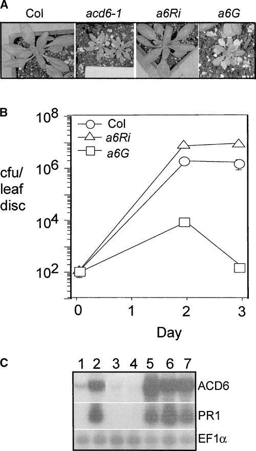
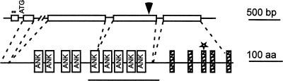
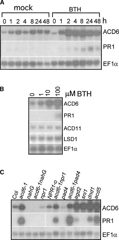
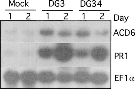
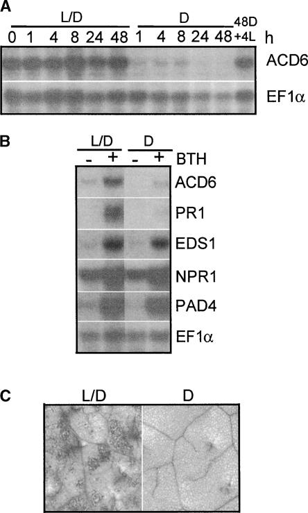
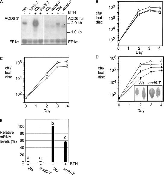
The previously reported Arabidopsis dominant gain-of-function mutant accelerated cell death6-1 (acd6-1) shows spontaneous cell death and increased disease resistance. acd6-1 also confers increased responsiveness to the major defense signal salicylic acid (SA). To further explore the role of ACD6 in the defense response, we cloned and characterized the gene. ACD6 encodes a novel protein with putative ankyrin and transmembrane regions. It is a member of one of the largest uncharacterized gene families in higher plants. Steady state basal expression of ACD6 mRNA required light, SA, and an intact SA signaling pathway. Additionally, ACD6 mRNA levels were increased in the systemic, uninfected tissue of Pseudomonas syringae-infected plants as well as in plants treated with the SA agonist benzothiazole (BTH). A newly isolated ACD6 loss-of-function mutant was less responsive to BTH and upon P. syringae infection had reduced SA levels and increased susceptibility. Conversely, plants overexpressing ACD6 showed modestly increased SA levels, increased resistance to P. syringae, and BTH-inducible and/or a low level of spontaneous cell death. Thus, ACD6 is a necessary and dose-dependent activator of the defense response against virulent bacteria and can activate SA-dependent cell death.


Figures

Figure 1.
Figure 1.
Defense Responses in the acd6-1 Mutant. (A) RNA gel blot analysis of defense gene induction. EF1α was used as a loading control. This experiment was repeated three times with similar results. (B) Staining of cell walls. Fourth and fifth leaves from the wild type (Columbia [Col]) and the acd6-1 mutant (a6) were examined for autofluorescence (top row) and callose (bottom row).
Figure 2.
Figure 2.
Suppression and Recapitulation of acd6-1 Mutant Phenotypes in Transgenic Plants. (A) Three-week-old wild-type and transgenic plants. a6Ri, acd6-1 transformed with an ACD6RNAi construct; a6G, Col transformed with an ACD6-1 genomic clone. At least 25 independent a6Ri and a6G lines behaved similarly to the lines shown. (B) P. syringae growth curve. Col, a6Ri, and a6G plants were infected with Pma DG3 (OD600 = 0.0001). Bars indicate standard errors; in some cases, the symbol obscures the error bars. The growth of bacteria in the three hosts was significantly different on days 2 and 3 (P < 0.001 [t test], n = 6). Similar results were obtained with several additional independent transformants (data not shown). This experiment was repeated three times with similar results. cfu, colony-forming units. (C) RNA gel blot analysis of the steady state accumulation of ACD6 and PR1 mRNAs. EF1α was used as a loading control. Total RNA was extracted from Col (lane 1), acd6-1 (lane 2), two a6Ri lines (lanes 3 and 4), and three a6G lines (lanes 5 to 7).
Figure 3.
Figure 3.
ACD6 Encodes a Putative Protein with Ankyrin and Transmembrane Regions. Structures of the ACD6 genomic DNA (top) and the predicted ACD6 protein (bottom). In the DNA structure, the boxes indicate exons, lines indicate untranslated regions and introns, and dots indicate the in-frame stop codons in exon 1. ATG is the putative translation start site. The arrowhead indicates the T-DNA insertion site in acd6-T. In the protein structure, boxes labeled ANK indicate ankyrin repeats, hatched boxes indicate transmembrane helices, and the star indicates the Leu-to-Phe mutation in acd6-1. The line below the structure indicates the fragment used for the ACD6 RNAi construct. The broken lines between the two structures connect the exons to their encoded protein regions. aa, amino acids.
Figure 4.
Figure 4.
SA-Dependent ACD6 Gene Expression. (A) Time-course induction by 100 μM BTH. This experiment was repeated three times with similar results. (B) BTH dose-dependent gene induction. Col leaves were collected 24 h after treatment with BTH at the indicated concentrations. This experiment was repeated three times with similar results. (C) Gene expression in different genotypes in the Col background. RNA samples were extracted from 20-day-old plants. This experiment was repeated twice with similar results.
Figure 5.
Figure 5.
ACD6 Gene Expression during Pathogen Infection. Col leaves were inoculated with 10 mM MgSO4 (mock treatment), Pma DG3, and Pma DG34 (carrying avrRpm1) at OD600 = 0.01. RNA was extracted at the indicated times from the uninfected leaves of inoculated plants. This experiment was repeated three times with similar results.
Figure 6.
Figure 6.
Light Is Required for ACD6 Expression and the acd6-1 Phenotype. (A) A time course of steady state ACD6 mRNA accumulation. Twenty-day-old Col plants grown in a 16-h-light/8-h-dark cycle were kept in these conditions (L/D) or shifted to continuous dark (D) 4 h after the lights normally came on (time 0) for the indicated times and then switched back to light for 4 h (4L). (B) Effect of light on BTH-induced defense gene expression. Col plants were treated with 100 μM BTH and subjected to the normal 16-h-light/8-h-dark cycle (L/D) or dark treatment (D) for 24 h. (C) Cell death staining. acd6-1-nahG plants were treated with 100 μM BTH or water and grown in the normal 16-h-light/8-h-dark (L/D; left) or 24-h dark (D; right) condition for 1 day. The fourth leaves were stained with trypan blue to detect cell death. Wild-type control and water-treated acd6-1-nahG tissue showed no cell death under the conditions used here (data not shown). These experiments were repeated three times with similar results.
Figure 7.
Figure 7.
Defense Response and Disease Susceptibility of an acd6-T Loss-of-Function Mutant. (A) Steady state accumulation of ACD6 mRNA using an ACD6-specific probe (ACD6 3′) (left) and the full-length ACD6 cDNA probe (ACD6 full) (right). EF1α served as a loading control. Wild-type Ws and the acd6-T mutant were treated with 300 μM BTH or water for 1 day. (B) and (C) Disease susceptibility and complementation of acd6-T plants. Wild-type Ws (circles), acd6-T (triangles), and/or acd6-T complemented with genomic ACD6 (squares) were infected with Pto DC3000 (B) or Pto DC3000 carrying avrRpt2 (C) at an OD600 = 0.0001. The growth of Pto DC3000 in acd6-T was significantly different from that in the wild type and complemented on days 2, 3, and 4 (P < 0.02 [t test], n = 6). The growth of Pto DC3000 carrying avrRpt2 in acd6-T was significantly different from that in the wild type on days 3 and 4 (P < 0.007 [t test], n = 6). These experiments were repeated twice with similar results. cfu, colony-forming units. (D) Reduced disease resistance of acd6-T treated with BTH. Wild-type Ws and acd6-T were pretreated with 300 μM BTH or water for 2 days and then subjected to infection by Pto DC3000 (OD600 = 0.0001). Open circles, water-treated Ws; closed circles, BTH-treated Ws; open triangles, water-treated acd6-T; closed triangles, BTH-treated acd6-T. Bars indicate standard errors; in some cases, the error bars are obscured by the symbols. For water-treated plants, the growth of bacteria in acd6-T was significantly different from that in the wild type on days 3 and 4 (P < 0.001 [t test], n = 6). For BTH-treated plants, the growth of bacteria in acd6-T was significantly different from that in the wild type on days 2, 3, and 4 (P < 0.001 [t test]). The inset shows BTH-treated and infected leaves taken 3 days after the infection. Note the increased lesion numbers on the acd6-T plants. This experiment was repeated three times with similar results. (E) Quantitation of the relative abundance of PR1 transcript in Ws and acd6-T plants after BTH treatment. Plants were treated as in (A). PR1 mRNA levels were normalized to the level of EF1α mRNA. Different letters indicate that the values are significantly different from each other (P < 0.01). The data were averaged from four independent experiments.
Figure 8.
Figure 8.
Reduced Defenses in acd6-T Plants. (A) Reduced SA levels in P. syringae–infected acd6-T plants. Plants were inoculated with 10 mM MgSO4 or Pto DC3000 at OD600 = 0.01. Samples were analyzed in triplicate. Asterisks indicate P < 0.05 at 12 h. FW, fresh weight. (B) Reduced PR1 expression in P. syringae–infected acd6-T plants. Plants treated as in (A) were used for RNA isolation. This experiment was repeated twice with similar results.
Figure 9.
Figure 9.
Growth of P. syringae in Plants with Extra Copies of ACD6. Wild-type (Col) and T2 plants from two independent transformants with extra copies of ACD6 (lines 12 and 14) were infected with Pto DC3000 at OD600 = 0.0001. Bars indicate standard errors (n = 6); in some cases, the error bars are obscured by the symbols. Bacteria grew significantly more in the wild type than in all of the lines carrying extra copies of ACD6 (P < 0.01). This experiment was repeated twice with similar results. cfu, colony-forming units.

References

    1. Adam, L., and Somerville, S.C. (1996). Genetic characterization of five powdery mildew disease resistance loci in Arabidopsis thaliana. Plant J. 9, 341–356. - PubMed
    1. Alvarez, M.E., Pennell, R.I., Meijer, P.J., Ishikawa, A., Dixon, R.A., and Lamb, C. (1998). Reactive oxygen intermediates mediate a systemic signal network in the establishment of plant immunity. Cell 92, 773–784. - PubMed
    1. Aviv, D.H., Rusterucci, C., Iii, B.F., Dietrich, R.A., Parker, J.E., and Dangl, J.L. (2002). Runaway cell death, but not basal disease resistance, in lsd1 is SA- and NIM1/NPR1-dependent. Plant J. 29, 381–391. - PubMed
    1. Bowling, S.A., Clarke, J.D., Liu, Y., Klessig, D.F., and Dong, X. (1997). The cpr5 mutant of Arabidopsis expresses both NPR1-dependent and NPR1-independent resistance. Plant Cell 9, 1573–1584. - PMC - PubMed
    1. Boyes, D., Nam, J., and Dangl, J. (1998). The Arabidopsis thaliana RPM1 disease resistance gene product is a peripheral plasma membrane protein that is degraded coincident with the hypersensitive response. Proc. Natl. Acad. Sci. USA 95, 15849–15854. - PMC - PubMed

Publication types

Associated data