. 2003 Oct 14;100(21):12331-6.
doi: 10.1073/pnas.1933740100. Epub 2003 Sep 25.

Induction of pulmonary hypertension by an angiopoietin 1/TIE2/serotonin pathway

Affiliations

Induction of pulmonary hypertension by an angiopoietin 1/TIE2/serotonin pathway

Christopher C Sullivan et al. Proc Natl Acad Sci U S A. .

Abstract

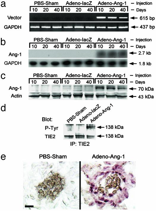
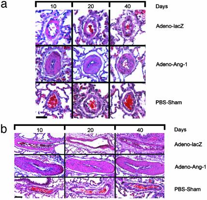
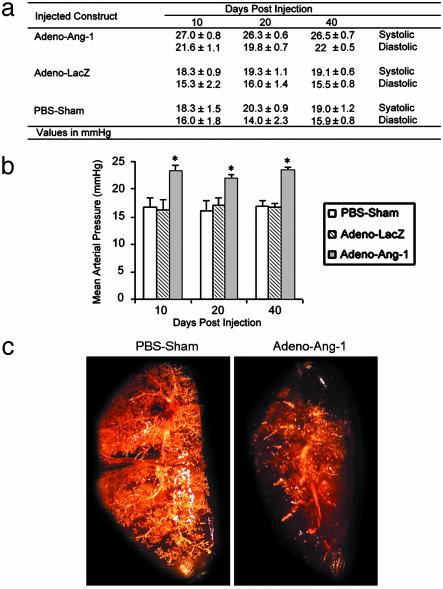
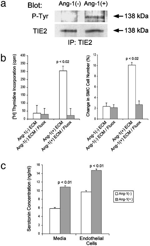
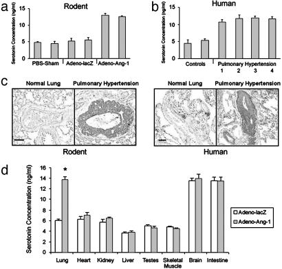
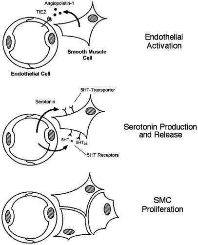
Smooth muscle cell proliferation around small pulmonary vessels is essential to the pathogenesis of pulmonary hypertension. Here we describe a molecular mechanism and animal model for this vascular pathology. Rodents engineered to express angiopoietin 1 (Ang-1) constitutively in the lung develop severe pulmonary hypertension. These animals manifest diffuse medial thickening in small pulmonary vessels, resulting from smooth muscle cell hyperplasia. This pathology is common to all forms of human pulmonary hypertension. We demonstrate that Ang-1 stimulates pulmonary arteriolar endothelial cells through a TIE2 (receptor with tyrosine kinase activity containing IgG-like loops and epidermal growth factor homology domains) pathway to produce and secrete serotonin (5-hydroxytryptamine), a potent smooth muscle mitogen, and find that high levels of serotonin are present both in human and rodent pulmonary hypertensive lung tissue. These results suggest that pulmonary hypertensive vasculopathy occurs through an Ang-1/TIE2/serotonin paracrine pathway and imply that these signaling molecules may be targets for strategies to treat this disease.


Figures

Fig. 1.
Fig. 1.
Constitutive lung expression of Ang-1 in animals whose pulmonary circulations were perfused with Adeno-Ang-1. (a) The pattern of Ang-1 expression in rodent lung samples was studied by PCR using primers specific to the virally transduced Ang-1 mRNA (Vector). Tissue was analyzed 10, 20, and 40 days after injection of Adeno-Ang-1, Adeno-lacZ, or PBS alone (PBS-Sham). (b) Northern blot demonstrating that lungs of animals injected with the Adeno-Ang-1 construct had detectable steady-state levels of Ang-1 mRNA, whereas control lung specimens did not. (c) Western blot showing Ang-1 protein in lung tissue infected with Adeno-Ang-1 virus. Ang-1 protein was undetectable in animals injected with either Adeno-lacZ or PBS. (d) Immunoblot analysis of anti-TIE2 immunoprecipitates from rodent lung tissue, demonstrating that constitutive Ang-1 expression induced TIE2 receptor tyrosine phosphorylation but did not modulate TIE2 protein levels. (Upper) Immunoprecipitated (IP) with anti-TIE2 antibody, blotted with anti-phosphotyrosine (P-Tyr) antibody. (Lower) IP with anti-TIE2, blotted with anti-TIE2. (e) Detection of recombinant viral mRNA by in situ hybridization. Lung tissue from rats injected with PBS demonstrated no staining when a virus-specific antisense RNA probe was used. Conversely, virus-specific mRNA was detected in animals infected with Adeno-Ang-1 in lung vessel walls. (Scale bar represents 10 μm.)
Fig. 2.
Fig. 2.
Lung vascular pathology in Ang-1-induced rat pulmonary hypertension. (a) Pulmonary arterioles 40 μm in diameter from animals 10, 20, and 40 days after injection with Adeno-Ang-1, Adeno-lacZ, or PBS alone (PBS-Sham). Adeno-Ang-1 animal lungs (middle horizontal series of micrographs in a and b) showed diffuse pulmonary medial arteriolar thickening. (Scale bar represents 25 μm.) (b) Pulmonary arterioles 200 μm in diameter from animals 10, 20, and 40 days after injection with Adeno-Ang-1, Adeno-lacZ, or PBS. (Scale bar represents 150 μm.)
Fig. 3.
Fig. 3.
Pulmonary artery pressures and angiography in animals with Ang-1-induced pulmonary hypertension. (a) Averaged pulmonary arterial pressures in rats injected with Adeno-Ang-1, Adeno-lacZ, or PBS at 10, 20, and 40 day intervals after gene transfer (four readings for each animal over a 10-min period, five animals for each group at each time point). Values are reported as number ± SEM. (b) Mean pulmonary arterial pressures in rats injected with Adeno-Ang-1, Adeno-lacZ, or PBS at 10, 20, and 40 day intervals after gene transfer. Each bar represents four measurements taken in each of five animals per group ± SEM. *, P < 0.01 versus the two control animal groups. (c) Pulmonary angiograms using the MicroFil cast technique of rodent lungs treated previously with either PBS carrier or Adeno-Ang-1. Note the absence of peripheral vessel staining in the Adeno-Ang-1-treated lung with large-vessel blunting.
Fig. 4.
Fig. 4.
Ang-1-treated human pulmonary endothelial cells release a potent smooth muscle cell growth factor: serotonin. (a) Immunoblot analysis of anti-TIE2 immunoprecipitates from subcultured pulmonary arteriolar endothelial cells either treated or not treated with Ang-1 protein. Cells incubated with Ang-1 protein demonstrated TIE2 receptor phosphorylation, whereas untreated cells did not (Upper). Protein levels of TIE2 were similar in the two groups (Lower). (Upper) Immunoprecipitated (IP) with anti-TIE2 antibody, blotted with anti-phosphotyrosine (P-Tyr) antibody. (Lower) IP with anti-TIE2, blotted with anti-TIE2. (b) Stimulation of pulmonary smooth muscle cell proliferation by serum-free medium from Ang-1-treated endothelial cells is blocked by a serotonin transporter inhibitor. (Left)[3H]Thymidine incorporation in smooth muscle cells treated with (i) medium from endothelial cells not treated with Ang-1 [Ang-1(–)ECM], (ii) medium from endothelial cells not treated with Ang-1 to which 1 μM fluoxetine was added [Ang-1(–)ECM/Fluox], (iii) medium from endothelial cells treated with Ang-1 [Ang-1(+)ECM], or (iv) medium from endothelial cells treated with Ang-1 to which 1 μM fluoxetine was added [Ang-1(+)ECM/Fluox]. (Right) Percent change in smooth muscle cell count after treatment with the same media combinations as in Left.(c) Pulmonary endothelial cells treated with 50 ng/ml Ang-1 produce and secrete serotonin. Graph depicts results of ELISA quantitation of serotonin from cells treated with or without Ang-1 (right bars) and from media taken from cells treated with or without Ang-1 (left bars).
Fig. 5.
Fig. 5.
High levels of serotonin in rodent and human pulmonary hypertensive lung tissue. (a) ELISA quantitation of serotonin in lungs from animals injected with Adeno-Ang-1 (pulmonary hypertension group) compared with serotonin levels in lungs from rodents injected with Adeno-lacZ or PBS (normotensive control groups). (b) ELISA quantitation of serotonin in lung samples taken from humans with pulmonary hypertension resulting from chronic thromboembolism (bar 1), pulmonary overcirulation from ventricular septal defects (bar 2), scleroderma (bar 3), and idiopathic nonfamilial disease (bar 4), compared with lung specimens obtained from patients without pulmonary hypertension (controls). (c) Immunohistochemical staining of lung tissue with anti-serotonin antibody shows that rodents with Ang-1-induced pulmonary hypertension have serotonin within the vascular wall of pulmonary vessels ≤500 μm in diameter (Left), whereas serotonin staining is not detected in normal rat lung tissue. Human tissue stained with anti-serotonin antibody demonstrates serotonin in the wall of small pulmonary vessels in pulmonary hypertensive lung tissue only (Right), without staining in lung specimens from patients without pulmonary hypertension. (Scale bars represent 50 μm.) (d) ELISA quantitation of serotonin in organs of animals with pulmonary hypertension induced by Adeno-Ang-1, compared with animals without pulmonary hypertension injected with control Adeno-lacZ virus. *, P < 0.01 compared with lung control group.
Fig. 6.
Fig. 6.
Proposed model for vascular smooth muscle cell (SMC) hyperplasia in pulmonary hypertension. Ang-1 released by SMCs binds the TIE2 receptor and induces endothelial cells (Top) to synthesize and release serotonin (Middle). Endothelial-derived serotonin acts as a mitogen by binding a specific transporter protein on the surface of SMCs, inducing the SMCs to proliferate (Bottom). In normotensive lung, Ang-1 is not produced.

References

    1. Folkman, J. & D'Amore, P. A. (1996) Cell 87, 1153–1155. - PubMed
    1. Hayes, A. J., Huang, W. Q., Mallah, J., Yang, D., Lippman, M. E. & Li, L. Y. (1999) Microvasc. Res. 58, 224–237. - PubMed
    1. Davis, S., Aldrich, T. H., Jones, P. F., Acheson, A., Compton, D. L., Jain, V., Ryan, T. E., Bruno, J., Radziejewski, C., Maisonpierre, P. C., et al. (1996) Cell 87, 1161–1169. - PubMed
    1. Jones, N. & Dumont, D. J. (2000) Cancer Metastasis Rev. 19, 13–17. - PubMed
    1. Davis, S. & Yancopoulos, G. D. (1999) Curr. Top. Microbiol. Immunol. 237, 173–185. - PubMed

Publication types

MeSH terms