. 2003 Oct;77(20):10799-807.
doi: 10.1128/jvi.77.20.10799-10807.2003.

Aichi virus leader protein is involved in viral RNA replication and encapsidation

Affiliations

Aichi virus leader protein is involved in viral RNA replication and encapsidation

Jun Sasaki et al. J Virol. 2003 Oct.

Abstract

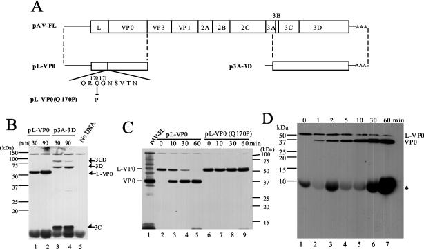
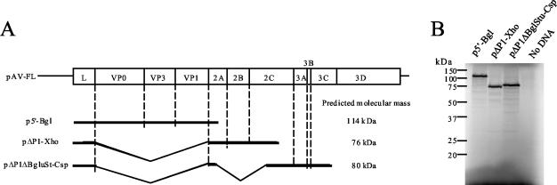
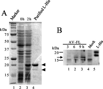
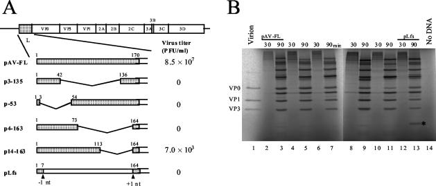
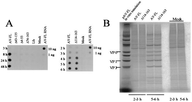
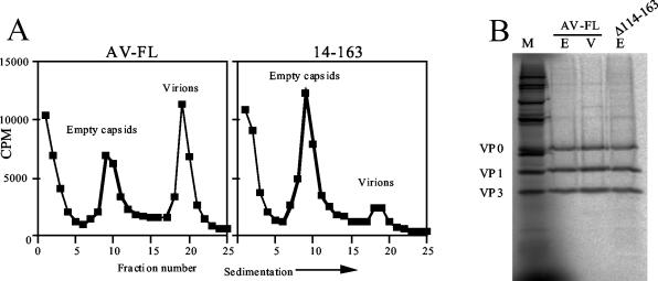
Aichi virus, a member of the family Picornaviridae, encodes a leader (L) protein of 170 amino acids (aa). The Aichi virus L protein exhibits no significant sequence homology to those of other picornaviruses. In this study, we investigated the function of the Aichi virus L protein in virus growth. In vitro translation and cleavage assays indicated that the L protein has no autocatalytic activity and is not involved in polyprotein cleavage. The L-VP0 junction was cleaved by 3C proteinase. Immunoblot analysis showed that the L protein is stably present in infected cells. Characterization of various L mutants derived from an infectious cDNA clone revealed that deletion of 93 aa of the center part (aa 43 to 135), 50 aa of the N-terminal part (aa 4 to 53), or 90 aa of the C-terminal part (aa 74 to 163) abolished viral RNA replication. A mutant (Delta114-163) in which 50 aa of the C-terminal part (aa 114 to 163) were deleted exhibited efficient RNA replication and translation abilities, but the virus yield was 4 log orders lower than that of the wild type. Sedimentation analysis of viral particles generated in mutant Delta114-163 RNA-transfected cells showed that the mutant has a severe defect in the formation of mature virions, but not in that of empty capsids. Thus, the data obtained in this study indicate that the Aichi virus L protein is involved in both viral RNA replication and encapsidation.


Figures

FIG. 1.
FIG. 1.
Analysis of cleavage at the L-VP0 junction. (A) Diagrams of an infectious cDNA clone of Aichi virus, pAV-FL, and the constructs used for in vitro translation and cleavage assays. The open box and lines indicate coding and noncoding regions, respectively. Below the diagram of pL-VP0, the predicted amino acid sequence around the L-VP0 junction is shown with the myristoylation motif. In pL-VP0(Q170P), a glutamine residue at aa 170 is mutated to a proline. In pAV-FL, pL-VP0, and pL-VP0(Q170P), the Aichi virus sequence is located downstream of the T7 promoter sequence, and in p3A-3D, the Aichi virus sequence is under the control of the SP6 promoter. (B) In vitro transcription-translation of pL-VP0 and p3A-3D in RRL. The plasmids were subjected to in vitro transcription-translation reactions in the presence of biotinylated lysyl-tRNA for 30 or 90 min at 30°C. Proteins were analyzed by SDS-PAGE and then blotted onto a polyvinylidene difluoride membrane. The biotin-labeled translation products were detected by chemiluminescence. The positions of translation products or cleavage products and molecular mass markers are indicated on the right and left, respectively. (C) In vitro cleavage assay. Biotin-labeled translation products of pL-VP0 and pL-VP0(Q170P) were incubated with unlabeled translation products of p3A-3D for various times at 37°C in the presence of RNase A. Then, biotin-labeled proteins were analyzed by SDS-PAGE and detected by chemiluminescence. As markers, in vitro transcription-translation products of pAV-FL were used. The positions of L-VP0 and VP0 and molecular mass markers are indicated on the left and right, respectively. (D) In vitro cleavage assay in the presence of protease inhibitors. The biotin-labeled translation product of pL-VP0 was reacted with the unlabeled translation product of p3A-3D in the presence of 1 mM PMSF, 2 μg of aprotinin/ml, 1 μg of pepstatin/ml, and 2 mM EDTA. Aliquots were taken at the indicated times and analyzed by SDS-15% PAGE. The asterisk indicates nonspecific signal, which was also observed in the sample containing no DNA (Fig. 1B, lane 5). The bromophenol blue dye front corresponds to the bottom of the blot.
FIG. 2.
FIG. 2.
Investigation of the ability of the Aichi virus L protein to process polyproteins. (A) Diagrams of pAV-FL and deletion mutants. In pAV-FL, the open box and lines indicate coding and noncoding regions, respectively. In deletion mutants, only the regions to be translated are shown as thick lines. Internal deletions are represented by angled lines. The predicted molecular masses of products of deletion mutants are shown. (B) In vitro transcription-translation of p5′-Bgl, pΔP1-Xho, and pΔP1ΔBglStu-Csp in RRL. The plasmids were subjected to in vitro transcription-translation reactions in the presence of [35S]methionine-cysteine for 90 min at 30°C. The translation products were analyzed by SDS-PAGE, and the gel was dried. Radioactive signals were detected with a phosphorimager. The positions of molecular mass markers are indicated on the left.
FIG. 3.
FIG. 3.
(A) Expression of the His-tagged L protein in E. coli and its purification. Lysates of E. coli cells transformed with pET-L were prepared at 0 and 2 h after induction with IPTG and then analyzed by SDS-PAGE. A fraction purified using an Ni affinity column was also analyzed (lane 4). After electrophoresis, the gel was stained with Coomassie blue. The positions of the 20- and 18-kDa proteins are indicated by arrowheads. (B) Synthesis of the L protein in Vero cells transfected with the AV-FL RNA. At the indicated times after transfection by electroporation, cell lysates were prepared and subjected to SDS-PAGE, and then the L protein was detected by Western blotting using antiserum raised against the purified His-tagged L protein. A fraction purified using an Ni affinity column was also analyzed (lane 5). The positions of molecular mass markers are indicated on the left. An arrow indicates the position of the 17-kDa protein.
FIG. 4.
FIG. 4.
(A) Schematic diagrams of pAV-FL and the L mutants and their abilities to produce viable viruses. The open boxes and lines indicate coding and noncoding regions, respectively. Vertical lines within the box represent putative cleavage sites for viral proteinase. L protein sequences are shaded, and deleted regions are represented by angled lines. Amino acid numbers are indicated above the boxes. In pLfs, the positions where a nucleotide was deleted (−1 nt) and added (+1 nt) are indicated. RNA transcripts synthesized from these plasmids were transfected into Vero cells by lipofection, and the virus titers in the cells 72 h after transfection were determined by plaque assay. The number of plaques was determined 72 h after infection. (B) In vitro transcription-translation of pAV-FL and the L mutants in RRL. The plasmids were reacted in an in vitro transcription-translation system in the presence of [35S]methionine-cysteine for 30 or 90 min at 30°C. The translation products were analyzed by SDS-PAGE, and the gel was dried. Radioactive signals were detected with a phosphorimager. To show the mobility of capsid proteins, 35S-labeled virions were analyzed. The asterisk indicates the unique protein produced from pLfs.
FIG. 5.
FIG. 5.
(A) RNA replication of AV-FL and the L mutants. Vero cells were electroporated with the RNA transcripts, and then total RNAs were extracted from the cells at the indicated times after electroporation. The total-RNA samples were dotted and probed with digoxigenin-labeled negative-sense viral RNA (nt 4790 to 5253). As controls, 10 and 1 ng of AV-FL transcripts were dotted. (B) Protein synthesis in Vero cells electroporated with the AV-FL or Δ114-163 RNA. At 2 or 5 h after electroporation, cells were labeled with [35S]methionine-cysteine for 1 h. The cells were lysed, and then the proteins were analyzed by SDS-PAGE. Radioactive signals were detected with a phosphorimager. As markers, in vitro transcription-translation products of pAV-FL were used. Mock, mock transfected.
FIG. 6.
FIG. 6.
(A) Sedimentation analysis of viral and subviral particles generated in Vero cells electroporated with AV-FL or Δ114-163 RNA. Vero cells transfected with the RNAs were labeled with [35S]methionine-cysteine. At 6 h after electroporation, the labeled viral and subviral particles were collected and centrifuged through a 10 to 30% sucrose gradient. The gradient was fractionated, and the radioactivity in each fraction was counted with a liquid scintillation counter. (B) Proteins constructing virions and empty capsids. Parts of fractions containing virions (lane V) of the AV-FL virus and empty capsids (lane E) of the AV-FL virus and the Δ114-163 virus were analyzed by SDS-PAGE, and 35S-labeled proteins were visualized with a phosphorimager. As markers, in vitro transcription-translation products of pAV-FL were used (lane M).

Similar articles

Cited by

References

    1. Badshah, C., M. A. Calenoff, and K. Rundell. 2000. The leader polypeptide of Theiler's murine encephalomyelitis virus is required for the assembly of virions in mouse L cells. J. Virol. 74:875-882. - PMC - PubMed
    1. Banerjee, R., A. Echeverri, and A. Dasgupta. 1997. Poliovirus-encoded 2C polypeptide specifically binds to the 3′-terminal sequences of viral negative-strand RNA. J. Virol. 71:9570-9578. - PMC - PubMed
    1. Belsham, G. J. 1992. Dual initiation sites of protein synthesis on FMDV RNA are selected following internal entry and scanning of ribosomes in vivo. EMBO J. 11:1105-1110. - PMC - PubMed
    1. Bienz, K., D. Egger, and L. Pasamontes. 1987. Association of polioviral proteins of the P2 genomic region with the viral replication complex and virus-induced membrane synthesis as visualized by electron microscopic immunocytochemistry and autoradiography. Virology 160:220-226. - PubMed
    1. Bienz, K., D. Egger, M. Troxler, and L. Pasamontes. 1990. Structural organization of poliovirus RNA replication is mediated by viral proteins of the P2 genomic region. J. Virol. 64:1156-1163. - PMC - PubMed

Publication types

LinkOut - more resources