. 2003 Oct;77(20):11244-59.
doi: 10.1128/jvi.77.20.11244-11259.2003.

Purification, characterization, and immunogenicity of a soluble trimeric envelope protein containing a partial deletion of the V2 loop derived from SF162, an R5-tropic human immunodeficiency virus type 1 isolate

Affiliations

Purification, characterization, and immunogenicity of a soluble trimeric envelope protein containing a partial deletion of the V2 loop derived from SF162, an R5-tropic human immunodeficiency virus type 1 isolate

Indresh K Srivastava et al. J Virol. 2003 Oct.

Abstract

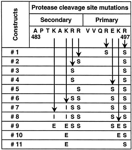
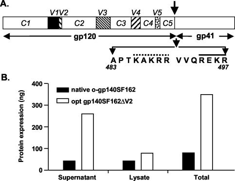
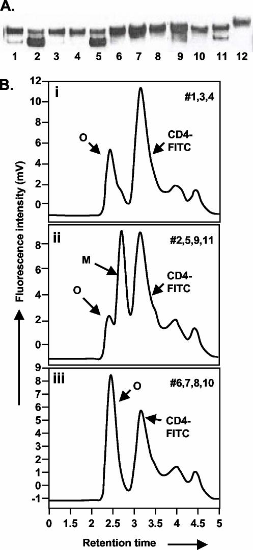
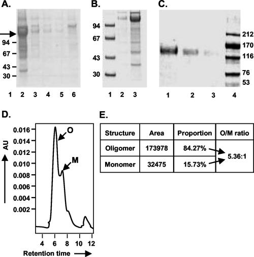
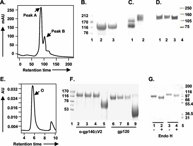
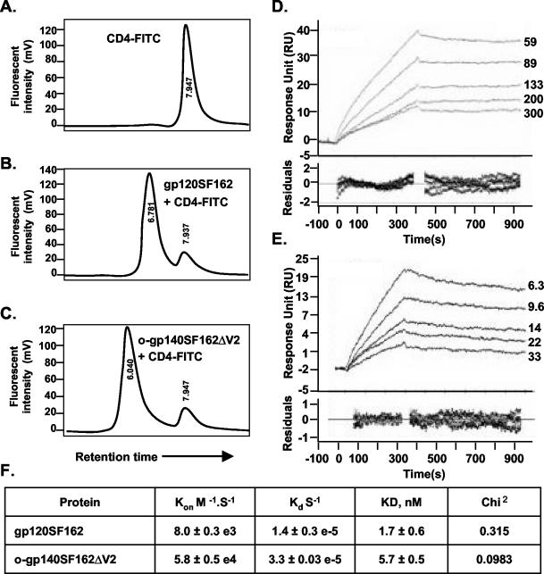
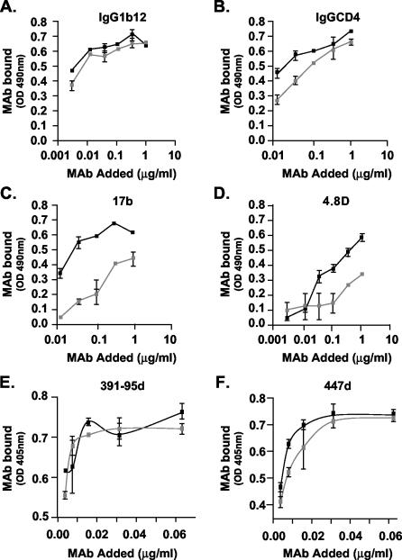
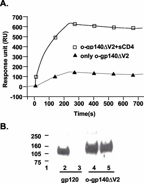
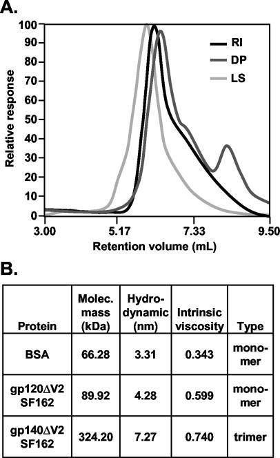
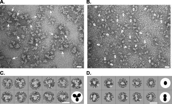
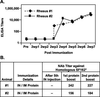
The envelope (Env) glycoprotein of human immunodeficiency virus type 1 (HIV-1) is the major target of neutralizing antibody responses and is likely to be a critical component of an effective vaccine against AIDS. Although monomeric HIV envelope subunit vaccines (gp120) have induced high-titer antibody responses and neutralizing antibodies against laboratory-adapted HIV-1 strains, they have failed to induce neutralizing antibodies against diverse heterologous primary HIV isolates. Most probably, the reason for this failure is that the antigenic structure(s) of these previously used immunogens does not mimic that of the functional HIV envelope, which is a trimer, and thus these immunogens do not elicit high titers of relevant functional antibodies. We recently reported that an Env glycoprotein immunogen (o-gp140SF162DeltaV2) containing a partial deletion in the second variable loop (V2) derived from the R5-tropic HIV-1 isolate SF162, when used in a DNA priming-protein boosting vaccine regimen in rhesus macaques, induced neutralizing antibodies against heterologous subtype B primary isolates as well as protection to the vaccinated animals upon challenge with pathogenic SHIV(SF162P4) virus. Here we describe the purification of this protein to homogeneity, its characterization as trimer, and its ability to induce primary isolate-neutralizing responses in rhesus macaques. Optimal mutations in the primary and secondary protease cleavage sites of the env gene were identified that resulted in the stable secretion of a trimeric Env glycoprotein in mammalian cell cultures. We determined the molecular mass and hydrodynamic radius (R(h)) using a triple detector analysis (TDA) system. The molecular mass of the oligomer was found to be 324 kDa, close to the expected M(w) of a HIV envelope trimer protein (330 kDa), and the hydrodynamic radius was 7.27 nm. Negative staining electron microscopy of o-gp140SF162DeltaV2 showed that it is a trimer with considerable structural flexibility and supported the data obtained by TDA. The structural integrity of the purified trimeric protein was also confirmed by determinations of its ability to bind the HIV receptor, CD4, and its ability to bind a panel of well-characterized neutralizing monoclonal antibodies. No deleterious effect of V2 loop deletion was observed on the structure and conformation of the protein, and several critical neutralization epitopes were preserved and well exposed on the purified o-gp140SF162DeltaV2 protein. In an intranasal priming and intramuscular boosting regimen, this protein induced high titers of functional antibodies, which neutralized the vaccine strain, i.e., SF162. These results highlight a potential role for the trimeric o-gp140SF162DeltaV2 Env immunogen in a successful HIV vaccine.


Figures

FIG. 1.
FIG. 1.
Mutations in the primary (REKR) and secondary protease (KAKRR) cleavage sites in the Env polypeptide.
FIG. 2.
FIG. 2.
Structure, expression, and stabilization of HIV-1 SF162ΔV2 envelope glycoprotein in oligomeric conformation. (A) Linear map of the HIV-1 gp120ΔV2 and gp41 envelope glycoproteins. The gp120 variable regions (V1 to V5) are indicated as solid squares, and arginine 483-to-serine 497 mutation in the protease cleavage site between gp120 and gp41 is indicated by an arrow. The primary (solid line) and secondary cleavage (dotted line) sites are indicated. The gp41 portion includes the N and C α helical regions known as oligomerization domain. (B) Effect of sequence modification upon expression of gp140ΔV2. Total protein expression obtained from native and sequence-modified constructs by using capture ELISA in different fractions is presented as nanograms/well.
FIG. 3.
FIG. 3.
Protease cleavage site scanning for optimum mutations required for stabilization of HIV-1 SF162ΔV2 oligomers. (A) Immunoprobing data. Lanes 1 to 11 correspond to constructs 1 to 11 in Fig. 1; lane 12 is o-gp140 US4. (B) A representative CD4 binding profile obtained for protease cleavage site mutations constructs. Profile i was obtained for constructs 1, 3, and 4; profile ii was obtained for constructs 2, 5, 9, and 11; profile iii was obtained for constructs 6, 7, 8, and 10. Peaks representing oligomer (O), monomer (M), and CD4 are indicated. Based on the structure and expression data, we selected construct no. 7 for developing stable CHO cell lines.
FIG. 4.
FIG. 4.
Analysis of gp140SF162ΔV2 containing fractions obtained at every step during purification. SDS-PAGE analysis of fractions obtained from GNA column (A) (lanes 2 to 6, elutions 1 to 5 from GNA column), from DEAE column (B) (lanes: 1, molecular weight standards; 2, unconcentrated flowthrough; 3, 10× concentrated DEAE flowthrough), and from CHAP column (C) (lanes: 1, 2, and 3, 10× concentrated flowthrough at 2, 1, and 0.5 μg, respectively; 4, molecular weight standards). o-gp140ΔV2 SF162 protein is indicated by an arrow. Also shown are size exclusion HPLC profile of gp140SF162ΔV2 after the CHAP column (D) and summary of the data (E).
FIG. 5.
FIG. 5.
Purification and glycosylation linkage analysis of o-gp140SF162ΔV2. (A) gp140SF162ΔV2 obtained after the CHAP column was further fractionated on a precalibrated Superdex-200 sizing column to separate oligomers from the dimers and/or monomers of gp140. Peaks corresponding to oligomer (peak A) and dimer or monomer (peak B) are indicated. (B and C) Polyacrylamide gel analysis of the sizing fractions in reducing and denaturing conditions (B) (lanes: 1, molecular weight standards; 2, o-gp140SF162ΔV2; 3, gp140SF162ΔV2 dimer/monomer) and native conditions (C) (lanes: 1, dimer and monomer; 2, oligomer). (D) Immunodetection of o-gp140 using a MAb (20-2-C8.5F3) directed against the C4 domain of gp120 SF2 (lanes: 1, o-gp140SF162ΔV2;2, gp140SF162ΔV2 monomer; 3, gp120SF2). (E) Size exclusion-HPLC profile of the purified o-gp140SF162ΔV2. (F) Carbohydrate linkage analysis of purified o-gp140SF162ΔV2. Lanes: 3 and 7, o-gp140SF162ΔV2 and gp120SF162 digested with NANase; 4 and 8, O-glycosidase; 5 and 9, PNGF; 2 and 6, o-gp140SF162ΔV2 and gp120SF162 without any enzyme, used as controls; 1, molecular weight standards. (G) Endo-H digestion of gp120SF162 (lanes 1 and 2) and o-gp140SF162ΔV2 (lanes 3 and 4). + and −, presence or absence of Endo-H.
FIG. 6.
FIG. 6.
(A to C) Shown is binding of purified gp120SF162 (B) and o-gp140SF162ΔV2 (C) to CD4, as determined by an HPLC-based assay, and an unbound CD4 profile (A). (D and E) Shown is the kinetics analysis of gp120SF162 (D) and o-gp140SF162ΔV2 (E) binding to immobilized sCD4. Different sensograms in panels D and E were generated by using a range of concentrations of gp120SF162 (59 to 300 nM) (D) and o-gp140SF162ΔV2 (6.3 to 33 nM) (E). Sensor data were prepared for kinetic analysis by subtracting binding responses collected from a blank reference surface. The association and dissociation phase data were fitted simultaneously to a single-site binding model by using Biaevaluation 3.2. (F) Summary of the kinetic data.
FIG. 7.
FIG. 7.
Immunochemical characterization of purified gp120SF162 (gray squares) and o-gp140SF162ΔV2 (black squares) using a panel of MAbs, namely, IgG1b12 (A), IgGCD4 (B), 17b (C), 4.8d (D), 391-95d (E), and 447d (F).
FIG. 8.
FIG. 8.
Increased exposure of CD4i epitope recognized by the MAb 4.8d in o-gp140SF162ΔV2 upon binding to CD4, as determined by Biacore. (A) For this experiment, a reactive surface was prepared by capturing the MAb 4.8d on CM5 Bia-chip, and o-gp140SF162ΔV2 with (□) and without (▴) incubation with CD4 was flowed over this surface. Data were analyzed by using Biaevaluation 3.2. (B) Recognition of gp120SF162 and o-gp140SF162ΔV2 by MAbs 2F5 and 2G12 in Farr-Western assay. Lanes: 2 and 3, reactivity of gp120SF162 with 2G12 and 2F5, respectively; 4 and 5, recognition of o-gp140SF162ΔV2 by 2G12 and 2F5, respectively.
FIG. 9.
FIG. 9.
Biophysical characterization of purified o-gp140SF162ΔV2 using TDA system. (A) Relative responses obtained for light scattering (LS), refractive index detector (RI), and viscometer (DP) analyses for o-gp140SF162ΔV2. The majority of the purified protein is homogeneous. (B) Summary of biophysical properties, including molecular weight, intrinsic viscosity and hydrodynamic radius, of o-gp140SF162ΔV2 compared to gp120SF162ΔV2.
FIG. 10.
FIG. 10.
EMs of o-gp140SF162ΔV2 obtained from preparative SEC (Fig. 5A). (A) EM of peak A (Fig. 5A) containing trilobed molecules (trimers) indicated with arrows. (B) EM of peak B (Fig. 5A) preparation with presumptive monomers and dimers indicated with arrowhead-balls and arrows, respectively. (C) Selected trimers from peak A. (D) Selected monomers (top) and dimers (bottom) from peak B. To aid interpretation, the molecules in panels C and D were enlarged, masked, and rotated such that one of the lobes of the dimers and trimers points downward. Graphic representations of a trimer and monomer and dimer (not drawn to scale) are shown in panels C and D, respectively. Bars, 20 nm.
FIG. 11.
FIG. 11.
Binding (A) (determined by ELISA) and neutralizing (B) antibodies induced in rhesus macaques following intranasal priming and intramuscular boosting regimen using o-gp140SF162ΔV2 in combination with LTK63 or LTR72. Briefly, two rhesus macaques were immunized five times at 3-week intervals with o-gp140SF162ΔV2 (300 μg) and LTK63 or LTR72 (100 μg) in a total volume of 800 μl (400 μl per nostril). After a resting period of 4 months, both the animals were immunized twice with 100 μg of o-gp140SF162ΔV2 in MF59 intramuscularly at 4-week intervals. For determining the neutralizing activity, samples were assayed at threefold dilutions in triplicate in M7-luc cells. Neutralization antibody (NAb) titers are the serum dilutions at which luciferase activity was reduced to 80% relative to the virus control wells (cells and virus).

References

    1. Back, N. K., L. Smit, J. J. De Jong, W. Keulen, M. Schutten, J. Goudsmit, and M. Tersmette. 1994. An N-glycan within the human immunodeficiency virus type 1 gp120 V3 loop affects virus neutralization. Virology 199:431-438. - PubMed
    1. Barnett, S. W., S. Lu, I. Srivastava, S. Cherpelis, A. Gettie, J. Blanchard, S. Wang, I. Mboudjeka, L. Leung, Y. Lian, A. Fong, C. Buckner, A. Ly, S. Hilt, J. Ulmer, C. T. Wild, J. R. Mascola, and L. Stamatatos. 2001. The ability of an oligomeric human immunodeficiency virus type 1 (HIV-1) envelope antigen to elicit neutralizing antibodies against primary HIV-1 isolates is improved following partial deletion of the second hypervariable region. J. Virol. 75:5526-5540. - PMC - PubMed
    1. Barnett, S. W., S. Rajasekar, H. Legg, B. Doe, D. H. Fuller, J. R. Haynes, C. M. Walker, and K. S. Steimer. 1997. Vaccination with HIV-1 gp120 DNA induces immune responses that are boosted by a recombinant gp120 protein subunit. Vaccine 15:869-873. - PubMed
    1. Barr, P. J., K. S. Steimer, E. A. Sabin, D. Parkes, C. George-Nascimento, J. C. Stephans, M. A. Powers, A. Gyenes, G. A. Van Nest, E. T. Miller, et al. 1987. Antigenicity and immunogenicity of domains of the human immunodeficiency virus (HIV) envelope polypeptide expressed in the yeast Saccharomyces cerevisiae. Vaccine 5:90-101. - PubMed
    1. Binley, J. M., R. W. Sanders, B. Clas, N. Schuelke, A. Master, Y. Guo, F. Kajumo, D. J. Anselma, P. J. Maddon, W. C. Olson, and J. P. Moore. 2000. A recombinant human immunodeficiency virus type 1 envelope glycoprotein complex stabilized by an intermolecular disulfide bond between the gp120 and gp41 subunits is an antigenic mimic of the trimeric virion-associated structure. J. Virol. 74:627-643. - PMC - PubMed

Publication types

Substances