. 2003 Dec 15;553(Pt 3):881-93.
doi: 10.1113/jphysiol.2003.049700. Epub 2003 Sep 26.

Migrating motor complexes do not require electrical slow waves in the mouse small intestine

Affiliations

Migrating motor complexes do not require electrical slow waves in the mouse small intestine

Nick J Spencer et al. J Physiol. .

Abstract

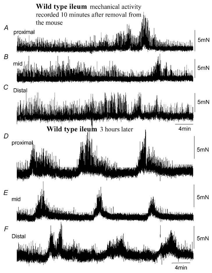
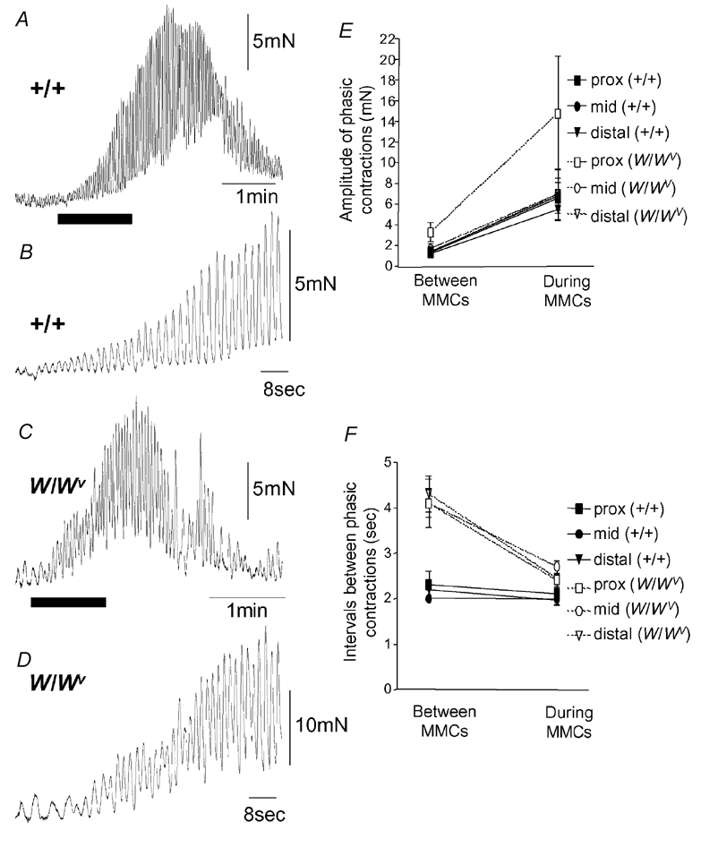
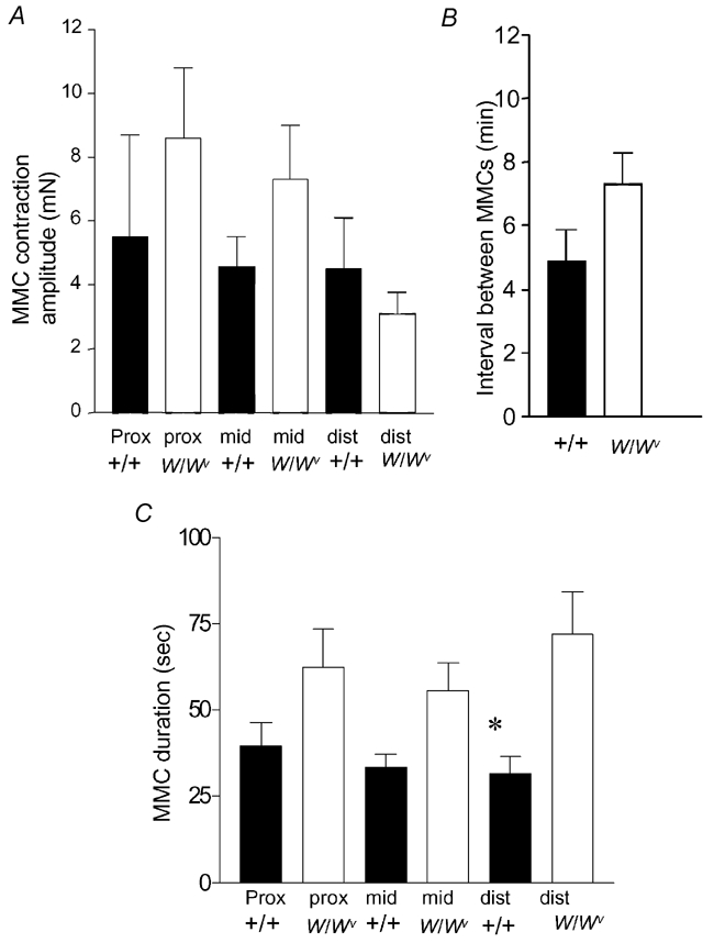
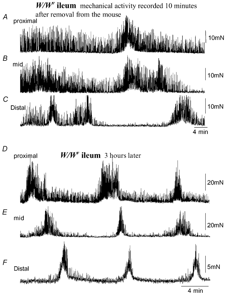
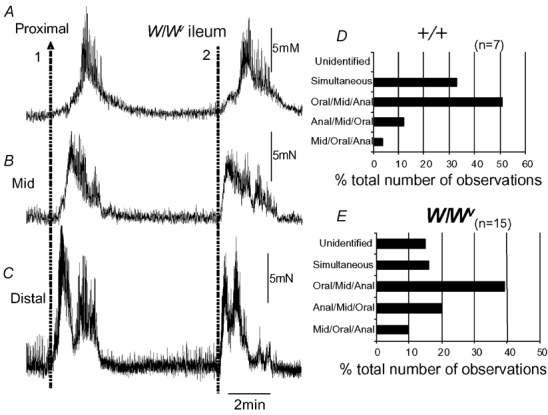
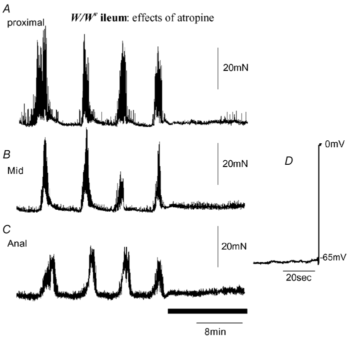
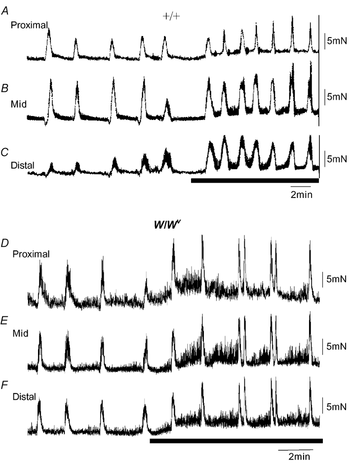
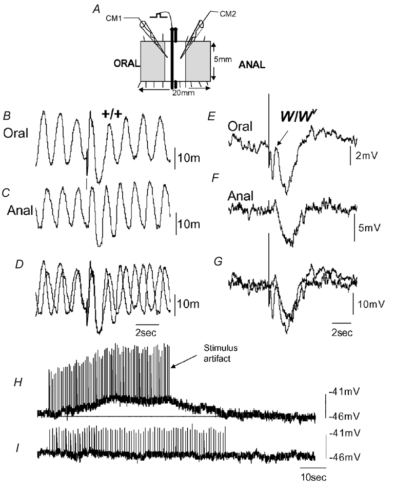
We have investigated whether migrating motor complexes (MMCs) are impaired or absent in the small intestine of W/Wv mutant mice, which lack pacemaker interstitial cells of Cajal (ICC) and electrical slow waves. The intracellular electrical and mechanical activities of the small intestines of wild-type (+/+) and W/Wv mutant mice were recorded. Electrical recordings from circular muscle cells confirmed the absence of slow waves in W/Wv mice, whereas slow waves were always recorded from +/+ muscle cells. Spontaneous phasic contractions were recorded from W/Wv muscles in the absence of slow waves, but these events occurred at a lower frequency than in +/+ tissues. MMC activity was recorded consistently from the ileum of +/+ mice, and normal MMCs were also recorded from W/Wv mice. MMCs in both +/+ and W/Wv mice were abolished by tetrodotoxin (1 microM), hexamethonium (300 microM) or atropine (1 microM), suggesting that the neural control mechanisms responsible for MMCs in +/+ mice are intact and are responsible for MMCs in W/Wv mice. Transmural nerve stimulation demonstrated intact inhibitory and excitatory neural regulation of W/Wv intestinal muscles. Prolonged trains of cholinergic motor nerve stimulation failed to activate slow waves in the intestinal muscles of W/Wv mice. Our findings show that the generation and directional propagation of MMC activity in mouse small intestine does not require slow-wave activity or an intact network of myenteric ICC. The generation and propagation of MMCs appear to be an intrinsic capability of the enteric nervous system and are not related to slow waves or the gradient in slow-wave frequency.


Figures

Figure 1
Figure 1. Mechanical recordings made simultaneously from three sites of the ileum: proximal, mid and distal
A, B and C, mechanical recordings made simultaneously from the ileum at three sites: proximal, mid and distal, respectively. Immediately upon removal from the animal, spontaneous phasic contractions were recorded that were temporally uncoordinated between each recording site. No clear MMCs were present. D, E and F, in the same preparation, approximately 3 h after the recordings in A–C were made, spontaneous MMCs were observed. MMCs originated in the proximal ileum and propagated to the terminal ileum.
Figure 3
Figure 3. Comparison of the amplitudes and intervals between phasic contractions in +/+ and W/Wv mice
A, a single MMC contraction recorded from a +/+ mouse ileum. The recording represented by the black bar is shown on expanded time scale in B. B, the increase in amplitude of phasic contractions during the onset of the MMC. Note, the interval between phasic contractions does not change. C, a single MMC contraction recorded from a W/Wv mouse. The black bar is shown on an expanded time scale in D. D, the increase in amplitude and decrease in interval between phasic contractions during the onset of the MMC. E, a graphical representation of the changes in phasic contraction amplitude from the period between MMCs to the peak of the MMC contraction. Both +/+ and W/Wv mice exhibited a significant increase in amplitude of phasic contractions during the MMC in the proximal, mid and distal ileum. F, in the +/+ mouse, the intervals between phasic contractions remained relatively constant from the transition between MMCs to the peak of the MMC (every ≈2 s). These intervals were not significantly different from one another (proximal, P = 0.64; mid, P = 0.74; distal, P = 0.50). However, in the W/Wv mouse, the proximal, mid and distal ileum exhibited a significant decrease in phasic contraction intervals from the transition between MMCs to the peak of the MMC (P = 0.006, P < 0.001 and P < 0.001, respectively).
Figure 2
Figure 2. Statistical comparison of the similarities of MMCs recorded from +/+ and W/Wv mouse ileum
A, there was no significant difference in MMC peak contraction amplitudes in either the proximal (prox), mid or distal (dist) regions of ileum obtained from +/+ or W/Wv mice. B, no significant difference was noted in the interval between MMCs in +/+ or W/Wv mice. C, the mean half-durations between MMC contractions in the proximal and mid ileum were not significantly different between +/+ and W/Wv mice. The half-durations of MMC contractions in the distal ileum of W/Wv mice were found to be significantly longer than in +/+ controls.
Figure 4
Figure 4. Mechanical activity recorded from isolated W/Wv mouse ileum
A, B and C, simultaneous recordings from the proximal (A), mid (B) and distal regions (C) of the W/Wv mouse ileum. Immediately upon removal from the mouse, spontaneous phasic contractions were recorded that were temporally uncoordinated between each recording site. No distinct MMC activity was present. D, E and F, approximately 3 h after the recording was made in A, B and C, spontaneous MMCs were observed, despite the absence of intestinal slow waves. MMCs in this example propagate from the proximal to distal ileum without slow waves.
Figure 5
Figure 5. Comparison of the direction of propagation of MMCs recorded from the ileum of +/+ and W/Wv mice
A, B and C, two MMC contractions, one that propagates from the anal to the oral region of the ileum (orally migrating, represented by arrow 1), while another (represented by the dotted line 2) appears to have occurred simultaneously in the proximal, mid and distal regions. D, the proportion of MMCs in +/+ mice where the direction of propagation was noted. E, shows the same graphical representation of MMCs in W/Wv mice and the direction of MMC propagation. In both types of mice, the predominant direction of propagation was from the proximal to mid to distal ileum.
Figure 6
Figure 6. Effects of atropine on MMCs recorded from the W/Wv mouse ileum
Anally migrating MMCs were recorded. Addition of atropine (1 μm; see black bar) immediately abolished all MMC activity. Intracellular recordings were made from CM at different regions of the ileum, proximal (A), mid (B) and distal (C) from the same preparation. Slow waves were confirmed to be absent from the preparation where MMCs were present. The electrical recording (D) shows withdrawal of the electrode.
Figure 7
Figure 7. Effects of inhibition of NO synthesis on MMCs recorded from +/+ and W/Wv mouse ileum
A, B and C, a recording of mechanical activity in the proximal, mid and distal ileum, respectively, in a +/+ mouse. MMCs occurred about every 4 min. The period represented by the hatched bar shows the point of application of l-NA (100 μm), where the resting tone increased and the interval between MMCs decreased at each point in the ileum. D, E and F, a typical example recording of mechanical activity from W/Wv mouse ileum. l-NA also increased the resting tension at each site along the ileum and decreased the interval between MMCs.
Figure 8
Figure 8. Intracellular electrical recordings from the CM and responses to transmural nerve stimulation in +/+ and W/Wv mouse ileum
A, schematic of the flat-sheet preparation used. Two recording electrodes were impaled simultaneously in two CM cells, one oral and one anal of the transmural wires. Recordings were made 1.2 mm oral and anal from the stimulating wires. B and C, simultaneous recordings from two CM cells shows spontaneous slow-wave activity. A brief train of stimuli (10 Hz, 0.5 s, 50 V) was applied to the enteric nerves, and an IJP was evoked both oral and anal to the stimulating wires. D, the same simultaneous recordings shown in B and C superimposed. E and F, simultaneous recordings from two CM cells in the W/Wv mouse ileum. Note the absence of slow waves. A train of stimuli (10 Hz, 0.5 s, 50 V) delivered to the ileum also evoked slow IJPs both oral and anal to the stimulus. At the oral recording site, a small EJP can be seen to truncate the IJP (see arrow in E). G, recordings shown in E and F superimposed. H, in the presence of l-NA (100 μm) and apamin (500 nm) to block inhibitory neurotransmission, a prolonged train of nerve stimuli (2 Hz, 40 s, 0.4 ms, 40 V) evoked a slow membrane depolarization of the CM, but no slow waves were induced. I, the effects of atropine (1 μm) on the response to nerve stimulation in the same animal. It can be seen that the depolarization is abolished.

References

    1. Andrews JM, O'Donovan DG, Hebbard GS, Malbert CH, Doran SM, Dent J. Human duodenal phase III migrating motor complex activity is predominantly antegrade, as revealed by high-resolution manometry and colour pressure plots. Neurogastroenterol Motil. 2002;14:331–338. - PubMed
    1. Beckett EAH, Horiguchi K, Khoyi M, Sanders KM, Ward SM. Loss of enteric motor neurotransmission in the gastric fundus of Sl/Sld mice. J Physiol. 2002;543:859–858. - PMC - PubMed
    1. Burns AJ, Lomax AE, Torihashi S, Sanders KM, Ward SM. Interstitial cells of Cajal mediate inhibitory neurotransmission in the stomach. Proc Natl Acad Sci U S A. 1996;93:12008–12013. - PMC - PubMed
    1. Bush TG, Spencer NJ, Watters N, Sanders KM, Smith TK. Spontaneous migrating motor complexes occur in both the terminal ileum and colon of the C57BL/6 mouse in vitro. Auton Neurosci. 2000;84:162–168. - PubMed
    1. Bush TG, Spencer NJ, Watters N, Sanders KM, Smith TK. Effects of alosetron on spontaneous migrating motor complexes in the murine small and large bowel. Am J Physiol. 2001;281:G974–983. - PubMed

Publication types

LinkOut - more resources