

Wnt ligand expression in malignant melanoma: pilot study indicating correlation with histopathological features
- PMID: 14514922
- PMCID: PMC1187339
- DOI: 10.1136/mp.56.5.280
Wnt ligand expression in malignant melanoma: pilot study indicating correlation with histopathological features
Abstract
Aims: Secreted Wnt ligands are key proteins regulating cell-cell interactions and cell growth and differentiation. These proteins, along with other components of the Wnt signalling pathway, are involved in the malignant transformation of various human cancers, including malignant melanoma. This study defines the expression of several members of the Wnt ligand family and correlates their expression with histological characteristics.
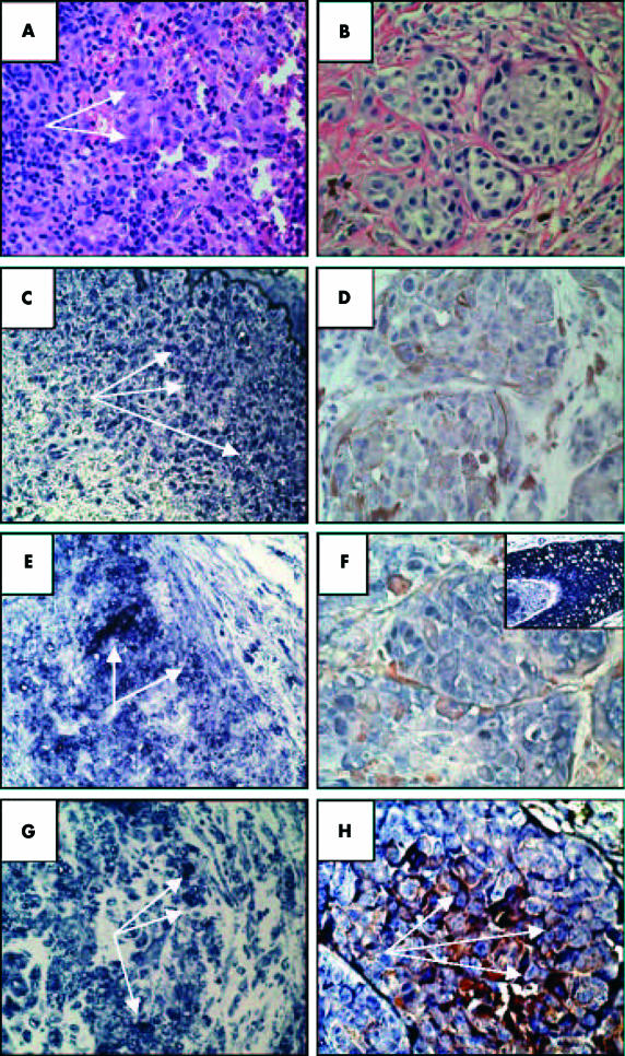
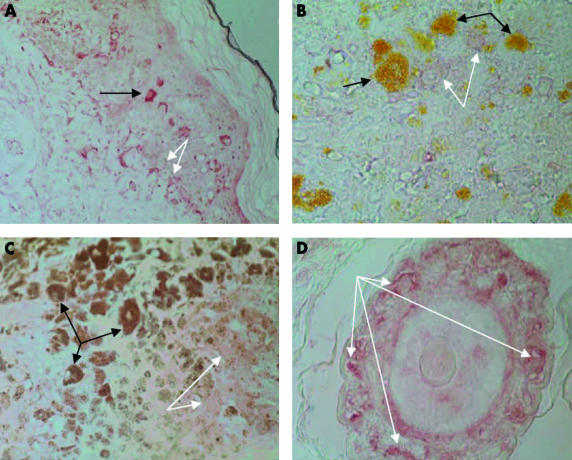
Methods: The expression of Wnt2, Wnt5a, Wnt5b, Wnt7b, and Wnt10b was defined by in situ, antisense RNA hybridisation of paraffin wax embedded sections of benign naevi and malignant melanoma. Immunoperoxidase based antibody staining was used to define the expression of frizzled (Fz) receptors.
Results: All naevi tested strongly expressed Wnt2, Wnt5a, Wnt7b, and Wnt10b. Melanomas characterised by small, uniform cells expressed each of the Wnts in a pattern similar to that seen for benign naevi. In contrast, melanomas characterised by large, pleomorphic cells expressed Wnt10b but did not express Wnt2 and had low levels of expression of Wnt5a. Expression of Wnt7b was variable in these melanomas. Fz receptor expression was present at a low level in normal epithelium and all naevi and melanomas.
Conclusions: The expression pattern of Wnt ligands in malignant melanoma correlates with histopathological features and may provide a basis for the molecular classification of this disease.
Figures

References
-
- Polakis P. Wnt signaling and cancer. Genes Dev 2000;14:1837–51. - PubMed
-
- Theisen H, Purcell J, Bennett M, et al. Dishevelled is required during wingless signaling to establish both cell polarity and cell identity. Development 1994;120:347–60. - PubMed
-
- Behrens J, von Kries JP, Kuhl M, et al. Functional interaction of β-catenin with the transcription factor LEF1. Nature 1996;382:638–42. - PubMed
-
- He TC, Sparks AB, Rago C, et al. Identification of c-MYC as a target of the APC pathway. Science 1998;281:1509–12. - PubMed
-
- Howe LR, Subbaramaiah K, Chung WJ, et al. Transcriptional activation of cyclooxygenase-2 in Wnt-1-transformed mouse mammary epithelial cells. Cancer Res 1999;59:1572–7. - PubMed
Publication types
MeSH terms
Substances
Grants and funding
LinkOut - more resources
Full Text Sources
Other Literature Sources
Medical