. 2003 Nov 3;466(1):80-90.
doi: 10.1002/cne.10880.

AMPA receptors mediate acetylcholine release from starburst amacrine cells in the rabbit retina

Affiliations

AMPA receptors mediate acetylcholine release from starburst amacrine cells in the rabbit retina

Sally I Firth et al. J Comp Neurol. .

Abstract

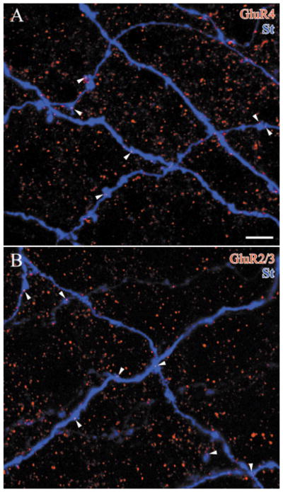
The light response of starburst amacrine cells is initiated by glutamate released from bipolar cells. To identify the receptors that mediate this response, we used a combination of anatomical and physiological techniques. An in vivo, rabbit eyecup was preloaded with [(3)H]-choline, and the [(3)H]-acetylcholine (ACh) released into the superfusate was monitored. A photopic, 3 Hz flashing light increased ACh release, and the selective AMPA receptor antagonist, GYKI 53655, blocked this light-evoked response. Nonselective AMPA/kainate agonists increased the release of ACh, but the specific kainate receptor agonist, SYM 2081, did not increase ACh release. Selective AMPA receptor antagonists, GYKI 53655 or GYKI 52466, also blocked the responses to agonists. We conclude that the predominant excitatory input to starburst amacrine cells is mediated by AMPA receptors. We also labeled lightly fixed rabbit retinas with antisera to choline acetyltransferase (ChAT), AMPA receptor subunits GluR1, GluR2/3, or GluR4, and kainate receptor subunits GluR6/7 and KA2. Labeled puncta were observed in the inner plexiform layer with each of these antisera to glutamate receptors, but only GluR2/3-IR puncta and GluR4-IR puncta were found on the ChAT-IR processes. The same was true of starburst cells injected intracellularly with Neurobiotin, and these AMPA receptor subunits were localized to two populations of puncta. The AMPA receptors are expected to desensitize rapidly, enhancing the sensitivity of starburst amacrine cells to moving or other rapidly changing stimuli.


Figures

Fig. 1
Fig. 1
A: [3H]-acetylcholine (ACh) released from a single superfused rabbit retina. These are responses to either photopic 3 Hz flashing light (*) for 4 minutes or kainate (KA) 15 μM for 1 minutes, with peak-to-base ratios of 2.8 and 15.9, respectively. These responses were blocked by the selective AMPA antagonist GYKI 53655 20 μM (GY53655) given for 1 minute prior and during the stimulus. B: Photopic 3 Hz light stimuli increased [3H]-ACh release (P = 0.0085). The change in [3H]-ACh release following flashing light (n = 4 rabbits) was significantly suppressed by GYKI 53655 (P = 0.0013), to [3H]-ACh release not significantly different from applying GYKI 53655 alone (P = 0.74). C: Kainate 15 μM significantly increased [3H]-ACh release (P = 0.025). The change in [3H]-ACh release following kainate 15 μM was significantly suppressed GYKI 53655 (P = 0.0045) to [3H]-ACh release not significantly different from GYKI 53655 alone (P = 0.99).
Fig. 2
Fig. 2
A: Change in [3H]-ACh release in response to bromowillardiine 0.5, 1, 2, 5, 10, and 20 μM (n = 3 rabbits, except 0.5 and 20 μM where n = 2). B: Bromowillardiine 5 μM significantly increased [3H]-ACh (P = 0.04). The change in [3H]-ACh release following bromowillardiine 5 μM (n = 4 rabbits) was significantly suppressed by a specific AMPA antagonist, GYKI 52466, 20 μM (P = 0.014) to [3H]-ACh release not significantly different from GYKI 52466 20 μM given alone (P = 0.76). SYM 2081 did not significantly change [3H]-ACh release at 100 μM (P = 0.14, n = 4 rabbits).
Fig. 3
Fig. 3
GluR4-IR puncta (red) are localized on the starburst amacrine cell dendrites labeled with antibodies to choline acetyltransferase (ChAT, blue). Labeling with the synaptic ribbon antibody kinesin II (KII, green) is included as a control. These optical sections are from sublamina b of the starburst amacrine cell plexus in peripheral whole-mounted retina. A: In this stack of optical sections (5 × 0.5 μm), many GluR4-IR puncta are localized on ChAT processes, while many other GluR4-IR puncta are not. The area within the box is shown at higher magnifications in B, C, D. B: A single optical section shows the triple label of GluR4, KII, and ChAT. C: In the same image as B, with the green channel hidden, some of the GluR4-IR puncta were found on the ChAT-IR processes (arrows). D: The same section as B, C is shown with the blue channel hidden. This demonstrates that most of the GluR4-IR puncta are associated with synaptic ribbons from bipolar cells (arrows). E–G: Signal averaging analysis shows the peak of GluR4 signal (E) correlated with a broader peak of ChAT signal (G). The KII signal (F) is deviated from the center of GluR4 peak (E), indicating the ribbons are adjacent, but not colocalized with GluR4. Scale bar = 5 μm in B (applies to B–D); 10 μm for A.
Fig. 4
Fig. 4
GluR2/3 glutamate receptor-IR puncta (red) are also localized on the ChAT-IR processes. As a control, bipolar cell synaptic ribbons are labeled with antibodies to kinesin II (green). These optical sections are from sublamina b of the starburst amacrine cell plexus in peripheral whole-mounted retina. A: GluR2/3-IR puncta are detected in the vicinity of the ChAT-IR processes. There are also GluR2/3-IR puncta that are not associated with the ChAT processes, but many GluR2/3-IR puncta are localized on ChAT-IR processes (arrows). B: The association of many of the GluR2/3-IR puncta with the kinesin-IR puncta suggests that most, if not all, of the GluR2/3-IR puncta are present at synaptic densities. Scale bars = 5 μm. C–E: Signal averaging analysis shows the peak of GluR2/3 signal (C) correlated with a broader peak of ChAT signal (E). The KII signal (D) is deviated from the center of GluR2/3 peak, indicating the ribbons are adjacent, but not colocalized with GluR4.
Fig. 5
Fig. 5
A: The GluR1-IR puncta (red) are very rarely detected on the ChAT-IR starburst amacrine cell dendrites (blue) in this stack of two optical sections (0.5 μm). There are not sufficient numbers of GluR1-IR puncta seen on the starburst amacrine cell processes to differentiate these puncta from chance superposition of puncta from neighboring dendrites. B: GluR6/7-IR puncta are rare in the strata containing ChAT immunoreactivity and colocalized puncta were not detected. C: Kainate receptor subunits, labeled with antibodies to KA2, are detected in the vicinity of the ChAT-IR processes. However, there was no convincing colocalization. D,E: Signal averaging analysis shows the peak of GluR1 signal (D) correlated to the depression of ChAT signal (E), indicating that GluR1 is not colocalized with dendrites of starburst cells. Scale bars = 10 μm.
Fig. 6
Fig. 6
Overlapping dye-injected starburst amacrine cell dendrites (St, blue) are shown in each confocal image. Each of the glutamate receptor-IR puncta localized on the starburst dendrites (arrowheads) could also be demonstrated on a single optical section and was on a swelling, branch point, or spine-like structure. These pieces of rabbit retina are double-labeled and show: A: GluR4-IR puncta (red, 4 × 0.5 μm), and B: GluR2/3-IR puncta (red, 3 × 0.5 μm). Scale bar = 5 μm.
Fig. 7
Fig. 7
A comparison of the distribution of GluR4-IR puncta (green) and GluR2 (3)-IR puncta (red) on a starburst amacrine cell dendrite (St, blue) in a confocal stack (3 × 0.5 μm, right). A: Examples of GluR2 puncta were marked by the white arrowheads and the clear arrowheads indicate the locations of GluR4 puncta in B. B: Examples of GluR4 puncta are marked by the clear arrowheads and the white arrowheads indicate the locations of GluR2 puncta in A. Scale bar = 5 μm.

References

    1. Ames A, Nesbett FB. In vitro retina as an experimental model of the central nervous system. J Neurochem. 1981;37:867–877. - PubMed
    1. Bleakman D, Ballyk BA, Schoepp DD, Palmer AJ, Bath CP, Sharpe EF, Woolley ML, Bufton HR, Kamboj RK, Tarnawa I, et al. Activity of 2,3-benzodiezepines at native rat and recombinant human glutamate receptors in vitro: stereospecificity and selectivity profiles. Neuropharmacology. 1996;35:1689–1702. - PubMed
    1. Bloomfield SA, Miller RF. A functional organization of ON and OFF pathways in the rabbit retina. J Neurosci. 1986;6:1–13. - PMC - PubMed
    1. Brandon C. Cholinergic neurons in the rabbit retina: dendritic branching and ultrastructural connectivity. Brain Res. 1987;426:119–130. - PubMed
    1. Brown SP, Masland RH. Costratification of a population of bipolar cells with direction-selective circuitry of the rabbit retina. J Comp Neurol. 1999;408:97–106. - PubMed

Publication types

MeSH terms

LinkOut - more resources