. 2003 Oct 15;74(2):227-39.
doi: 10.1002/jnr.10759.

Blood-spinal cord barrier after spinal cord injury: relation to revascularization and wound healing

Affiliations

Blood-spinal cord barrier after spinal cord injury: relation to revascularization and wound healing

William D Whetstone et al. J Neurosci Res. .

Abstract

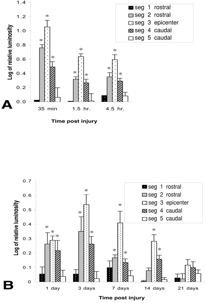
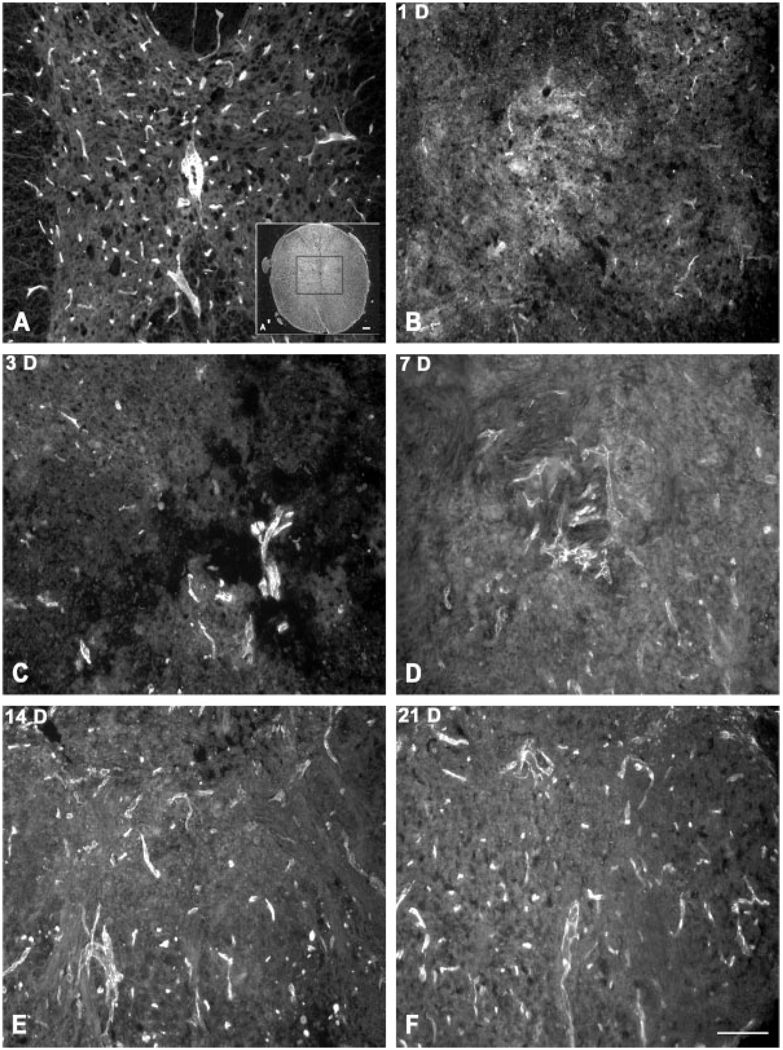
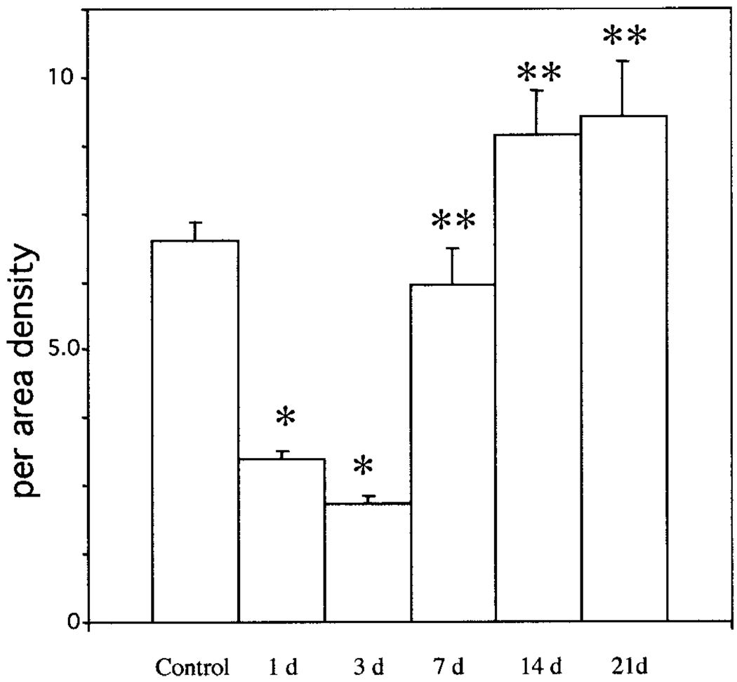
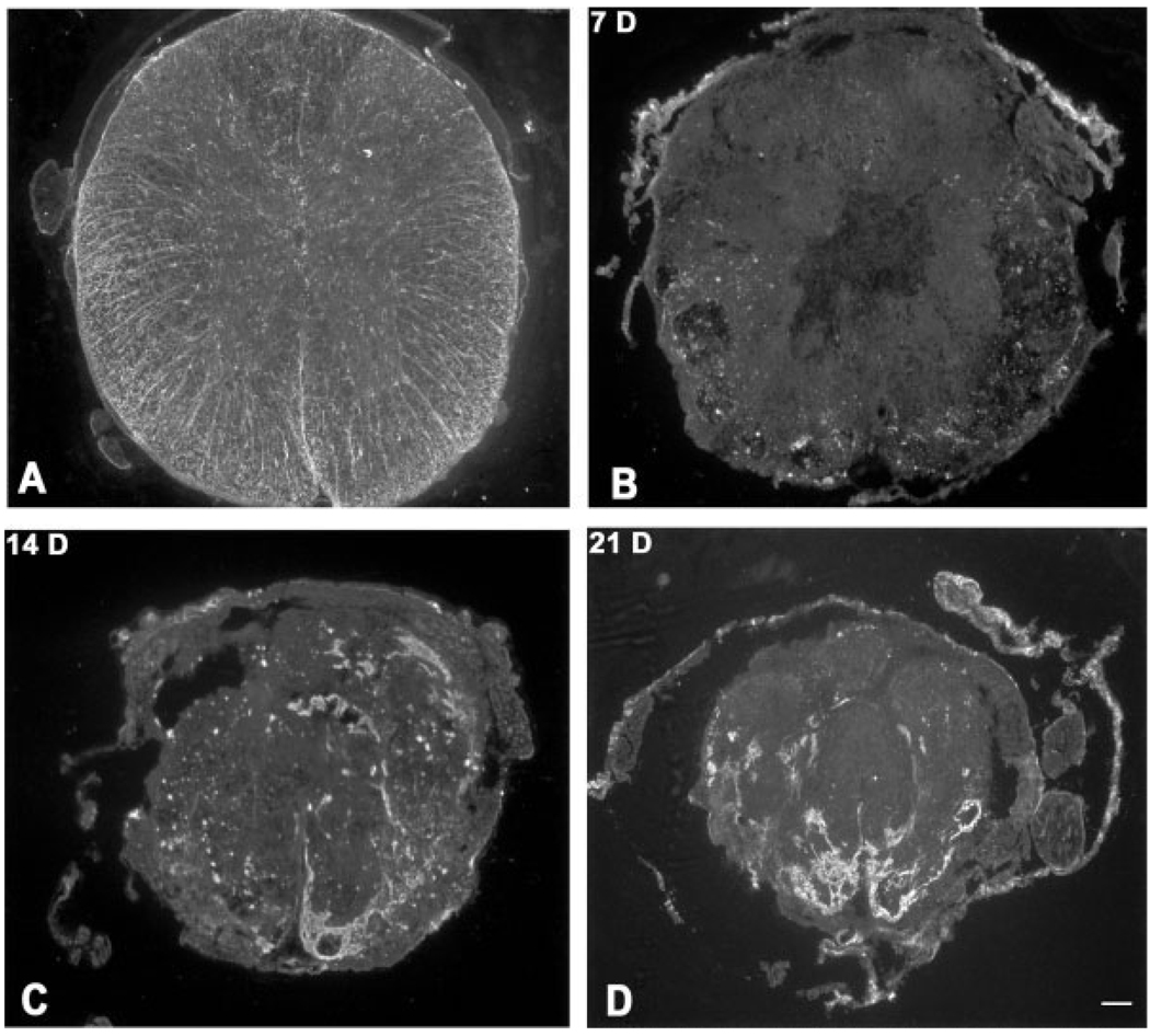
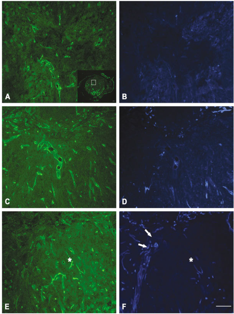
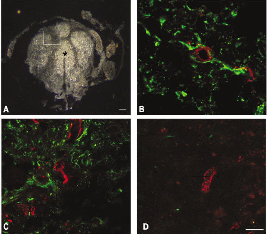
Spinal cord injury produces prominent disruption of the blood-spinal cord barrier. We have defined the blood-spinal cord barrier breakdown to the protein luciferase (61 kDa) in the acutely injured murine spinal cord and during revascularization. We show that newly formed and regenerating blood vessels that have abnormal permeability exhibit differential expression of the glucose-1 transporter (Glut-1), and that its expression is dependent on astrocytes. There was overt extravasation of luciferase within the first hour after injury, a period that coincided with marked tissue disruption within the epicenter of the lesion. Although there was a significant reduction in the number of blood vessels relative to controls by 24 hr after injury, abnormal barrier permeability remained significantly elevated. A second peak of abnormal barrier permeability at 3-7 days postinjury coincided with prominent revascularization of the epicenter. The barrier to luciferase was restored by 21 days postinjury and vascularity was similar to that of controls. During wound-healing process, the cord was reorganized into distinct domains. Between 14 and 21 days postinjury, each domain consisted primarily of nonneuronal cells, including macrophages. Astrocytes were limited characteristically to the perimeter of each domain. Only blood vessels affiliated closely with astrocytes in the perimeter expressed Glut-1, whereas blood vessels within each domain of the repairing cord did not express it. Together, these data demonstrate that both injured and regenerating vessels exhibit abnormal permeability and suggest that Glut-1 expression during revascularization is dependent on the presence of astrocytes.


Figures

Fig. 1
Fig. 1
Distribution of Evans blue albumin (EBA, A–E) and immuno-localization of CD11b (F) after spinal cord injury. Dark-field image of a control (injured) spinal cord (A). Enclosed area denotes pericentral gray matter used for assessment of EBA extravasation in uninjured cord (B) and at 1 (C), 3 (D), and 7 (E) days after spinal cord injury. No extravasation is noted in control, uninjured spinal cord (B). There is both a diffuse and a cellular pattern of EBA fluorescence within the epicenter at 1 (C), 3 (D), and 7 (E) days postinjury. Inset (E) is shown at higher magnification in F, which has been immunolabeled for macrophages. Scale bars = 100 µm.
Fig. 2
Fig. 2
Time course of blood-spinal cord barrier leakage to luciferase. A 15-mm length of cord, centered over impact site, was divided into five segments of equal lengths. Luciferase activity was determined in rostral segments (seg 1 and seg 2), epicenter (epi), and caudal segments (seg 4 and seg 5). Epicenter of injury demonstrates biphasic permeability. The most overt leakage occurs in the first 35 min after injury. Although there is a decline in leakage by 24 hr postinjury, a significant increase in permeability, relative to the 24-hr point, occurs by 3 days after injury**. Values decline thereafter but remain significantly elevated, relative to controls, up to 14 days after injury*. Significant leakage to luciferase occurs in both rostral and caudal segments, adjacent to the epicenter, within the first 3 days after injury. Whereas the barrier to luciferase is restored in rostral segments between 7 and 14 days postinjury, the caudal segment, adjacent to the epicenter, remains significantly permeable until 21 days after injury. Values represent mean ± SEM. *P < 0.05 compared to controls. F-value, 9.64; df = 5. **P < 0.05 as compared to 24-hr point.
Fig. 3
Fig. 3
Immunolocalization of PECAM-1-labeled blood vessels within the lesioned epicenter. PECAM-1-labeled vessels are distributed throughout uninjured spinal cord (A). The central gray matter, as outlined in the inset (A′), was selected for analysis of PECAM-1-labeled vessels after spinal cord injury (B–F). There is an overt loss in PECAM-1-labeled vessels by 1 day after injury. Large, PECAM-1-positive vessels are apparent, although limited in number, by 3 (C) and 7 (D) days postinjury. Both large and small diameter vessels are identified at 14 (E) and 21 (F) days after injury. Scale bar = 100 µm.
Fig. 4
Fig. 4
Revascularization after spinal cord injury. PECAM-1-positive blood vessels were quantified in uninjured and injured spinal cord. There is a significant loss of blood vessels in the first 3 days after injury*. Revascularization is most prominent between 3 and 7 days postinjury, with a significant increase in the number of vessels by 7 days compared to the 3-day point**. Although there is a modest increase in the number of vessels at 14 and 21 days postinjury, these values are not statistically different from that of controls. Values represent mean per-area densities ± SEM. *P < 0.01 as compared to controls; **P < 0.01 as compared to the 3-day point.
Fig. 5
Fig. 5
Formation of heterodomains during wound healing after spinal cord injury. Dark-field optics define the unusual reorganization pattern within the lesioned epicenter. Distinct organization of gray and white matter in control cord (A) contrasts dissolution of tissue at 1 (B) and 3 days (C) after injury. Between 3 (C) and 7 (D) days after injury, dark central homogenous zones become evident. Between 14 (E) and 21 (F) days after injury, the cord is reorganized into bright zones that are surrounded by dark bands. Scale bar = 100 µm.
Fig. 6
Fig. 6
Evolution of glial scarring after injury. Immunolocalization of GFAP-positive astrocytes in the control cord (A) and at 7 (B), 14 (C), and 21 (D) days postinjury. There is an overt loss of GFAP-labeled astrocytes in the first week after injury. Thereafter, astrocytes reorganize into distinct dense bands that segregate the cord into distinct domains. Scale bar = 100 µm.
Fig. 7
Fig. 7
Double immunolabeling for PECAM-1 (A,C,E) and Glut-1 (B,D,F) at 7 (A,B), 14 (C,D), and 21 (E,F) days postinjury. Inset is higher magnification of area in 21-day postinjury cord labeled for GFAP. There seems to be an increase in PECAM-1-labeled vessels between 7 and 14 days after injury. Glut-1-labeled vessels also seem to increase with time but are evident in a more restricted distribution. Only limited numbers of vessels are labeled with both PECAM-1 and Glut-1. By 21 days after injury, PECAM-1-labeled vessels are noted throughout a domain (E, asterisk) whereas Glut-1 labeling is restricted to the perimeter (arrows) surrounding the domain (F, asterisk). Scale bar = 100 µm.
Fig. 8
Fig. 8
Relationship of vessels to astrocytes by confocal microscopy at 21 days postinjury. Heterodomains are evident by dark-field microscopy (A). Enclosed area and the center of a domain (asterisk) are shown at higher magnification in C and D, respectively. In a section rostral to the epicenter, a PECAM-1-labeled vessel (red) is surrounded by astrocytic processes (green, B). Similarly, at the injury epicenter, Glut-1-labeled vessels (red) are in close proximity to astrocytes (C) within the perimeter of the domain (dark band of inset, A). These findings contrast the center of a domain (asterisk in A) mostly void of astrocytes and is composed of vessels that are predominately PECAM-1 positive (red, D). Scale bars = 100 µm (A); 10 µm (B–D).

References

    1. Beck DW, Roberts RL, Olson JJ. Glial cells influence membrane-associated enzyme activity at the blood-brain barrier. Brain Res. 1986;381:131–137. - PubMed
    1. Beck DW, Vinters HV, Hart MN, Cancilla PA. Glial cells influence polarity of the blood-brain barrier. J Neuropathol Exp Neurol. 1984;43:219–224. - PubMed
    1. Bilgen M, Abbe R, Narayana PA. Dynamic contrast-enhanced MRI of experimental spinal cord injury: in vivo serial studies. Magn Reson Med. 2001;45:614–622. - PubMed
    1. Bush TG, Puvanachandra N, Horner CH, Polito A, Ostenfeld T, Svendsen CN, Mucke L, Johnson MH, Sofroniew MV. Leukocyte infiltration, neuronal degeneration, and neurite outgrowth after ablation of scar-forming, reactive astrocytes in adult transgenic mice. Neuron. 1999;23:297–308. - PubMed
    1. Cancilla PA, Berliner J. Brain endothelial-astrocyte interactions. New York: Raven Press; 1993.

Publication types

MeSH terms