Review
. 2003 Sep 30;169(7):681-93.

Diagnosis and management of benign paroxysmal positional vertigo (BPPV)

Affiliations
Review

Diagnosis and management of benign paroxysmal positional vertigo (BPPV)

Lorne S Parnes et al. CMAJ. .

Abstract

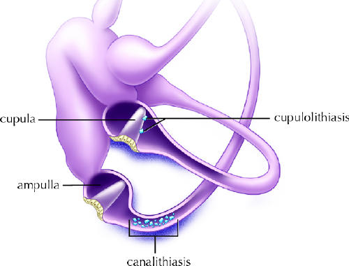
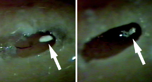
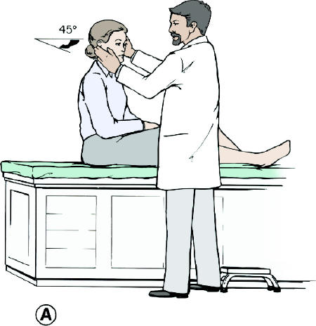
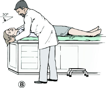
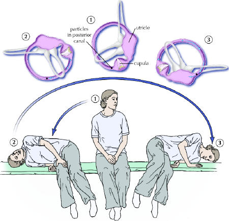
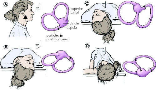
There is compelling evidence that free-floating endolymph particles in the posterior semicircular canal underlie most cases of benign paroxysmal positional vertigo (BPPV). Recent pathological findings suggest that these particles are otoconia, probably displaced from the otolithic membrane in the utricle. They typically settle in the dependent posterior canal and render it sensitive to gravity. Well over 90% of patients can be successfully treated with a simple outpatient manoeuvre that moves the particles back into the utricle. We describe the various techniques for this manoeuvre, plus treatments for uncommon variants of BPPV such as that of the lateral canal. For the rare patient whose BPPV is not responsive to these manoeuvres and has severe symptoms, posterior canal occlusion surgery is a safe and highly effective procedure.


Figures

None
Fig. 1: Spatial orientation of the semicircular canals. Note how the posterior canal on 1 side is in the same plane as the contralateral superior canal. Both lateral canals are in the same plane, 30ο above the horizontal. Photo: Christine Kenney
None
Fig. 2: Osseous (grey/white) and membranous (lavender) labyrinth of the left inner ear. Perilymph fills the osseous labyrinth external to the membranous labyrinth, whereas endolymph fills the membranous labyrinth. Photo: Christine Kenney
None
Fig. 3: Schematic drawing of the physiology of the left posterior semicircular canal. In the image on the right, note the excitatory response (increased neural firing) with utriculofugal cupular displacement. The same excitatory response would occur in the superior (anterior) canal with utriculofugal cupular displacement, whereas the opposite (inhibitory) response would occur with utriculofugal cupular displacement in the lateral canal. The same rules would apply to the image on the left. CNVIII = vestibular nerve, ms = millisecond. Photo: Christine Kenney
None
Fig. 4: Left inner ear. Depiction of canalithiasis of the posterior canal and cupulolithiasis of the lateral canal. Photo: Christine Kenney
None
Fig. 5: Sequential computer-regenerated photographs taken from an intra-operative video of a fenestrated posterior semicircular canal. Note the single white conglomerate mass within the membranous duct (arrow) (left). Note how the mass has fragmented into tiny particles 2–3 minutes later, after the membranous duct has been probed (right).
None
Fig. 6: Dix–Hallpike manoeuvre (right ear). The patient is seated and positioned so that the patient's head will extend over the top edge of the table when supine. The head is turned 45ο toward the ear being tested (position A). The patient is quickly lowered into the supine position with the head extending about 30ο below the horizontal (position B). The patient's head is held in this position and the examiner observes the patient's eyes for nystagmus. In this case with the right side being tested, the physician should expect to see a fast-phase counter-clockwise nystagmus. To complete the manoeuvre, the patient is returned to the seated position (position A) and the eyes are observed for reversal nystagmus, in this case a fast-phase clockwise nystagmus. Photo: Christine Kenney
None
Fig. 7: Liberatory manoeuvre of Semont (right ear). The top panel shows the effect of the manoeuvre on the labyrinth as viewed from the front and the induced movement of the canaliths (from blue to black). This manoeuvre relies on inertia, so that the transition from position 2 to 3 must be made very quickly. Photo: Christine Kenney
None
Fig. 8: Particle repositioning manoeuvre (right ear). Schema of patient and concurrent movement of posterior/ superior semicircular canals and utricle. The patient is seated on a table as viewed from the right side (A). The remaining parts show the sequential head and body positions of a patient lying down as viewed from the top. Before moving the patient into position B, turn the head 45° to the side being treated (in this case it would be the right side). Patient in normal Dix–Hallpike head-hanging position (B). Particles gravitate in an ampullofugal direction and induce utriculofugal cupular displacement and subsequent counter-clockwise rotatory nystagmus. This position is maintained for 1–2 minutes. The patient's head is then rotated toward the opposite side with the neck in full extension through position C and into position D in a steady motion by rolling the patient onto the opposite lateral side. The change from position B to D should take no longer than 3–5 seconds. Particles continue gravitating in an ampullofugal direction through the common crus into the utricle. The patient's eyes are immediately observed for nystagmus. Position D is maintained for another 1–2 minutes, and then the patient sits back up to position A. D = DIRECTION OF VIEW OF LABYRINTH, DARK CIRCLE = POSITION OF PARTICLE CONGLOMERATE, OPEN CIRCLE = PREVIOUS POSITION. ADAPTED FROM PARNES AND ROBICHAUD (Otolaryngol Head Neck Surg 1997;116: 238-43).45 Photo: Christine Kenney

References

    1. Katsarkas A. Benign paroxysmal positional vertigo (BPPV): idiopathic versus post-traumatic. Acta Otolaryngol 1999;119(7):745-9. - PubMed
    1. Bárány R. Diagnose von Krankheitserschernungen in Bereiche des Otolithenapparates. Acta Otolaryngol (Stockh) 1921;2:434-7.
    1. Dohlman G. Investigators in the function of the semicurcular canals. Acta Otolaryngol Suppl (Stockh) 1944;51:211.
    1. Hall SF, Ruby RR, McClure JA. The mechanics of benign paroxysmal vertigo. J Otolaryngol 1979;8(2):151. - PubMed
    1. Parnes LS, McClure JA. Free-floating endolymph particles: a new operative finding during posterior semicircular canal occlusion. Laryngoscope 1992; 102 (9):988-92. - PubMed