. 2003 Oct 1;22(19):4910-21.
doi: 10.1093/emboj/cdg500.

High-resolution structure of the E.coli RecQ helicase catalytic core

Affiliations

High-resolution structure of the E.coli RecQ helicase catalytic core

Douglas A Bernstein et al. EMBO J. .

Abstract

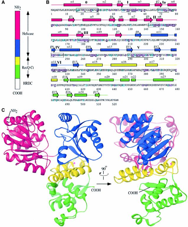
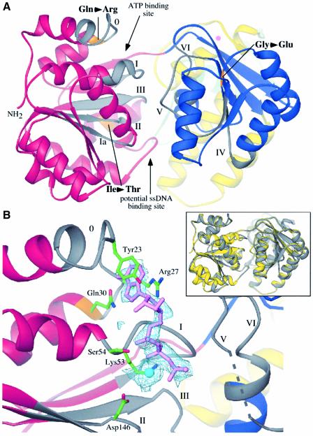
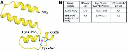
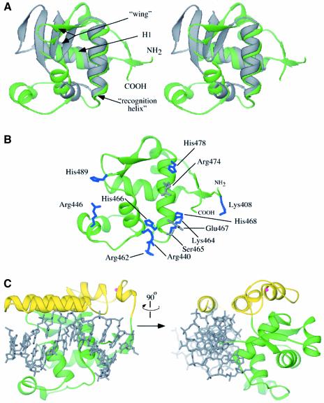
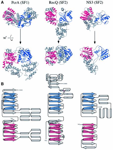
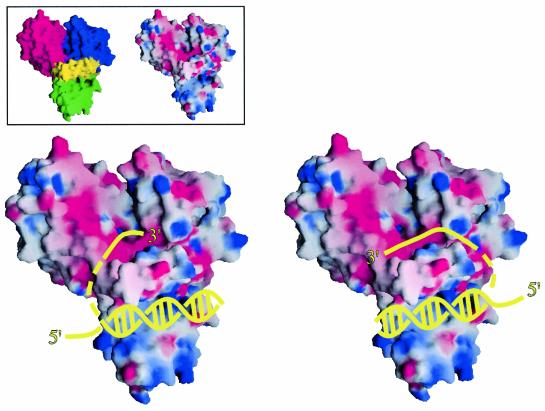
RecQ family helicases catalyze critical genome maintenance reactions in bacterial and eukaryotic cells, playing key roles in several DNA metabolic processes. Mutations in recQ genes are linked to genome instability and human disease. To define the physical basis of RecQ enzyme function, we have determined a 1.8 A resolution crystal structure of the catalytic core of Escherichia coli RecQ in its unbound form and a 2.5 A resolution structure of the core bound to the ATP analog ATPgammaS. The RecQ core comprises four conserved subdomains; two of these combine to form its helicase region, while the others form unexpected Zn(2+)-binding and winged-helix motifs. The structures reveal the molecular basis of missense mutations that cause Bloom's syndrome, a human RecQ-associated disease. Finally, based on findings from the structures, we propose a mechanism for RecQ activity that could explain its functional coordination with topoisomerase III.


Figures

None
Fig. 1. Structure of the E.coli RecQ catalytic core. (A) Schematic diagram of E.coli RecQ. Three conserved regions, helicase, RecQ-conserved (RecQ-Ct), and Helicase-and-RNaseD-C-terminal (HRDC) (Morozov et al., 1997), are labeled. The catalytic core of E.coli RecQ (RecQΔC) includes only the helicase and RecQ-Ct regions, and comprises four apparent subdomains in the structure: residues 1–208 in red, 209–340 in blue, 341–406 in yellow, and 407–516 in green. (B) Sequence and secondary structure of RecQΔC. Helices (boxes) and β-strands (arrows) are shown above the sequence and labeled sequentially. Color coding is the same as in (A). Conserved helicase motifs (motif 0, Bernstein and Keck, 2003; and motifs I-VI, Gorbalenya and Koonin, 1993) are labeled and enclosed in boxes. Residues that are invariant among 65 bacterial RecQ proteins are underlined, and residues that are invariant or highly conserved with a subset of eukaryotic RecQ proteins (human WRN, BLM, and S.cerevisiae Sgs1) are highlighted in purple or light-blue boxes, respectively. (C) Orthogonal views of a ribbon diagram of the crystal structure of RecQΔC, color-coded as in (A). A bound Zn2+ ion is shown as a magenta sphere.
None
Fig. 2. Features of the helicase region of RecQΔC. (A) View into the cleft formed by the two helicase subdomains. Color coding is the same as Figure 1A, except that helicase motifs are labeled and colored in grey, and residues corresponding to known BLM missense mutations are labeled and colored in orange. Sites where nucleotide and ssDNA have been observed to bind in other helicase structures are indicated. (B) Structure of ATPγS/Mn2+-bound RecQΔC. (Inset) overlay of the apo- and ATPγS-bound RecQΔC structures in yellow and grey, respectively. A slight relative rotation of the second helicase lobe is seen in the structure with bound nucleotide. (Main panel) FoFc difference electron density (light blue) is contoured at 1.5 σ. Motif I is in an open conformation relative to its position in Figure 2A. The ATPγS (lavender) adenine moiety is sandwiched between Tyr23 and Arg27, and hydrogen bonds are formed between the N6 and N7 atoms of the adenine and the side chain of Gln30. The triphosphate is bound by interactions with Lys53 and backbone amides from motif I. A Mn2+ ion (cyan) is bound by Ser54 from motif I and Asp146 from motif II. Helicase motif labels and color-coding are the same as in Figure 2A. Electron density was not observed for residues 296 to 299 (dashed line).
None
Fig. 3. Features of the Zn2+-binding subdomain in RecQΔC. (A) View of the Zn2+-binding subdomain (residues 341–406) extracted from the rest of RecQΔC. Four highly conserved cysteine residues (Cys380, Cys397, Cys400, Cys403) are arrayed around a Zn2+ ion (magenta). Two BLM missense mutations alter individual cysteines in this array as labeled. The N- and C-terminal residues that connect to the helicase and WH subdomains, respectively, are labeled. (B) Biochemical evidence that full-length E.coli RecQ binds Zn2+ in a site that includes 3–4 cysteine residues. [Zn2+] and [thiolfree] were determined for E.coli RecQ and E.coli RecQ that was dialyzed against 10 mM EDTA, 1 mM DTT to extract bound Zn2+.
None
Fig. 4. Features of the WH subdomain in RecQΔC. (A) Stereo diagram of the superposition of the WH subdomain extracted from the RecQΔC structure (green, residues 407–516) with the DNA binding WH domain of CAP (Schultz et al., 1991) (grey). The two major helices of the fold (H1 and recognition helix) and the wing elements are labeled. The N-terminal residue of the RecQΔC WH fold, which connects to the Zn2+ binding region, and C-terminal residue are labeled. (B) Potential DNA binding residues in the RecQΔC WH subdomain. Residues that are structurally conserved with DNA binding residues in CAP (Schultz et al., 1991) are shown in grey and other residues with positive charge potential on the same putative DNA binding face are shown in blue. (C) Orthogonal views of a model of dsDNA binding to the RecQ-Ct region of RecQ. dsDNA from the CAP/DNA complex structure (grey) (Schultz et al., 1991) is overlayed onto the RecQ-Ct region. A minor opening of the helical Zn2+-binding region relative to the WH region would create a cleft sufficiently wide for dsDNA binding.
None
Fig. 5. Structural comparison of RecQΔC to representative helicases belonging to the SF1 and SF2 helicase superfamilies. (A) Structures of PcrA (Subramanya et al., 1996) and NS3 (Yao et al., 1997), SF1 and SF2 helicases, respectively, were superimposed on RecQΔC using the structurally related Cα atoms in their N-terminal most helicase subdomains (in red) and then translated to allow visual comparison. Red and blue colors mark the conserved RecA-like folds found in the helicase subdomains of each protein, while grey coloration indicates sequences outside of the RecA fold. An orthogonal view is shown of each protein below the top row. (B) Topology diagrams of PcrA, RecQΔC and NS3. Colors are shown as in (A), with RecQΔC’s Zn2+ in magenta. Helices and β-strands in RecQΔC are labeled as in Figure 1B. Diagrams of PcrA and NS3 were adapted from Caruthers and McKay (2002).
None
Fig. 6. Models for DNA binding to E.coli RecQΔC. Surface representations of E.coli RecQΔC are shown (color-coded as in Figure 1, inset left) bound to cartoon diagrams of a partially unwound DNA molecule in two possible orientations (yellow). The RecQΔC surface is colored by its electrostatic surface potential (inset right and main panel) at + or –6 kBT/e for positive (blue) or negative (red) charge potential using the program GRASP (Nicholls et al., 1991). Since E.coli RecQ may unwind DNA as an oligomer (Harmon and Kowalczykowski, 2001), it is not clear whether ssDNA bound by the helicase region would come from dsDNA bound by the same protomer or another protomer in the oligomer during unwinding. As such, the connection linking the dsDNA to the 3′ ssDNA is shown as a dashed line. In addition, relative subdomain orientations could change in the DNA-bound form.

References

    1. Bahr A., De Graeve,F., Kedinger,C. and Chatton,B. (1998) Point mutations causing Bloom’s syndrome abolish ATPase and DNA helicase activities of the BLM protein. Oncogene, 17, 2565–2571. - PubMed
    1. Barakat A., Ababou,M., Onclercq,R., Dutertre,S., Chadli,E., Hda,N., Benslimane,A. and Amor-Gueret,M. (2000) Identification of a novel BLM missense mutation (2706T>C) in a Moroccan patient with Bloom’s syndrome. Hum. Mutat., 15, 584–585. - PubMed
    1. Berg J.M. and Shi,Y. (1996) The galvanization of biology: a growing appreciation for the roles of zinc. Science, 271, 1081–1085. - PubMed
    1. Bernstein D.A. and Keck,J.L. (2003) Domain mapping of Escherichia coli RecQ defines the roles of conserved amino- and carboxyl-terminal regions in the RecQ family. Nucleic Acids Res., 31, 2778–2785. - PMC - PubMed
    1. Caruthers J.M., Johnson,E.R. and McKay,D.B. (2000) Crystal structure of yeast initiation factor 4A, a DEAD-box RNA helicase. Proc. Natl Acad. Sci. USA, 97, 13080–13085. - PMC - PubMed

Publication types

MeSH terms

LinkOut - more resources