. 2003 Oct 1;22(19):4922-32.
doi: 10.1093/emboj/cdg494.

Crystal structure and snapshots along the reaction pathway of a family 51 alpha-L-arabinofuranosidase

Affiliations

Crystal structure and snapshots along the reaction pathway of a family 51 alpha-L-arabinofuranosidase

Klaus Hövel et al. EMBO J. .

Abstract

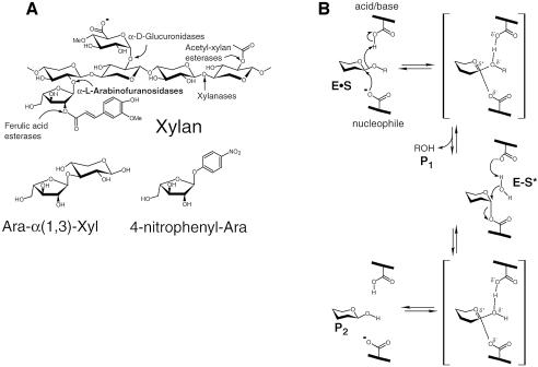
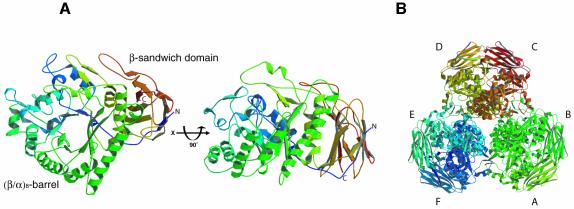
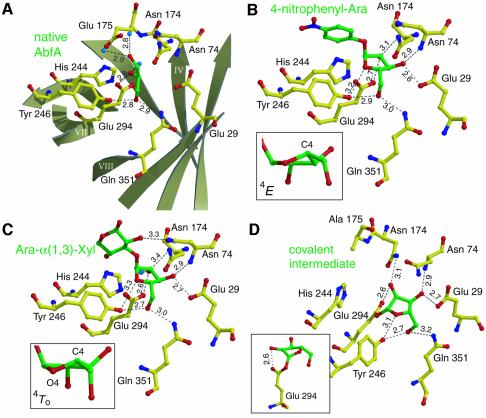
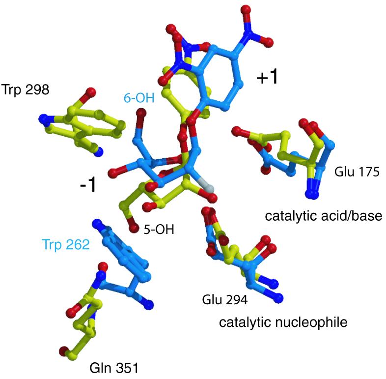
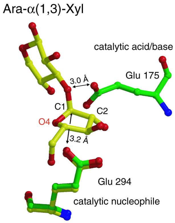
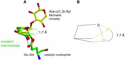
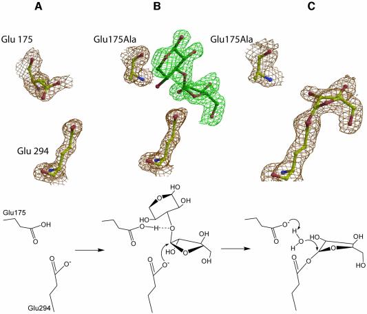
High-resolution crystal structures of alpha-L-arabinofuranosidase from Geobacillus stearothermophilus T-6, a family 51 glycosidase, are described. The enzyme is a hexamer, and each monomer is organized into two domains: a (beta/alpha)8-barrel and a 12-stranded beta sandwich with jelly-roll topology. The structures of the Michaelis complexes with natural and synthetic substrates, and of the transient covalent arabinofuranosyl-enzyme intermediate represent two stable states in the double displacement mechanism, and allow thorough examination of the catalytic mechanism. The arabinofuranose sugar is tightly bound and distorted by an extensive network of hydrogen bonds. The two catalytic residues are 4.7 A apart, and together with other conserved residues contribute to the stabilization of the oxocarbenium ion-like transition state via charge delocalization and specific protein-substrate interactions. The enzyme is an anti-protonator, and a 1.7 A electrophilic migration of the anomeric carbon takes place during the hydrolysis.


Figures

None
Fig. 1. (A) Upper, the basic structural components of xylan, and the hemicellulases responsible for its degradation; lower, the natural and synthetic substrates of α-l-arabinofuranosidases used in this work: Ara-α(1,3)-Xyl and 4-nitrophenyl-Ara. (B) The double-displacement reaction mechanism for retaining glycosidases (the Koshland mechanism).
None
Fig. 2. Stereo view of the 2DFo – mFc electron density maps of a representative portion of the native AbfA at 1.75 Å, contoured at 2.0 σ. The area shown is that of the active-site residues Glu29 and Glu294, and the non-proline cis-peptide bond between residues Ala350 and Gln351. Selected hydrogen bonds between the protein and the water molecule are shown in dotted lines. Color coding: red, oxygen; blue, nitrogen; yellow, carbon.
None
Fig. 3. Overall structure of AbfA. (A) Two views of the AbfA monomer related by a 90° rotation. (B) The hexameric enzyme is shown along the crystallographic 3-fold axis. All non-schematic figures were prepared with Molscript (Kraulis, 1991), Bobscript (Esnouf, 1997) and Raster3D (Merritt and Bacon, 1997).
None
Fig. 4. Interactions between ligands and key residues in the active site. Hydrogen bonds are shown as dotted black lines, their length in Ångströms is indicated. Color coding: yellow, carbon atoms of protein residues; green, carbon atoms of ligands; red, oxygen; blue, nitrogen. (A) Native AbfA with a glycerol molecule in the active site. The β-strands of the (β/α)8-barrel are shown schematically in light grey. (B) The Michaelis complex of the E175A mutant with 4-nitrophenyl-Ara. (C) The Michaelis complex of the E175A mutant with Ara-α(1,3)-Xyl. The insets in (B) and (C) show the two energetically close conformations of the arabinofuranose ring at the –1 subsite. (D) The covalent arabinofuranosyl–enzyme complex, obtained by using the E175A mutant with 2,5-dinitrophenyl-Ara. The inset shows the strong hydrogen bond between the sugar 2-hydroxyl and the nucleophile (Glu294).
None
Fig. 5. Substrate specificity of AbfA and Cel5A. Superposition of the Michaelis complexes of AbfA with 4-nitrophenyl-Ara and Cel5A endoglucanase with 2,4-dinitrophenyl-2-deoxy-2-fluoro-β-d-cellobioside (PDB code: 1H2J). Only the catalytic residues and the residues responsible for the discrimination between the d-glucopyranosidic and l-arabinofuranosidic substrates are shown. Color coding: red, oxygen; blue, nitrogen; white, fluorine; yellow, carbon atoms of AbfA; light blue, carbon atoms of Cel5A.
None
Fig. 6. Superposition of the structures of the native enzyme (green) with that of the Michaelis complex (yellow). Glu175 is located 3.0 Å from the glycosidic oxygen of Ara-α(1,3)-Xyl, which is a hydrogen-bonding distance, allowing the direct protonation of the departing aglycon. Glu294 is in an appropriate distance (3.2 Å) for a nucleophilic attack on the anomeric carbon.
None
Fig. 7. Electrophilic migration of C1. (A) Overlay of the two critical states of the glycosylation step: Michaelis complex (yellow) and covalent intermediate (green). C1 migrates ∼1.7 Å to form the covalent bond with the nucleophilic oxygen. (B) Simplified schematic representation of the electrophilic migration of C1.
None
Fig. 8. Snapshots along the reaction pathway obtained from the X-ray structures (upper), and a schematic representation of the corresponding stages of the glycosylation step (lower). (A) The catalytic residues of the native AbfA. (B) Michaelis complex of the intact substrate Ara-α(1,3)-Xyl located in subsites –1 and +1. The absence of electron density at this contour level for the 4-OH of the xylose at the +1 subsite results from the heterogeneity of the substrate. The second xylose connected to only part of the substrate molecules probably distorts the O4 of the first xylose. (C) Structure of the trapped covalent arabinofuranosyl–enzyme intermediate. The 2DFo – mFc electron density maps are contoured at 1.5 σ.

References

    1. Bourne Y. and Henrissat,B. (2001) Glycoside hydrolases and glycosyltransferases: families and functional modules. Curr. Opin. Struct. Biol., 11, 593–600. - PubMed
    1. Collins P. and Ferrier,R. (1995) Monosaccharides, their Chemistry and their Roles in Natural Products. John Wiley and Sons Ltd, Chichester, UK.
    1. Davies G., Sinnott,M.L. and Withers,S.G. (1998a) Glycosyl Transfer. In Sinnott,M.L. (ed.), Comprehensive Biological Catalysis. Academic Press Limited, London, UK, Vol. 1, pp. 119–209.
    1. Davies G.J., Mackenzie,L., Varrot,A., Dauter,M., Brzozowski,A.M., Schulein,M. and Withers,S.G. (1998b) Snapshots along an enzymatic reaction coordinate: analysis of a retaining β-glycoside hydrolase. Biochemistry, 37, 11707–11713. - PubMed
    1. Debeche T., Bliard,C., Debeire,P. and O’Donohue,M.J. (2002) Probing the catalytically essential residues of the α-l-arabinofuranosidase from Thermobacillus xylanilyticus. Protein Eng., 15, 21–28. - PubMed

Publication types

MeSH terms

Substances