. 2003 Oct 1;22(19):4991-5002.
doi: 10.1093/emboj/cdg481.

Clathrin-mediated endocytosis is essential in Trypanosoma brucei

Affiliations

Clathrin-mediated endocytosis is essential in Trypanosoma brucei

Clare L Allen et al. EMBO J. .

Abstract

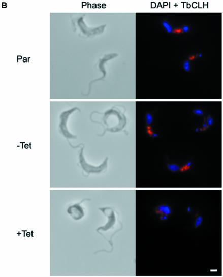
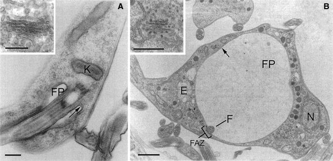
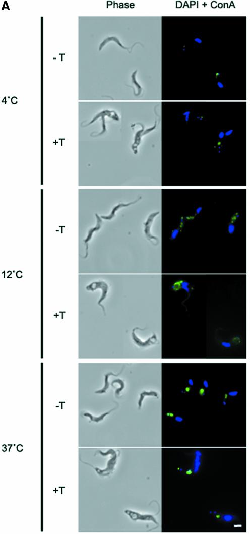
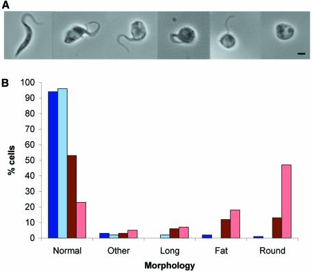
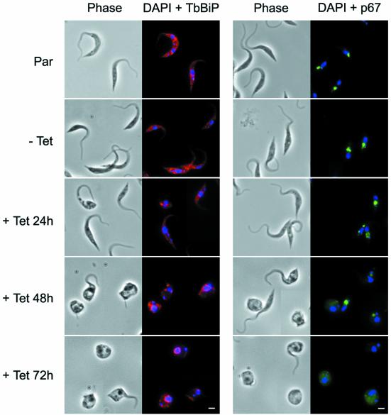
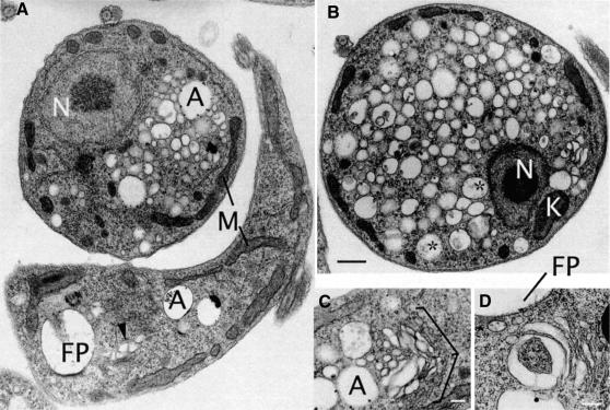
In Trypanosoma brucei, the plasma membrane is dominated by glycosylphosphatidylinositol (GPI)-anchored proteins. Endocytic activity correlates with expression levels of the clathrin heavy chain TbCLH, and additional evidence suggests that rapid endocytosis may play a role in evasion of the immune response. TbCLH is present on both endocytic vesicles and post-Golgi elements, suggesting a similar range of functions in trypanosomes to higher eukaryotes. We have assessed the role of TbCLH using RNA interference (RNAi). Suppression of TbCLH expression results in rapid lethality in the bloodstream stage, the form most active for endocytosis. The flagellar pocket, the site of both endocytosis and exocytosis, becomes massively enlarged, suggesting that membrane delivery is unaffected but removal is blocked. Endocytosis in TbCLHRNAi cells is essentially undetectable, suggesting that clathrin-mediated mechanisms are the major route for endocytosis in T.brucei and hence that GPI-anchored proteins are endocytosed by clathrin-dependent pathways in trypanosomes. In contrast, a massive internal accumulation of vesicles and significant alterations to trafficking of a lysosomal protein were observed in the procyclic stage, indicating developmental variation in clathrin function in trypanosomes.


Figures

None
Fig. 1. Depletion of clathrin heavy chain leads to BigEye morphology. (A) Gallery of images from cultures expressing clathrin RNAi. The top line shows interphase cells with increased abnormality running from left to right. The majority of cells in early cultures correspond to the four leftmost images, whilst more severe phenotypes, as shown on the right, emerge and increase in prevalence in older cultures. The bottom line shows examples of mitotic cells exhibiting BigEye phenotype. In this case, more than one vacuole is present, and these are frequently of very different sizes (BigEye/LittleEye cells). Scale bar: 2 µm. (B) Immunofluorescence analysis demonstrates knockdown of clathrin expression following induction in bloodstream form (BSF) cells. Left, phase-contrast image; right, DAPI (blue) and anti-TbCLH stain (red). Par, BSF 90-13 parental cells; BSFp2T7TiCLH, RNAi cells uninduced (–Tet) and induced (+Tet). Scale bar: 2 µm.
None
Fig. 2. BigEye is due to flagellar pocket enlargement. (A) Parental cells. The flagellar pocket is comparatively small, with a diameter of ∼0.5 µm and a flask shape due to tight association with the flagellum. (B) Induced clathrin RNAi cells. The flagellar pocket is enlarged. Other structures, including acidocalcisomes, the nucleus and endosomal membranous structures appear normal. Note also the presence of electron-dense material on either side of the flagellum where it contacts the pocket membrane, suggesting the structures responsible for flagellum/flagellar pocket association are still present in this cell. In both (A) and (B), the inset shows a Golgi complex. For both the parental and induced cell lines, the Golgi profile appears normal. E, endosome; F, flagellum; FP, flagellar pocket; FAZ, flagellar attachment zone; K, kinetoplast; N, nucleus. Arrows indicate the electron-dense variant surface glycoprotein (VSG) coat of the flagellar pocket. Scale bars: (A and A inset) 200 nm, (B) 1 µm and (B inset) 500 nm.
None
Fig. 3. Endocytosis is perturbed in BigEye cells. (A) In uninduced TbCLH RNAi cells at 4°C, lectin concanavalin A (Con A) is retained at the flagellar pocket, at 12°C the lectin is internalized to the endocytic system and at 37°C the lectin is delivered to the terminal lysosome. Induced cells demonstrate a severe defect in uptake. At 37°C, the lectin remains associated with the flagellar pocket. (B) Enlarged image of induced bloodstream form (BSF) clathrin RNAi cells (from A), showing that Con A is localized to the enlarged flagellar pocket at 37°C. (C) At 4°C, FM 4-64 accumulates in the flagellar pocket in both the parental and induced clathrin RNAi cell line. Note that the staining appears rather more extensive in the RNAi cells due to the enlargement of the pocket. At 37°C, the fluorophore has been internalized in the parental cells, but remains associated with the enlarged pocket structure in the RNAi cell line. (A) Left, phase; right, DAPI (DNA) in blue, lectin in green. (C) Left, phase; right, DAPI (DNA) in blue, FM 4-64 in red. Scale bar: 2 µm. Par, BSF 90-13 parental cells; –Tet, uninduced BSFp2T7TiCLH cells; +Tet, BSFp2T7TiCLH cells induced for 16 h.
None
Fig. 4. Preservation of normal morphology of internal membrane structures in BigEye cells. Immunofluorescence analysis for endoplasmic reticulum (TbBiP), recycling endosome (TbRAB11) and lysosomal (p67) proteins. Top, the parental BSF 90-13 cells (Par); middle, uninduced bloodstream form (BSF) RNAi line (–Tet); and bottom, induced (+Tet). Induction is confirmed by the presence of the BigEye. The distribution of marker proteins is unaltered between cell lines. Left, phase; right, DAPI (DNA) in blue, TbBiP or TbRAB11 in red, p67 in green. Scale bar: 2 µm.
None
Fig. 5. Variant surface glycoprotein location in BigEye cells. CryoimmunoEM images of parental (A and C) and clathrin-depleted cells (B and D). Variant surface glycoprotein (VSG) is observed on the plasma membrane, within the flagellar pocket and over a number of internal structures, tentatively assigned as endoplasmic reticulum (arrowheads) and TbRAB11 recycling endosomes (arrows), based on previous studies (Grunfelder et al., 2002, 2003). In the BigEye cells, VSG is also seen with similar distribution on the plasma membrane, endoplasmic reticulum and in recycling endosome figures. Hence, VSG remains associated with TbRAB11 transport intermediates, and these structures retain normal morphology. Scale bar: 200 nm.
None
Fig. 6. Depletion of clathrin heavy chain in procyclic culture form (PCF) cells leads to a rounded morphology. (A) Gallery of phase images from PCF cultures expressing clathrin RNAi. The leftmost image shows the ‘normal’ PCF phenotype, the next two images show the ‘fat’ phenotype, and the three images on the right show the ‘round’ phenotype. (B) Incidence of aberrant cell morphologies in induced PCF clathrin RNAi cell cultures. Blue bars, uninduced PCFp2T7TiCLH cells cultured for 48 h (dark blue) or 72 h (pale blue); red bars, induced PCFp2T7TiCLH cultures at 48 h (dark red) or 72 h (pale red). Morphologies were defined as follows: normal, average cell body length 15 µm, average width 1.54 µm; long, cell body length >20 µm, average cell body length 23.3 µm, average width 1.54 µm; fat, enlarged posterior of the cell, average cell body length 8.55 µm, average width 3.07 µm; round, cell body completely rounded up, with detached or no visible flagellum, average cell body length 4.66 µm, average width 4.26 µm; other, all other abnormal forms.
None
Fig. 7. Alteration of lysosomal protein targeting in procyclic culture form (PCF) BigEye cells. Clathrin RNAi cells were stained following 24, 48 and 72 h of induction as indicated. For TbBiP a disorganization was observed, suggesting that the endoplasmic reticulum has become deformed. In addition, the localization of p67 was altered, so that at 48 h the stain was less intense and defocused, and at 72 h this was more extreme with a major loss of integrity. Left image of each set, phase; right image, DAPI (DNA) in blue, TbBiP in red, p67 in green. Scale bar: 2 µm. Par, PCF 29-13 parental cells; –Tet, uninduced PCF 29-13p2T7TiCLH cells; +Tet, PCF29-13p2T7TiCLH cells induced for indicated time periods.
None
Fig. 8. A distinct ultrastructural defect accompanies TbCLH-depletion in procyclic culture form (PCF) trypanosomes. Electron micrographs of PCF cells following 48 h induction of clathrin dsRNA. (A) Two cells exhibiting normal (bottom) and abnormal (top) phenotypes. The major effects of clathrin double-stranded RNA expression is in the accumulation of vesicular profiles of variable diameter (∼50–400 nm) and a rounding up of the cell. Other structures (nucleus, mitochondria, acidocalcisomes) appear normal. (B) A more extreme example of a PCF clathrin RNAi cell. The plasma membrane has become smooth, possibly due to increased cytoplasmic pressure. In addition, the nucleus, kinetoplast and mitochondria appear normal. (C and D) Details of the Golgi region of PCF RNAi cells showing distortions to the trans-Golgi cisternae and membranes. The cell in (C) is in early mitosis, as the Golgi stack appears to be undergoing binary fission (Field et al., 2000). A, acidocalcisomes; K, kinetoplast; FP, flagellar pocket; M, mitochondria; N, nucleus; *, vesicles containing electron-dense matrix-like material (possibly FP-derived); arrowhead, distorted trans-Golgi. Scale bars: (A and B) 500 nm, (C) 200 nm and (D) 250 nm.

References

    1. Alexander D.L., Schwartz,K.J., Balber,A.E. and Bangs,J.D. (2002) Developmentally regulated trafficking of the lysosomal membrane protein p67 in Trypanosoma brucei. J. Cell Sci., 115, 3253–3263. - PubMed
    1. Bangs J.D., Andrews,N.W., Hart,G.W. and Englund,P.T. (1986) Posttranslational modification and intracellular transport of a trypanosome variant surface glycoprotein. J. Cell Biol., 103, 255–263. - PMC - PubMed
    1. Bangs J.D., Uyetake,L., Brickman,M.J., Balber,A.E. and Boothroyd,J.C. (1993) Molecular cloning and cellular localization of a BiP homologue in Trypanosoma brucei. Divergent ER retention signals in a lower eukaryote. J. Cell Sci., 105, 1101–1113. - PubMed
    1. Benting J.H., Rietveld,A.G. and Simons,K. (1999) N-glycans mediate the apical sorting of a GPI-anchored, raft-associated protein in Madin-Darby canine kidney cells. J. Cell Biol., 146, 313–320. - PMC - PubMed
    1. Boehm M. and Bonifacino,J.S. (2002) Genetic analyses of adaptin function from yeast to mammals. Gene, 286, 175–186. - PubMed

Publication types

MeSH terms