. 2003 Oct 1;22(19):5079-89.
doi: 10.1093/emboj/cdg474.

c-Jun N-terminal kinases (JNK) antagonize cardiac growth through cross-talk with calcineurin-NFAT signaling

Affiliations

c-Jun N-terminal kinases (JNK) antagonize cardiac growth through cross-talk with calcineurin-NFAT signaling

Qiangrong Liang et al. EMBO J. .

Abstract

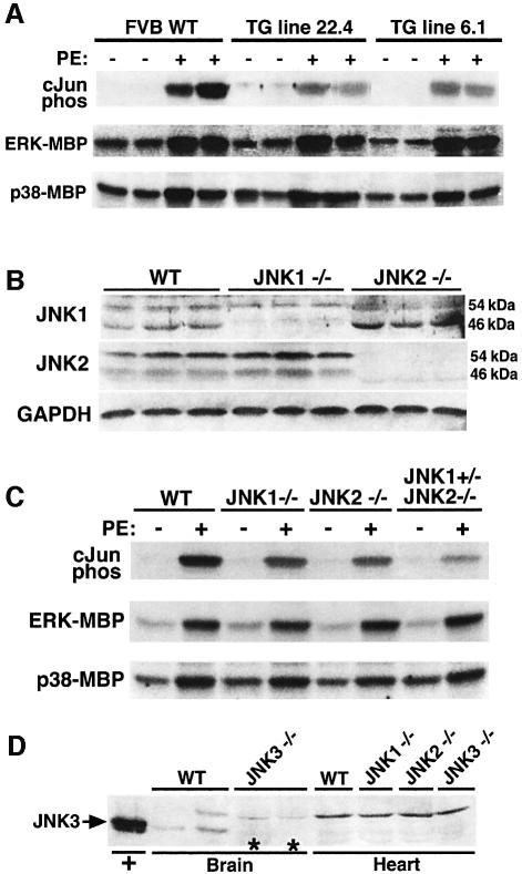
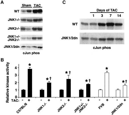
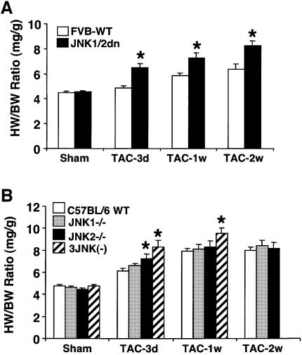
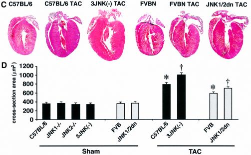
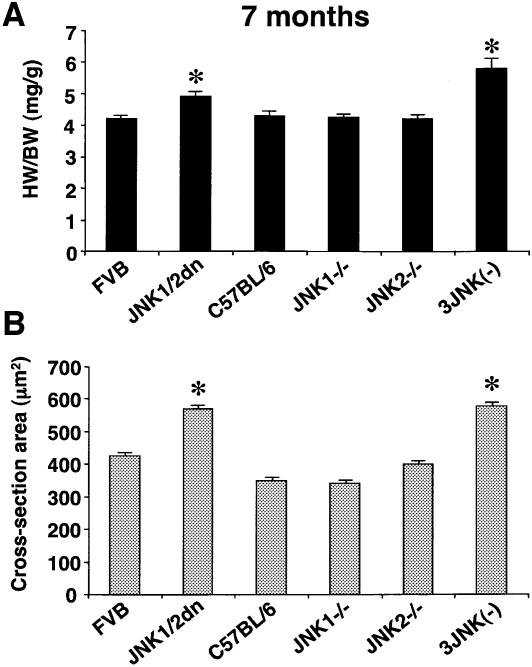
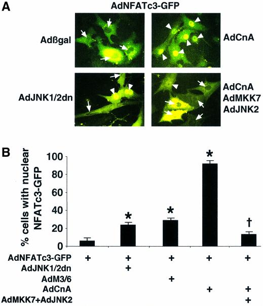
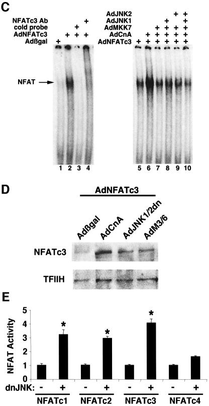
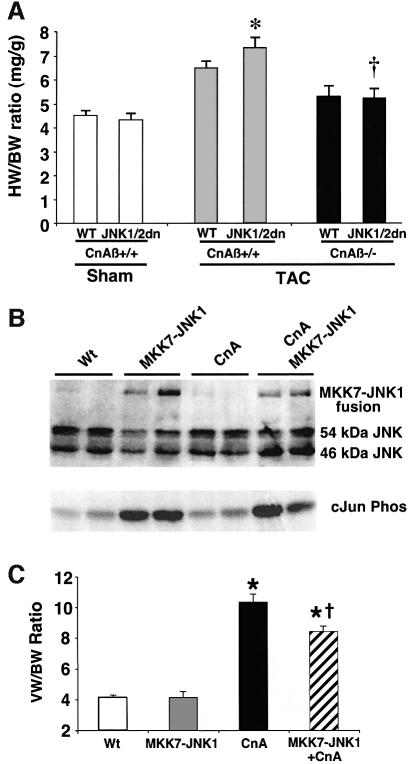
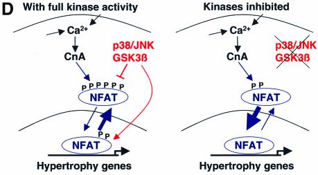
The c-Jun N-terminal kinase (JNK) branch of the mitogen-activated protein kinase (MAPK) signaling pathway regulates cellular differentiation, stress responsiveness and apoptosis in multicellular eukaryotic organisms. Here we investigated the functional importance of JNK signaling in regulating differentiated cellular growth in the post-mitotic myocardium. JNK1/2 gene-targeted mice and transgenic mice expressing dominant negative JNK1/2 were determined to have enhanced myocardial growth following stress stimulation or with normal aging. A mechanism underlying this effect was suggested by the observation that JNK directly regulated nuclear factor of activated T-cell (NFAT) activation in culture and in transgenic mice containing an NFAT-dependent luciferase reporter. Moreover, calcineurin Abeta gene targeting abrogated the pro-growth effects associated with JNK inhibition in the heart, while expression of an MKK7-JNK1 fusion protein in the heart partially reduced calcineurin-mediated cardiac hypertrophy. Collectively, these results indicate that JNK signaling antagonizes the differentiated growth response of the myocardium through direct cross-talk with the calcineurin-NFAT pathway. These results also suggest that myocardial JNK activation is primarily dedicated to modulating calcineurin-NFAT signaling in the regulation of differentiated heart growth.


Figures

None
Fig. 1. Characterization of mice with diminished JNK activity. (A) Non-transgenic littermates and dnJNK1/2 transgenic mice from lines 22.4 and 6.1 were injected with either PBS or PE (15 mg/kg), and cardiac JNK activity was measured 30 min later using GST-cJun (1–79) as a substrate. ERK and p38 kinase activities were also measured using myelin basic protein (MBP) as the substrate after immunoprecipitation. (B) Western blot analysis with antibodies partially specific for JNK1 and JNK2 from the hearts of JNK1 null and JNK2 null mice. (C) JNK activity assay showing diminished PE-induced JNK activity in the hearts of JNK1–/–, JNK2–/– and 3-allele JNK deleted mice, yet ERK and p38 activities were unaffected. (D) Western blot analysis with a JNK3-specific antibody showed no detectable JNK3 protein in the wild-type mouse heart and no upregulation of JNK3 in either JNK1–/– or JNK2–/– hearts. Controls consisted of HEK293 cell lysate after JNK3 plasmid transfection or brain protein lysate from wild-type and JNK3 targeted mice.
None
Fig. 2. TAC-induced cardiac JNK activation is reduced in dominant negative mice and gene-targeted mice. (A) Autoradiograph of 32P-labeled GST-cJun (1–79) phosphorylation from heart protein extracts of the indicated mice at baseline or 7 days after TAC stimulation (some lanes were spliced together for simplicity). (B) Quantitative analysis of JNK activity in each of the indicated groups (N = 6 each) (*P < 0.05 versus sham; †P < 0.05 versus C57BL/6 or FVB WT TAC). (C) Time course of cJun kinase activity in wild-type (WT) or JNK1/2 dn mice following TAC stimulation.
None
Fig. 3. JNK-inhibited mice show enhanced cardiac hypertrophy following TAC. (A) Two-month-old JNK1/2dn transgenic mice showed enhanced cardiac hypertrophy following 3, 7 and 14 days of TAC compared with FVB wild-type (FVB-WT) control mice (as assessed by wet heart-to-body weight ratio HW/BW) (N = 10–15 mice in each group; *P < 0.05 versus FVB-WT TAC). (B) TAC-induced increase in HW/BW ratio was significantly higher in JNK2 null and JNK1+/– JNK2–/– three-allele null mice compared with strain-matched wild-type controls (C57BL/6) at 3 days and/or 1 week (N = 6–15 for each group; *P < 0.05 versus C57BL/6 TAC). (C) Histological analysis of HE-stained hearts in frontal section from 1-week TAC- stimulated mice. (D) Assessment of cardiomyocyte cross-sectional areas at baseline or 7 days after TAC stimulation (N ≥ 100 cells in each group; *P < 0.05 versus respective sham; †P < 0.05 versus WT TAC).
None
Fig. 4. JNK-inhibited mice show spontaneous cardiac hypertrophy with aging. (A) Wet heart-to-body-weight ratios HW/BW were determined for 7-month-old mice C57BL/6 WT or three-allele JNK-targeted mice (N = 6 in each group; *P < 0.05) or in FVB WT and double-allele JNK1/2dn transgenic mice (N = 10–13; *P < 0.05). (B) Cross-sectional area measurements from wheat germ agglutinin–TRITC stained histological sections for each cohort. At least 100 muscle fibers on sections from two separate mice were measured for each group (*P < 0.05).
None
Fig. 5. MKK7/JNK pathway negatively regulates NFATc3 activation. (A) Representative fluorescent images showing the nuclear-cytoplasmic shuttling of NFATc3–GFP in neonatal rat cardiomyocytes infected with AdNFATc3–GFP, Adβgal, AdJNK1/2dn, AdCnA, AdMKK7 and/or AdJNK1/2. Arrows show cytoplasmic localization and arrowheads show nuclear localization of NFATc3–GFP. (B) Quantitation from three independent experiments of the percentage of cells with nuclear NFATc3–GFP by each of the indicated recombinant adenoviruses (*P < 0.05 versus AdNFATc3–GFP alone). (C) EMSA showing that MKK7/JNK signaling inhibits calcineurin-induced NFATc3 DNA binding activity in nuclear protein extracts from recombinant adenoviral infected neonatal rat cardiomyocytes. Nuclear protein extracts were generated 24 h after viral infection. (D) NFATc3 western blot from nuclear protein extracts derived from adenoviral-infected cardiomyocytes. All cells were infected with wild-type AdNFATc3 and Adβgal, AdCnA, AdJNK1/2dn or AdM3/6. TFIIH was used as a nuclear protein loading control. (E) Transient transfection experiment with an NFAT- dependent luciferase reporter in Cos-7 cells shows that dominant negative JNK1/2 augments the activity of NFATc1, NFATc2 and NFATc3 (*P < 0.05 versus no dnJNK1/2 cotransfection). The results represent the average of triplicate assays.
None
Fig. 6. JNK inhibition enhances NFAT activation in vivo. (A) dnJNK1/2 mice were crossed with the transgenic NFAT indicator line and activity in the heart was assessed in 2-month-old mice at baseline or after 7 days of TAC stimulation (N = 4–6 mice in each group). (B) The increased NFAT activity correlated with the enhanced hypertrophic response to TAC in the same dnJNK1/2 transgenic mice as assessed by heart-to-body weight ratio HW/BW. (*P < 0.05 versus sham NFAT-Luc; †P < 0.05 versus NFAT indicator TG alone after TAC).
None
Fig. 7. Calcineurin–NFAT signaling is interconnected with JNK signaling in vivo. (A) The dnJNK1/2 transgene was crossed with calcineurin Aβ gene-targeted mice to generate null mice (CnAβ–/–) or wild-type littermate controls (CnAβ+/+) for gravimetric analysis 1 week after TAC or sham operation. (N = 4–9 mice in each group; *P < 0.05 versus wild-type mice subjected to TAC; †P < 0.05 versus JNK1/2dn subjected to TAC). (B) Western for JNK1/2 protein from the hearts of MKK7–JNK1 fusion transgenic mice (upper band), wild-type mice, activated calcineurin transgenic mice or mice containing both the activated calcineurin and MKK7–JNK1 transgenes. The lower panel shows cJun kinase activity. (C) Analysis of wet ventricle-to-body weight ratios VW/BW from MKK7–JNK1 transgenic mice crossed with activated calcineurin transgenic mice (N = 4 mice in each group; *P < 0.05 versus WT; †P < 0.05 versus CnA transgenic. (D) Model whereby NFAT nuclear translocation and occupancy are controlled by the interplay between calcineurin-mediated dephosphorylation and kinase-mediated phosphorylation (left). Inhibition of a key NFAT kinase, such as JNK, shifts the equilibrium so that the same activation signal by calcineurin now results in greater NFAT translocation and nuclear occupancy.

References

    1. Antos C.L., McKinsey,T.A., Frey,N., Kutschke,W., McAnally,J., Shelton,J.M. Richardson,J.A. Hill,J.A. and Olson,E.N. (2002). Activated glycogen synthase-3 beta suppresses cardiac hypertrophy in vivo. Proc. Natl Acad. Sci. USA, 99, 907–912. - PMC - PubMed
    1. Braz J.C., Bueno,O.F., Liang,Q., Wilkins,B.J., Dai,Y.-S., Braunwart,J., Glascock,B.J., Klevitsky,R., Kimball,T.F. et al. (2003). Targeted inhibition of p38 MAPK promotes hypertrophic cardiomyopathy through upregulation of calcineurin–NFAT signaling. J. Clin. Invest., 111, 1475–1486. - PMC - PubMed
    1. Bueno O.F., Brandt,E.B., Rothenberg,M.E. and Molkentin,J.D. (2002a). Defective T cell development and function in Calcineurin A-deficient mice. Proc. Natl Acad. Sci. USA, 99, 9398–9403. - PMC - PubMed
    1. Bueno O.F., Wilkins,B.J., Tymitz,K.M., Glascock,B.J., Kimball,T.F., Lorenz,J.N. and Molkentin,J.D. (2002b). Impaired cardiac hypertrophic response in Calcineurin A beta-deficient mice. Proc. Natl Acad. Sci. USA, 99, 4586–4591. - PMC - PubMed
    1. Choukroun G., Hajjar,R., Kyriakis,J.M., Bonventre,J.V., Rosenzweig,A. and Force,T. (1998). Role of the stress-activated protein kinases in endothelin-induced cardiomyocyte hypertrophy. J. Clin. Invest., 102, 1311–1320. - PMC - PubMed

MeSH terms