. 2003 Oct 1;22(19):5163-74.
doi: 10.1093/emboj/cdg478.

Local action of the chromatin assembly factor CAF-1 at sites of nucleotide excision repair in vivo

Affiliations

Local action of the chromatin assembly factor CAF-1 at sites of nucleotide excision repair in vivo

Catherine M Green et al. EMBO J. .

Abstract

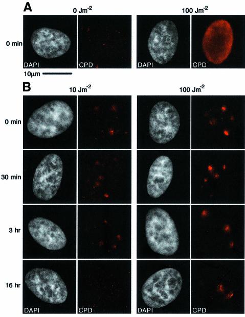
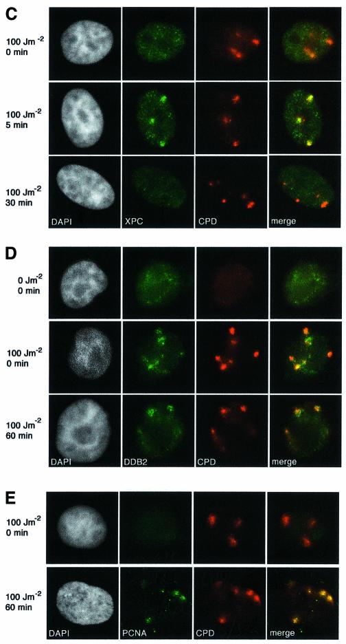
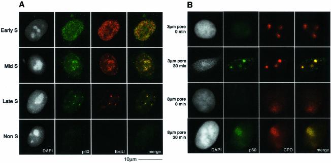
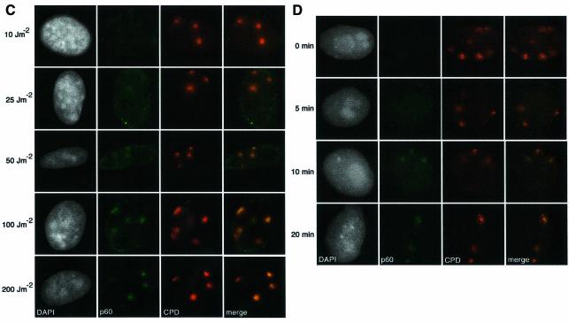
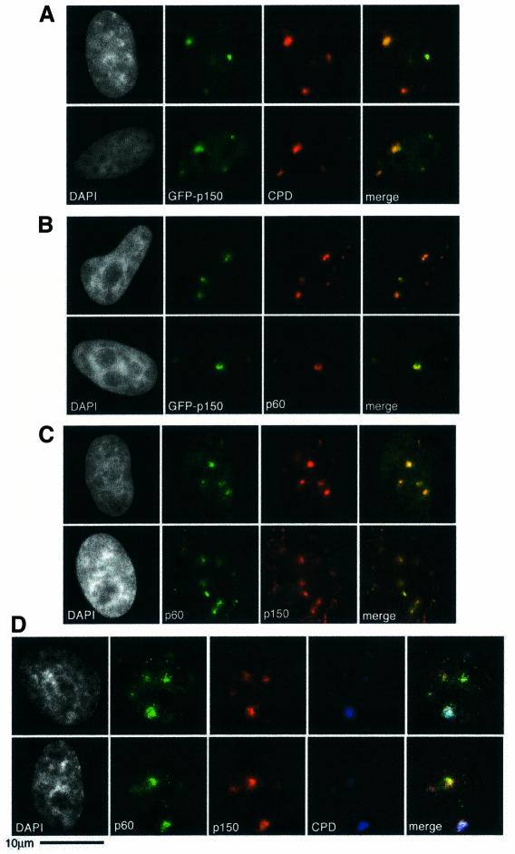
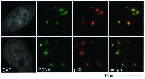
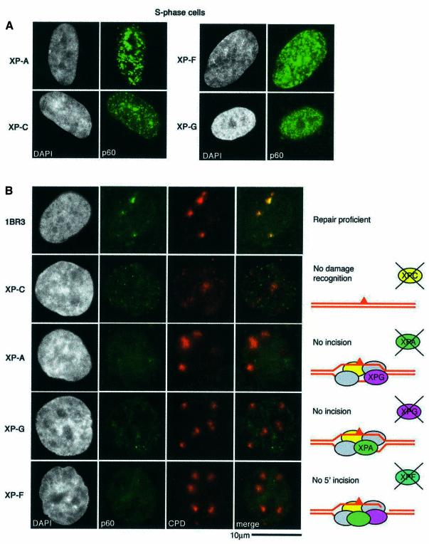
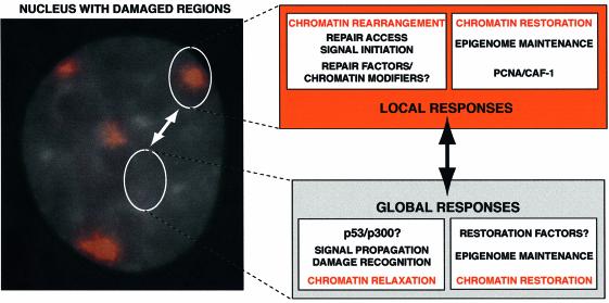
DNA damage and its repair can cause both local and global rearrangements of chromatin structure. In each case, the epigenetic information contained within this structure must be maintained. Using the recently developed method for the localized UV irradiation of cells, we analysed responses that occur locally to damage sites and global events triggered by local damage recognition. We thus demonstrate that, within a single cell, the recruitment of chromatin assembly factor 1 (CAF-1) to UV-induced DNA damage is a strictly local phenomenon, restricted to damage sites. Concomitantly, proliferating cell nuclear antigen (PCNA) locates to the same sites. This localized recruitment suggests that CAF-1 participates directly in chromatin structural rearrangements that occur in the vicinity of the damage. Use of nucleotide excision repair (NER)-deficient cells shows that the NER pathway--specifically dual incision--is required for recruitment of CAF-1 and PCNA. This in vivo demonstration of the local role of CAF-1, depending directly on NER, supports the hypothesis that CAF-1 ensures the maintenance of epigenetic information by acting locally at repair sites.


Figures

None
Fig. 1. In vivo formation and repair of localized DNA damage. (A) HeLa cells were UV-irradiated at 100 J/m2, or mock treated, and immediately fixed without detergent extraction. DNA damage was visualized by indirect immunofluorescence using a mouse monoclonal antibody against thymine dimers (CPD, red). (B) HeLa cells were locally irradiated through a polycarbonate UV-absorbing filter, at the doses indicated, followed by post- irradiation incubation for the times indicated on the left. DNA damage was detected by indirect immunofluorescence as in (A). (C) The recruitment of XPC protein to damage sites was visualized by indirect immunofluorescence following an irradiation dose of 100 J/m2 using a rabbit polyclonal antibody to XPC (green) and the mouse anti-thymine dimer monoclonal antibody (red). (D) The recruitment of stably expressed HA-tagged DDB2 protein to damage was visualized by indirect immunofluorescence following a dose of 100 J/m2 (or mock treatment) using a rat monoclonal antibody against the HA epitope (green) and the mouse anti-thymine dimer monoclonal antibody (red). (E) The local recruitment of PCNA, a protein involved in the repair synthesis stage of NER, to damage sites was visualized by indirect immunofluorescence after irradiation at 100 J/m2. Unless otherwise stated, all cells were treated with Triton prior to fixation to remove soluble nuclear proteins, and the DNA was visualized with DAPI (white). The scale bar in (A) represents 10 µm, the magnification used for all the images.
None
Fig. 2. Recruitment of CAF-1 p60 to replication and damage sites. (A) Characteristic patterns of CAF-1 p60 staining throughout the cell cycle. Cells were pulsed with BrdU followed by double labelling with a polyclonal against p60 (green) and a rat monoclonal against BrdU after denaturation with 4 M HCl (red). (B) CAF-1 p60 is locally recruited to sites of UV damage. Cells were irradiated with 100 J/m2 through filters with either 3 µm or 8 µm pores, with or without post-irradiation incubation, as indicated. Indirect immunofluorescence was performed with a polyclonal against p60 (green) and the anti-thymine dimer mouse monoclonal antibody (red). (C) Damage sites and CAF-1 p60 were visualized as in (B), 30 min after irradiation at the doses indicated. (D) Damage sites and CAF-1 p60 were visualized as in (B), at different times after a dose of 150 J/m2.
None
Fig. 3. Recruitment of CAF-1 p150 to damage sites. (A) Transient transfection of HeLa cells with a plasmid expressing the large subunit of CAF-1 tagged with GFP (GFP-p150) was used to visualize recruitment of GFP-p150 (green) to damage sites detected by indirect immunofluorescence (red) 30 min after irradiation at 100 J/m2. (B) Under the same conditions, GFP-p150 also colocalizes with endogenous p60 (red), detected by indirect immunofluorescence using the rabbit polyclonal antibody. (C) Endogenous p150 and p60 subunits were detected by indirect immunofluoresence 1 h after irradiation at 150 J/m2, using the rabbit polyclonal against p60 (green) and the mouse monoclonal against p150 (red). (D) A triple marking strategy was used to demonstrate the colocalization of endogenous p60 and p150 at damage sites 30 min after 150 J/m2. The proteins were detected by indirect immunofluorescence (p60 in green and p150 in red), with direct detection of the damage using the anti-damage antibody covalently coupled to AlexaFluor647 (blue); in this case a colocalization of all three antibodies results in a white signal in the merged view.
None
Fig. 4. PCNA recruitment concomitant with CAF-1. Locally recruited, detergent-insoluble PCNA, detected with a mouse monoclonal against PCNA (green), colocalized with p60, detected with a rabbit polyclonal antibody (red) 30 min after localized irradiation at 150 J/m2.
None
Fig. 5. Steps in repair processing required for CAF-1 recruitment. (A) XP fibroblasts show normal S-phase staining for CAF-1. Asynchronous cells were fixed and CAF-1 p60 was revealed by indirect immunofluorescence (green). (B) Repair-proficient primary human fibroblasts and those derived from XP patients defective at different stages of NER, as depicted in the cartoons on the right, were locally irradiated at 100 J/m2. Recovery was at 37°C for 1 h before processing for indirect immunofluorescence using the rabbit polyclonal antibody against p60 (green) and the mouse anti-CPD monoclonal (red).
None
Fig. 6. A chromatin-based view of the cellular response to UV damage. The local irradiation technique enables the independent analysis of local and global responses to UV damage. The image shows a nucleus stained with DAPI, with damage sites, visualized by immunofluorescence, in red. Although each damaged region contains many individual lesions, this can be used as a model for events that occur at a single lesion surrounded by undamaged DNA (the more physiologically relevant case). Here, we highlight that the damage response involves both local and global events and that there will be crosstalk between these two systems; a local event can initiate signals that propagate to have global effects, and such global effects can in turn impinge on events occurring local to the damage. Alterations of the chromatin structure, whether global or local, will need to be reversed once damage is complete to ensure stability of the epigenome. In this work we have demonstrated that CAF-1 is likely to perform this role at a local level.

References

    1. Aboussekhra A. and Wood,R.D. (1995) Detection of nucleotide excision repair incisions in human fibroblasts by immunostaining for PCNA. Exp. Cell Res., 221, 326–332. - PubMed
    1. Bakkenist C.J. and Kastan,M.B. (2003) DNA damage activates ATM through intermolecular autophosphorylation and dimer dissociation. Nature, 421, 499–506. - PubMed
    1. Batty D.P. and Wood,R.D. (2000) Damage recognition in nucleotide excision repair of DNA. Gene, 241, 193–204. - PubMed
    1. Bernard P., Maure,J.F., Partridge,J.F., Genier,S., Javerzat,J.P. and Allshire,R.C. (2001) Requirement of heterochromatin for cohesion at centromeres. Science, 294, 2539–2542. - PubMed
    1. Biggerstaff M. and Wood,R.D. (1999) Assay for nucleotide excision repair protein activity using fractionated cell extracts and UV-damaged plasmid DNA. Methods Mol. Biol., 113, 357–372. - PubMed

Publication types

MeSH terms