. 2003 Oct 1;22(19):5220-9.
doi: 10.1093/emboj/cdg509.

Unfolding pathways of native bacteriorhodopsin depend on temperature

Affiliations

Unfolding pathways of native bacteriorhodopsin depend on temperature

Harald Janovjak et al. EMBO J. .

Abstract

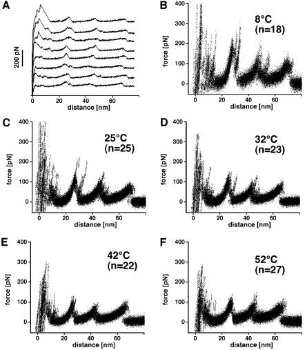
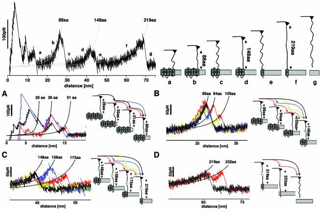
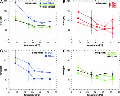
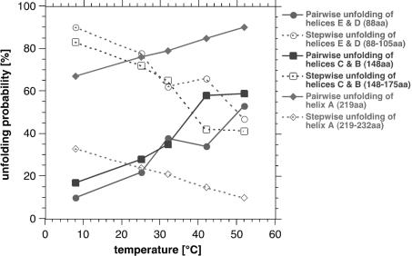
The combination of high-resolution atomic force microscopy (AFM) imaging and single-molecule force-spectroscopy was employed to unfold single bacteriorhodopsins (BR) from native purple membrane patches at various physiologically relevant temperatures. The unfolding spectra reveal detailed insight into the stability of individual structural elements of BR against mechanical unfolding. Intermittent states in the unfolding process are associated with the stepwise unfolding of alpha-helices, whereas other states are associated with the unfolding of polypeptide loops connecting the alpha-helices. It was found that the unfolding forces of the secondary structures considerably decreased upon increasing the temperature from 8 to 52 degrees C. Associated with this effect, the probability of individual unfolding pathways of BR was significantly influenced by the temperature. At lower temperatures, transmembrane alpha-helices and extracellular polypeptide loops exhibited sufficient stability to individually establish potential barriers against unfolding, whereas they predominantly unfolded collectively at elevated temperatures. This suggests that increasing the temperature decreases the mechanical stability of secondary structural elements and changes molecular interactions between secondary structures, thereby forcing them to act as grouped structures.


Figures

None
Fig. 1. Unfolding BR from native purple membrane at various temperatures. (A) Force curves of individual BR molecules recorded at 25°C. To show common unfolding patterns among single-molecule events, the force spectra recorded at different temperatures were superimposed. (BF) BR unfolded at 8°C (B), 25°C (C), 32°C (D), 42°C (E) and 52°C (F) in 300 mM KCl, 20 mM Tris–HCl with a pH of 7.8 being adjusted for each temperature.
None
Fig. 2. Unfolding pathways of BR. Top, pairwise unfolding pathways of transmembrane α-helices. The left curve shows a representative unfolding spectrum of a single BR, while the schematic drawing of the unfolding pathways is shown on the right. The first force peaks detected within a separation of 0–15 nm to the purple membrane surface indicate the unfolding of transmembrane α-helices F and G, and of loops FG and EF. The first peaks within this region are superimposed by non-specific surface interactions between purple membrane and AFM tip. After the unfolding event (a), the amount of aa stretched is increased to 88 and the cantilever relaxes. Further separating tip and sample stretches the polypeptide (b) thereby pulling on helix E. At a certain pulling force, the mechanical stability of helices E and D is insufficient and they unfold together with loops DE and CD (c). The available 148 aa are now stretched (d), the polypeptide being pulling on helix C. Helices B and C and loops BC and AB unfold within a single step, thereby relaxing the cantilever (e). By further separating tip and purple membrane, the cantilever pulls on helix A (f) until the polypeptide is completely extracted from the membrane (g). The force-spectroscopy curve was recorded in 300 mM KCl, 20 mM Tris–HCl pH 7.8 at room temperature. (AD) Unfolding events of individual secondary structures. (A) Occasionally the first major unfolding peak shows side peaks at about 26, 36 and 51 aa. The peak at 26 aa indicates the unfolding of the cytoplasmic half of α-helix G up to the covalently bound retinal, which is embedded in the hydrophobic membrane core. The peak at 36 aa indicates the G helix to be unfolded completely. At 51 aa, helix G and the loop connecting helices G and F are unfolded and the force pulls directly on helix F until this helix unfolds together with loop EF. (B) The side peaks of the second major peak indicate the stepwise unfolding of helices E and D and loop DE. The peak at 88 aa indicates the unfolding of helix E, that at 94 aa of the loop DE, and the peak at 105 aa indicates unfolding of helix D. (C) The side peaks of the third major peak indicate the stepwise unfolding of helices C and B and loop BC. The peak at 148 aa indicates the unfolding of helix C, that at 158 aa of the loop BC, and the peak at 175 aa indicates unfolding of helix B. (D) The side peak of the last major peak indicates the unfolding of helix A (219 aa) and of the pulling of the N-terminal end through the purple membrane (232 aa).
None
Fig. 3. Unfolding forces of secondary structural elements depend on temperature. (A) Rupture forces of main peaks, which exhibited no side peaks. The forces represent the pairwise unfolding of transmembrane α-helices E and D (88 aa), C and B (148 aa) and the unfolding of helix A (219 aa). The main peak representing the pairwise unfolding of helices G and F are not shown because unspecific surface interactions between the AFM tip and the purple membrane scatter the position and appearance of the force peaks significantly. (BD) Rupture forces of side peaks represent unfolding of single α-helices and of their connecting loops (see text). The thermally induced weakening of the unfolding forces was fitted (dotted lines) using equation (2).
None
Fig. 4. Probability of unfolding pathways depends on temperature. The occurrence of main force peaks exhibiting no side peaks (solid lines) increased with increasing temperature. As a consequence, the probability of the main peaks exhibiting side peaks (dashed lines) decreased significantly. Solid lines represent probabilities for the pairwise unfolding of transmembrane α-helices E and D (88 aa, red), C and B (148 aa, blue) and of helix A (219 aa, green). The probability of their stepwise unfolding is presented by the dotted lines. This indicates α-helices of BR unfold preferentially pairwise at elevated temperatures. The probability of single structural elements, such as helices or loops, to unfold in a separate event decreases with increasing temperature.

References

    1. Baldwin J.M. (1993) The probable arrangement of the helices in G protein-coupled receptors. EMBO J., 12, 1693–1703. - PMC - PubMed
    1. Bell G.I. (1978) Models for the specific adhesion of cells to cells. Science, 200, 618–627. - PubMed
    1. Belrhali H., Nollert,P., Royant,A., Menzel,C., Rosenbusch,J.P., Landau,E.M. and Pebay-Peyroula,E. (1999) Protein, lipid and water organization in bacteriorhodopsin crystals: a molecular view of the purple membrane at 1.9 Å resolution. Structure Fold. Des., 7, 909–917. - PubMed
    1. Blume A. (1983) Apparent molar heat capacities of phospholipids in aqueous dispersion: effects of chain length and head group structure. Biochemistry, 22, 5436–5442.
    1. Booth P.J., Templer,R.H., Meijberg,W., Allen,S.J., Curran,A.R. and Lorch,M. (2001) In vitro studies of membrane protein folding. Crit. Rev. Biochem. Mol. Biol., 36, 501–603. - PubMed

Publication types

Substances