

Pluripotency deficit in clones overcome by clone-clone aggregation: epigenetic complementation?
- PMID: 14517267
- PMCID: PMC204490
- DOI: 10.1093/emboj/cdg507
Pluripotency deficit in clones overcome by clone-clone aggregation: epigenetic complementation?
Abstract
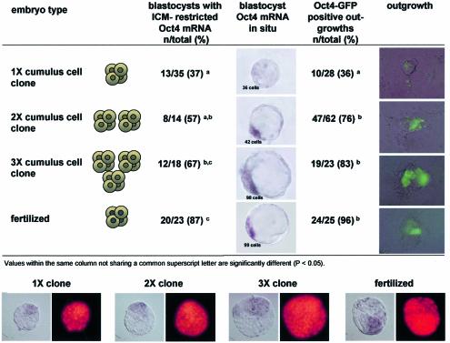
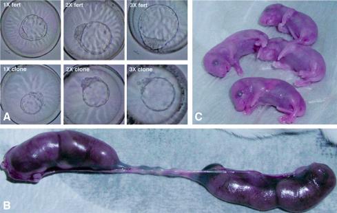
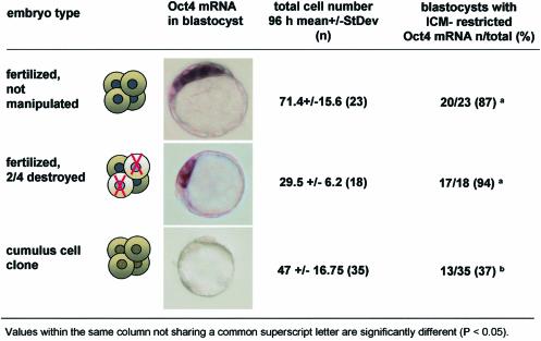
Abnormal gene expression patterns in somatic cell clones and their attrition in utero are commonly considered a consequence of errors in nuclear reprogramming. We observe that mouse clone blastocysts have less than half the normal cell number, and that higher cell number correlates with correct expression of Oct4, a gene essential for peri-implantation development and embryonic pluripotency. To increase the cell number, we aggregated genetically identical clones at the 4-cell stage. Clone-clone aggregates did not form more blastocysts, but the majority expressed Oct4 normally and had higher rates of fetal and postnatal development. Fertilized blastocysts with low cell numbers, induced by removal of two blastomeres at the 4-cell stage, did not exhibit abnormal Oct4 expression, indicating that improved gene expression and post-implantation development of clone-clone aggregates is not a consequence of increased cell number. Rather, we propose that complementation of non-cell-autonomous defects of genetically identical, but epigenetically different, embryos results in improved gene expression in clone-clone aggregates.
Figures

References
-
- Boni R., Tosti,E., Roviello,S. and Dale,B. (1999) Intercellular communication in in vivo- and in vitro-produced bovine embryos. Biol. Reprod., 61, 1050–1055. - PubMed
-
- Booth P.J., Viuff,D., Tan,S., Holm,P., Greve,T. and Callesen,H. (2003) Numerical chromosome errors in day 7 somatic nuclear transfer bovine blastocysts. Biol. Reprod., 68, 922–928. - PubMed
-
- Bortvin A., Eggan,K., Skaletsky,H., Akutsu,H., Berry,D.L., Yanagimachi,R., Page,D.C. and Jaenisch,R. (2003) Incomplete reactivation of Oct4-related genes in mouse embryos cloned from somatic nuclei. Development, 130, 1673–1680. - PubMed
-
- Buehr M. and McLaren,A. (1974) Size regulation in chimaeric mouse embryos. J. Embryol. Exp. Morphol., 31, 229–234. - PubMed
Publication types
MeSH terms
Substances
Grants and funding
LinkOut - more resources
Full Text Sources
Other Literature Sources
Medical
