. 2003 Oct;23(20):7108-21.
doi: 10.1128/MCB.23.20.7108-7121.2003.

Daxx silencing sensitizes cells to multiple apoptotic pathways

Affiliations

Daxx silencing sensitizes cells to multiple apoptotic pathways

Liuh-Yow Chen et al. Mol Cell Biol. 2003 Oct.

Abstract

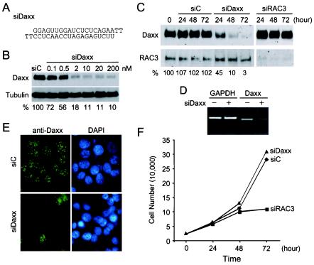
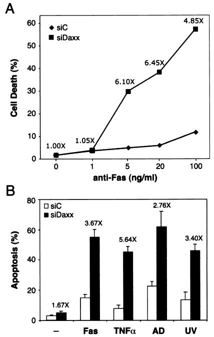
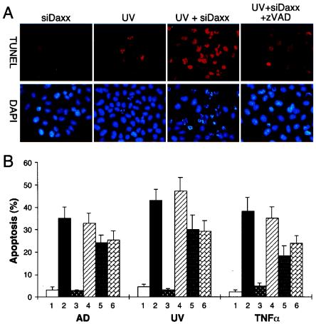
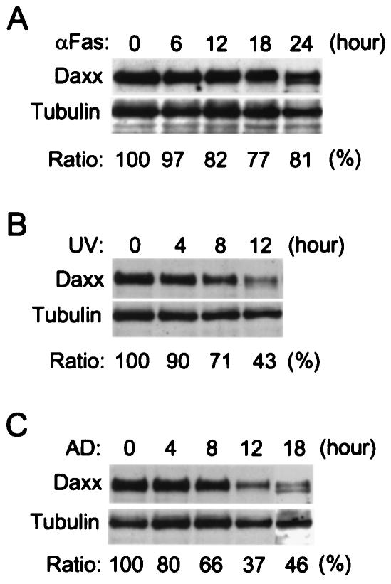
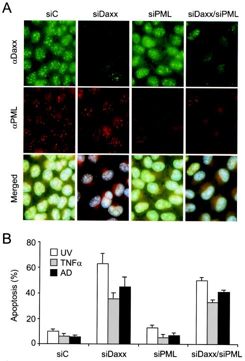
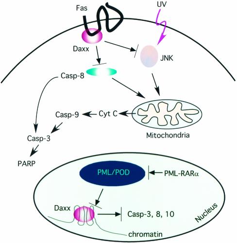
Daxx is a nuclear protein involved in apoptosis and transcriptional repression, and it interacts with the death receptor Fas, promyelocytic leukemia protein (PML), and several transcriptional repressors. The function of Daxx in apoptosis is controversial because opposite results were obtained in transient overexpression and genetic knockout studies. Furthermore, the roles of PML and transcriptional repression in Daxx-regulated apoptosis are currently unknown. In this study, we investigated the role of Daxx in Fas- and stress-induced apoptosis by small interfering RNA-mediated Daxx silencing in mammalian cells. Daxx silencing had no apparent cytotoxic effects on mammalian cells within 72 h. Intriguingly, Daxx silencing strongly sensitized cells to Fas- and stress-induced apoptosis, which was accompanied by caspase activation, cytochrome c release, and Jun N-terminal kinase activation. Consistently, endogenous Daxx was degraded rapidly upon induction of apoptosis by stress or anti-Fas antibody. Finally, PML silencing had no effect on Daxx silencing-mediated apoptotic events, while caspase gene expression was upregulated in the absence of Daxx. These data strongly suggest that Daxx may inhibit Fas and stress-mediated apoptosis by suppressing proapoptotic gene expression outside of PML domains.


Figures

FIG. 1.
FIG. 1.
Silencing of Daxx expression in mammalian cells by siRNA. (A) Nucleotide sequence of the double-stranded siRNA against Daxx mRNA (siDaxx). (B) Dose-dependent inhibition of Daxx expression by siDaxx. HeLa cells were transfected with control reagent (siC) or increasing concentrations of siDaxx. Total cell lysate were prepared 48 h after transfection and analyzed by SDS-PAGE and Western blot detection of Daxx and tubulin. The relative Daxx protein levels after normalization with tubulin are shown at bottom. (C) Time course-dependent silencing of Daxx by siDaxx. HeLa cells were transfected with siC, siDaxx, or the siRNA against coactivator RAC3 (siRAC3) at 50 nM concentration. Cells were harvested at indicated hours after transfection and analyzed for Daxx and RAC3 expression by Western blot. Relative Daxx levels in comparison with untransfected cells are shown at bottom. (D) RT-PCR analysis of Daxx mRNA in untransfected cells (−) and cells transfected with siDaxx (+). Total RNA was isolated 48 h after transfection and analyzed by RT-PCR for GAPDH and Daxx mRNAs. PCR products were analyzed on a 1% agarose gel followed by ethidium bromide staining and imaging with the Kodak EDAS system. (E) Immunofluorescence staining of Daxx in siDaxx-transfected cells. MCF-7 cells transfected with siC or siDaxx for 48 h were analyzed by immunofluorescence staining for Daxx with affinity-purified rabbit anti-Daxx polyclonal antibodies. Cell nuclei were visualized with 4′,6′-diamidino-2-phenylindole (DAPI) staining. (F) Effect of Daxx silencing on cell proliferation. HeLa cells were transfected with 50 nM siC (♦), siDaxx (▴), or siRAC3 (▪). Total cell numbers were counted on a hemacytometer at the indicated hours after transfection. Daxx silencing had no effect on cell growth and proliferation.
FIG. 2.
FIG. 2.
Daxx silencing sensitizes cells to Fas and stress-induced apoptosis. (A) Effect of siDaxx on Fas-induced cell death. HeLa cells transfected with control reagent siC (♦) or siDaxx (▪) for 36 h were treated with increasing concentrations of the agonistic anti-Fas antibody CH11. Percent cell death was determined at 24 h after the addition of antibody by trypan blue staining. The antibody concentrations are shown on a log scale, with 0 ng/ml placed at an arbitrary position. The increases in cell death are shown above the siDaxx data points at each antibody concentration, and the actual increases were, from left to right, 0, 0.2, 25, 33, and 46%, respectively. (B) Effect of siDaxx on stress-induced apoptosis. HeLa cells transfected with siC (open bars) or siDaxx (black bars) for 48 h were treated with the anti-Fas antibody (CH11, 200 ng/ml) for 12 h, TNF-α (10 ng/ml) or actinomycin D (AD, 100 nM) for 8 h, or UV (50 J/m2), followed by an 8-h incubation. Apoptosis was determined by TUNEL assay as described in Materials and Methods. Enhancement of apoptosis between siDaxx- and siC-treated cells were shown above the black bars.
FIG. 3.
FIG. 3.
Involvement of caspases in Daxx silencing-mediated apoptosis. (A) TUNEL assay showing synergistic induction of apoptosis by siDaxx and UV. HeLa cells were transfected with siDaxx or control reagent for 48 h. The caspase inhibitor z-VAD-fmk (z-VAD, 20 μM) was added 1 h prior to UV irradiation (50 J/m2). Apoptosis was determined by TUNEL (red) and DAPI (blue) staining 8 h after UV irradiation. (B) Effect of various caspase inhibitors on siDaxx-mediated apoptosis. HeLa cells were transfected with siC (lanes 1) or siDaxx (lanes 2 to 6), followed by treatment with actinomycin D (AD, 100 nM), UV irradiation (50 J/m2), or TNF-α (10 ng/ml) in the absence (lanes 2) or presence (lanes 3 to 6) of various caspase inhibitors. Cells were treated with the general caspase inhibitor z-VAD-fmk (lanes 3), the caspase 1 inhibitor z-YVAD-fmk (lanes 4), the caspase 8 inhibitor z-IETD-fmk (lanes 5), or the caspase 9 inhibitor z-LEHD-fmk (lanes 6). Apoptotic cells were detected by TUNEL assay and counting at least 200 cells in three different fields. The general caspase inhibitor z-VAD-fmk completely blocked siDaxx-enhanced apoptosis, while z-IETD-fmk and z-LEHD-fmk partially suppressed siDaxx-enhanced apoptosis.
FIG. 4.
FIG. 4.
Synergistic activation of caspases by Daxx silencing. (A) Effects of siDaxx on cleavage of caspase 8, caspase 9, and PARP. HeLa cells with (+) or without (−) siDaxx transfection were treated with anti-Fas antibody (200 ng/ml), UV (50 J/m2), or actinomycin D (AD, 100 nM). Proteolytic cleavage of caspase 8 (Casp-8), caspase 9 (Casp-9), and PARP was determined by Western blotting 6 h after the treatment. Arrows at the right mark the proteolytic fragments. Protein molecular size markers are shown at left (in kilodaltons). Daxx silencing synergistically activated caspase 8, caspase 9, and PARP cleavage with anti-Fas antibody, UV, and actinomycin D. These proteolytic cleavages were inhibited by the general caspase inhibitor z-VAD-fmk (z-VAD, 20 μM), but not by the caspase 1 inhibitor z-YVAD-fmk (z-YVAD, 20 μM). (B) Kinetics of caspase activation in siDaxx-treated cells. Control siC or siDaxx-transfected HeLa cells were treated with anti-Fas antibody (200 ng/ml), TNF-α (10 ng/ml), TNF-α (2 ng/ml) plus cycloheximide (Chx, 2 μg/ml), actinomycin D (100 nM), or UV (50 J/m2), and cells were harvested at the time points shown at the bottom. The harvested cells were analyzed by Western blotting for proteolytic processing of caspase 8 and caspase 9 and by TUNEL assay for percent apoptosis. Boxes at the bottom mark the apparent time points of caspase activation and apoptosis.
FIG. 5.
FIG. 5.
Daxx silencing stimulates early cytochrome c release. HeLa cells transfected with the siC control or siDaxx were treated with UV irradiation (50 J/m2), anti-Fas antibody (200 ng/ml), or actinomycin D (AD, 100 nM) for 1 h. Daxx (green) and cytochrome c (red) were double stained with the respective polyclonal and monoclonal antibodies along with DAPI staining for nuclei (blue). (A) Representative immunofluorescence images for mitochondrial cytochrome c release at 1 h after the indicated treatments. Release of cytochrome c from mitochondria caused diffuse cytochrome c staining in cells marked by arrows. (B) Quantitation of cytochrome c release after various treatments. The data represent three independent experiments with at least 300 cells scored from three different fields. Under the experimental conditions, siC, siDaxx, and the treatment alone did not cause significant cytochrome c release. However, siDaxx transfection in combination with anti-Fas antibody, UV, or actinomycin D synergistically enhanced cytochrome c release.
FIG. 6.
FIG. 6.
Daxx silencing affects JNK activation. (A) Effect of Daxx silencing on Fas-mediated JNK activation. HeLa cells were transfected with siC or siDaxx for 48 h, followed by anti-Fas antibody (200 ng/ml) treatment for the indicated periods of time. The JNK kinase assay was performed as described in Materials and Methods. Phosphorylated Jun (p-Jun) was analyzed by SDS-PAGE and autoradiography. The immunoprecipitated JNK1 and -2 proteins were detected by Western blot (W.B.). Daxx silencing slightly enhanced JNK activation at 120 and 240 min of anti-Fas antibody treatment. (B) Effect of Daxx silencing on UV-induced JNK activation. The JNK kinase assay (K.A.) and Western blot (W.B.) were performed as described above. Cells were harvested at the indicated time (in minutes) after UV irradiation (50 J/m2). A threefold-higher level of phosphorylated Jun remained in siDaxx- versus siC-treated cells at 120 min after UV irradiation. (C) Effect of Daxx silencing on JNK activation induced by TNF-α. The JNK kinase assay (K.A.) and Western blot (W.B.) are described above. There was a 33% increase in JNK kinase activity at 15 min after TNF-α treatment (10 ng/ml).
FIG. 7.
FIG. 7.
Reduction of Daxx protein levels during induction of apoptosis. The endogenous Daxx and tubulin protein levels in HeLa cells were analyzed by Western blot upon treatment of the cells with (A) anti-Fas antibody (200 ng/ml), (B) UV irradiation (50 J/m2), or (C) actinomycin D (AD, 100 nM). Total cell lysates from approximately equal numbers of cells were prepared at the indicated time points after treatment and analyzed by SDS-PAGE followed by Western blot detection for Daxx and tubulin. The relative Daxx protein levels are shown at the bottom as a ratio over the tubulin protein levels, which remained relatively unchanged during the treatment. The Daxx protein level declined to 82, 43, and 37% at 12 h after treatment with anti-Fas antibody, UV, and actinomycin D, respectively.
FIG. 8.
FIG. 8.
Role of PML in Daxx silencing-mediated apoptosis. (A) HeLa cells were transfected with siC, siDaxx, siPML, or siDaxx plus siPML, followed by coimmunofluorescence staining for Daxx (green) and PML (red) with anti-Daxx polyclonal and anti-PML monoclonal antibodies. (B) Apoptotic response of Daxx- and/or PML-depleted cells to UV, TNF-α, and actinomycin D treatment. Apoptosis was determined by the TUNEL assay, with at least 300 cells scored in three different fields.
FIG. 9.
FIG. 9.
Daxx silencing affects expression of apoptosis-regulatory genes. HeLa cells were transfected with the siC control (−) or siDaxx (+) for 48 h, and total RNAs were isolated. Three micrograms of total RNAs was analyzed by RT-PCR for the indicated genes. The PCR products were analyzed on a 2% agarose gel and ethidium bromide staining. The relative intensities of expected PCR bands were measured by the Kodak ID image analysis software. The increase or decrease (in parentheses) in expression for each gene is shown at the bottom. The upper band in the caspase 10 reaction was the expected PCR product, while the lower band may represent an alternatively spliced form.
FIG. 10.
FIG. 10.
Potential mechanisms of the antiapoptotic function of Daxx. Daxx might inhibit Fas-mediated caspase 8 activation and UV- or stress-induced JNK activation. This ability might be due to association of Daxx with the cytoplasmic domain of Fas. Consequently, depletion of Daxx stimulated caspase 8 and JNK activation, causing early mitochondrial cytochrome c release and activation of caspase 9 and caspase 3. In addition, PML may sequester Daxx to PODs, inhibiting its transcriptional repressor function. The oncogenic fusion protein PML-retinoic acid receptor alpha (RARα) may disrupt PODs, causing the release of Daxx from PODs and its association with condensed chromatin. We hypothesize that the association of Daxx with condensed chromatin might cause repression of selected caspase genes. Consequently, depletion of Daxx may increase the expression of apoptotic proteins, resulting in sensitization of cells to induction of apoptosis.

References

    1. Amin, H. M., S. Saeed, and S. Alkan. 2001. Histone deacetylase inhibitors induce caspase-dependent apoptosis and downregulation of daxx in acute promyelocytic leukaemia with t(15;17). Br. J. Haematol. 115:287-297. - PubMed
    1. Bernstein, E., A. M. Denli, and G. J. Hannon. 2001. The rest is silence. RNA 7:1509-1521. - PMC - PubMed
    1. Borden, K. L., E. J. CampbellDwyer, and M. S. Salvato. 1997. The promyelocytic leukemia protein PML has a proapoptotic activity mediated through its RING domain. FEBS Lett. 418:30-34. - PMC - PubMed
    1. Cermak, L., a. imova, A. Pintzas, V. Horeji, and L. Andera. 2002. Molecular mechanisms involved in CD43-mediated apoptosis of TF-1 cells: roles of transcription, Daxx expression and adhesion molecules. J. Biol. Chem. 277:7955-7961. - PubMed
    1. Chang, H. Y., H. Nishitoh, X. Yang, H. Ichijo, and D. Baltimore. 1998. Activation of apoptosis signal- regulating kinase 1 (ASK1) by the adapter protein daxx. Science 281:1860-1863. - PubMed

Publication types

MeSH terms

LinkOut - more resources