. 2003 Oct;23(20):7198-209.
doi: 10.1128/MCB.23.20.7198-7209.2003.

Nrf2 is a direct PERK substrate and effector of PERK-dependent cell survival

Affiliations

Nrf2 is a direct PERK substrate and effector of PERK-dependent cell survival

Sara B Cullinan et al. Mol Cell Biol. 2003 Oct.

Abstract

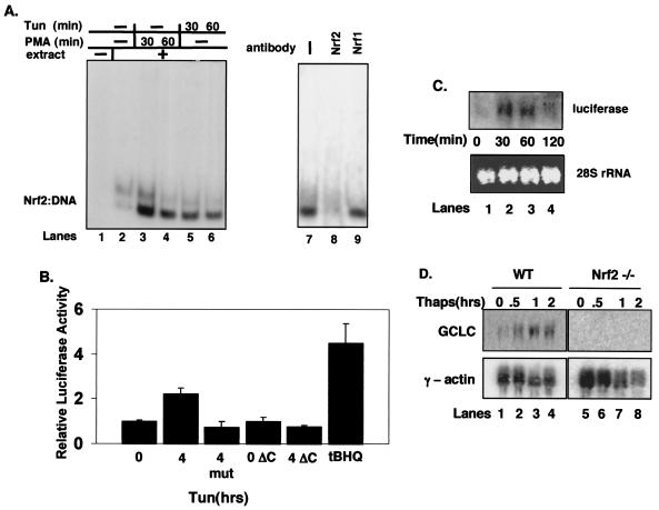
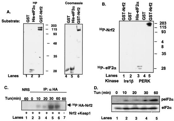
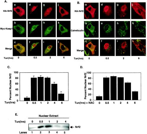
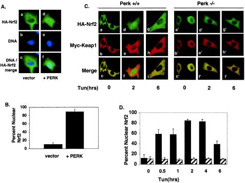
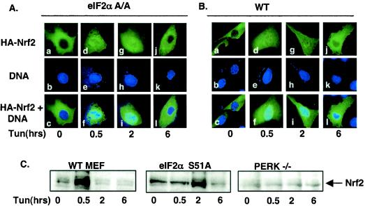
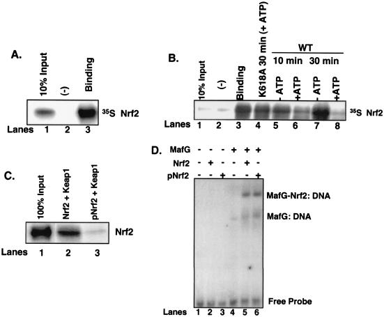
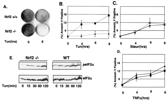
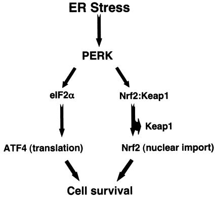
Activation of PERK following the accumulation of unfolded proteins in the endoplasmic reticulum (ER) promotes translation inhibition and cell cycle arrest. PERK function is essential for cell survival following exposure of cells to ER stress, but the mechanisms whereby PERK signaling promotes cell survival are not thoroughly understood. We have identified the Nrf2 transcription factor as a novel PERK substrate. In unstressed cells, Nrf2 is maintained in the cytoplasm via association with Keap1. PERK-dependent phosphorylation triggers dissociation of Nrf2/Keap1 complexes and inhibits reassociation of Nrf2/Keap1 complexes in vitro. Activation of PERK via agents that trigger the unfolded protein response is both necessary and sufficient for dissociation of cytoplasmic Nrf2/Keap1 and subsequent Nrf2 nuclear import. Finally, we demonstrate that cells harboring a targeted deletion of Nrf2 exhibit increased cell death relative to wild-type counterparts following exposure to ER stress. Our data demonstrate that Nrf2 is a critical effector of PERK-mediated cell survival.


Figures

FIG. 1.
FIG. 1.
Nrf2, and not Nrf1, is activated by ER stress. (A) Whole-cell extracts prepared from NIH 3T3 cells treated with PMA (lanes 3 and 4) or tunicamycin (tun) (1 μg/ml) (lanes 5 and 6) were mixed with a radiolabeled oligonucleotide containing a consensus ARE in an EMSA reaction. In lanes 7 to 9, tunicamycin-treated (60 min) whole-cell extracts were exposed to antibodies against Nrf1 (lane 9) or Nrf2 (lane 8) in the EMSA reactions. (B) NIH 3T3 cells were cotransfected with plasmids encoding the 4xARE reporter or a reporter lacking an ARE (mut) along with the cytomegalovirus-Renilla control (internal normalization control) with or without PERKΔC. Cells were treated with 5 μg of tunicamycin/ml or 100 μM tBHQ for the indicated time intervals (in hours), harvested, and assayed for luciferase and Renilla activities. Luciferase activity is shown as the ratio of reporter activity to Renilla luciferase activity; activity in untreated cells was set as 1. Error bars represent the standard deviation from three independent experiments. (C) NIH 3T3 cells were transfected with the 4xARE reporter and treated with tunicamycin (5 μg/ml) for the indicated intervals. RNA was extracted, and expression of luciferase was assessed by Northern blot analysis. (D) Wild-type (lanes 1 to 4) and Nrf2−/− (lanes 5 to 8) fibroblasts were treated with thapsigargin (1 μM) for the indicated intervals. RNA was extracted, and expression of GCLC and γ-actin was determined by Northern blot analysis.
FIG. 2.
FIG. 2.
Phosphorylation of Nrf2 by PERK and during the UPR. (A) Recombinant GST (lane 1), GST-Nrf2 (lane 3), or His-eIF2α (lane 2) was mixed with the catalytic domain of PERK along with [γ-32P]ATP and incubated at 30° for 30 min. Phosphorylated proteins were resolved on denaturing polyacrylamide gels and visualized by autoradiography. Lanes 4, 5, and 6 show Coomassie brilliant blue staining of GST, His-eIF2α, and GST-Nrf2, respectively. (B) Lysates prepared from NIH 3T3 cells infected with PERK or Ire1β were precipitated with an antibody against PERK or a C-terminal myc epitope, respectively. The precipitated protein was mixed with recombinant His-eIF2α, GST, or GST-Nrf2 along with [γ-32P]ATP. Phosphorylated proteins were resolved on denaturing gels and visualized by autoradiography. (C) NIH 3T3 cells cotransfected with HA-Nrf2 and myc-Keap1 were treated with 5 μg of tunicamycin/ml and [32P]orthophosphate for the indicated times. Lysates were precipitated with the 12CA5 antibody; complexes were resolved by SDS-PAGE and visualized by autoradiography. (D) Lysates prepared from NIH 3T3 cells were treated with 5 μg of tunicamycin/ml and resolved by SDS-PAGE, and membranes were probed with antibodies specific for total and phosphorylated eIF2α.
FIG. 3.
FIG. 3.
The UPR triggers PERK-dependent Nrf2 nuclear translocation. (A and B) NIH 3T3 cells proliferating on glass coverslips were cotransfected with plasmids encoding HA-Nrf2 and myc-Keap1. Transfected cells were treated with 5 μg of tunicamycin/ml for the indicated periods of time and fixed, and proteins were visualized by confocal microscopy with antibodies specific for the HA epitope, the myc epitope, and calreticulin. (C) Graphical representation of data in panels A and B. Error bars represent the standard deviations from three independent experiments. (D) NIH 3T3 cells cotransfected with plasmids encoding HA-Nrf2 and myc-Keap1 were pretreated with 5 mM NAC and then treated as described for panels A and B. Error bars represent the standard deviations from three independent experiments. (E) Nuclear extracts prepared from NIH 3T3 cells treated with 5 μg of tunicamycin/ml for the indicated periods of time were resolved by SDS-PAGE, transferred to a nitrocellulose membrane, and probed with an antibody specific for Nrf2.
FIG. 4.
FIG. 4.
PERK activity is necessary and sufficient for Nrf2 nuclear translocation. (A) NIH 3T3 cells proliferating on glass coverslips were cotransfected with plasmids encoding HA-Nrf2, Myc-Keap1 (panels a to c), or HA-Nrf2, myc-Keap1 and myc-PERK (panels d to f). Twenty-four hours posttransfection, cells were fixed and proteins were detected by indirect immunofluorescence with an antibody specific for the HA epitope. DNA was stained with Hoechst dye. (B) Graphical representation of data in panel A. Error bars represent standard deviations from three independent experiments. (C) Wild-type (PERK+/+) or PERK−/− murine fibroblasts proliferating on glass coverslips were cotransfected with plasmids encoding HA-Nrf2 and myc-Keap1 and treated with 5 μg of tunicamycin/ml for the indicated time intervals. Proteins were detected with antibodies specific for the HA and myc epitopes and visualized by confocal microscopy. (D) Graphical representation of data presented in panel C. Solid bars represent wild-type cells, and hatched bars represent PERK−/− cells. Error bars represent the standard deviations from three independent experiments.
FIG. 5.
FIG. 5.
Nrf2 nuclear translocation occurs independently of eIF2α phosphorylation. (A and B) Wild-type fibroblasts (B) or eIF2α(S51A) fibroblasts (A) were cotransfected with plasmids encoding HA-Nrf2 and myc-Keap1 and treated with 5 μg of tunicamycin/ml for the indicated time intervals. HA-Nrf2 was detected with the 12CA5 monoclonal antibody and visualized by confocal microscopy. (C) Nuclear extracts were prepared from wild-type (WT), eIF2α(S51A), or PERK−/− MEFs treated with 5 μg of tunicamycin/ml for the indicated intervals, resolved by SDS-PAGE, transferred to a nitrocellulose membrane, and probed with an antibody specific for Nrf2.
FIG. 6.
FIG. 6.
Phosphorylation-dependent dissociation of Nrf2 from Keap1. (A) Nrf2 in vitro transcribed and translated in the presence of [35S]methionine was mixed with recombinant Keap1 (lane 3) or eIF2α (lane 2). Complexes were affinity purified by nickel chromatography, resolved by SDS-PAGE, and visualized by autoradiography. Lane 1 is 10% Nrf2 input. (B) Nrf2/Keap1 complexes prepared as described for panel A and mixed with the catalytic domain of PERK in the absence (lanes 5 and 7) or presence (lanes 6 and 8) of ATP (500 μM) or a kinase-dead PERK in the presence of ATP (lane 4) for the indicated intervals. Lane 1 is 10% Nrf2 input, and lane 3 is Nrf2-Keap1 binding. Lane 2 is binding to the negative control, eIF2α. Upon completion of the kinase reactions, the complexes were washed, resolved by SDS-PAGE, and visualized by autoradiography. (C) Nrf2 was mixed with the catalytic domain of PERK in the absence (lane 2) or presence (lane 3) of ATP and then mixed with recombinant Keap1 as described above. After binding, the complexes were washed extensively and proteins were resolved by SDS-PAGE, transferred to a nitrocellulose membrane, and probed with an antibody specific for Nrf2. Lane 1 shows 100% Nrf2 input. (D) Nrf2 (lanes 2 and 5) or PERK-phosphorylated Nrf2 (lanes 3 and 6) was mixed with in vitro-transcribed and -translated MafG (lanes 5 and 6) along with a radiolabeled oligonucleotide containing a consensus ARE in an EMSA reaction. Also shown are EMSA reactions with MafG alone (lane 4) or no protein (lane 1) mixed with the radiolabeled oligonucleotide.
FIG. 7.
FIG. 7.
Nrf2 promotes increased cell survival after chronic ER stress. (A) Wild-type and Nrf2−/− MEFs were left untreated or treated with 1 μg of tunicamycin/ml for 8 h and then allowed to recover for 7 days in normal medium. Colonies were visualized by using Giemsa stain. (B to D) Wild-type (▴) and Nrf2−/− (▪) MEFs proliferating on glass coverslips were left untreated or treated with 5 μg of tunicamycin/ml (B), 4 μM staurosporine (C), or 5 ng of TNF-α/ml plus 10 μg of cycloheximide/ml (D) for the indicated intervals (in hours). Apoptotic cells were visualized with a FITC-conjugated annexin V antibody. Error bars represent the standard deviations from three independent experiments. (E) Wild-type and Nrf2−/− MEFs were treated with 5 μg of tunicamycin/ml for the indicated intervals. Proteins were resolved by SDS-PAGE, and membranes were probed with antibodies specific for total and phosphorylated eIF2α.
FIG. 8.
FIG. 8.
Model depicting the bifurcation of PERK-dependent survival signals (details in text).

References

    1. Andrews, N. C., H. Erdjument-Bromage, M. B. Davidson, P. Tempst, and S. H. Orkin. 1993. Erythroid transcription factor NF-E2 is a haematopoietic-specific basic-leucine zipper protein. Nature 362:722-728. - PubMed
    1. Balachandran, S., C. N. Kim, W.-C. Yeh, T. W. Mak, K. Bhalla, and G. N. Barber. 1998. Activation of the dsRNA-dependent protein kinase, PKR, induces apoptosis through FADD-mediated death signaling. EMBO J. 17:6888-6902. - PMC - PubMed
    1. Berlanga, J. J., J. Santoyo, and C. Haro. 1999. Characterization of a mammalian homolog of the GCN2 eukaryotic initiation factor 2α kinase. Eur. J. Biochem. 265:754-762. - PubMed
    1. Bertolotti, A., Y. Zhang, L. M. Hendershot, H. P. Harding, and D. Ron. 2000. Dynamic interaction of BiP and ER stress transducers in the unfolded-protein response. Nat. Cell Biol. 2:326-332. - PubMed
    1. Brewer, J. W., and J. A. Diehl. 2000. PERK mediates cell-cycle exit during the mammalian unfolded protein response. Proc. Natl. Acad. Sci. USA 97:12625-12630. - PMC - PubMed

Publication types

MeSH terms