. 2003 Oct;23(20):7230-42.
doi: 10.1128/MCB.23.20.7230-7242.2003.

Myostatin signals through a transforming growth factor beta-like signaling pathway to block adipogenesis

Affiliations

Myostatin signals through a transforming growth factor beta-like signaling pathway to block adipogenesis

A Rebbapragada et al. Mol Cell Biol. 2003 Oct.

Abstract

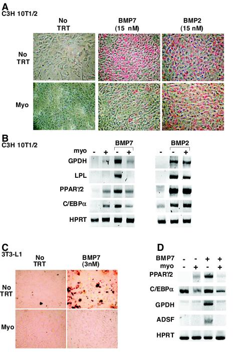
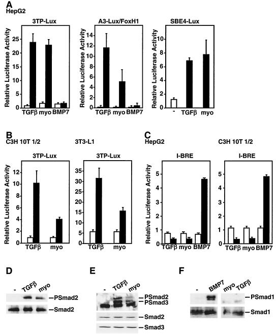
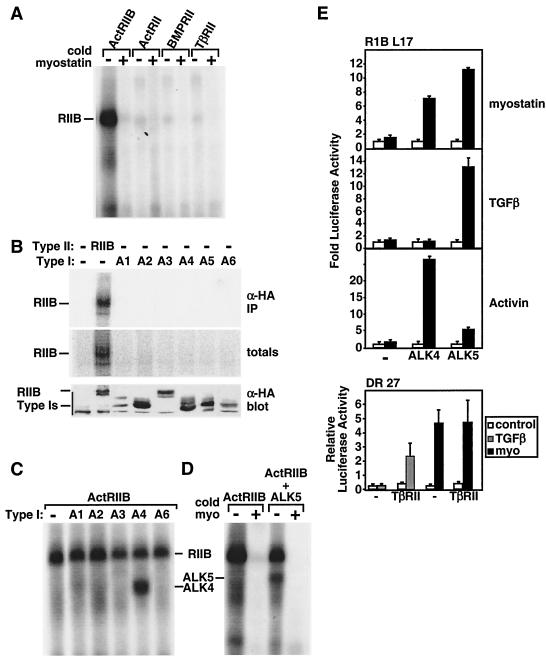
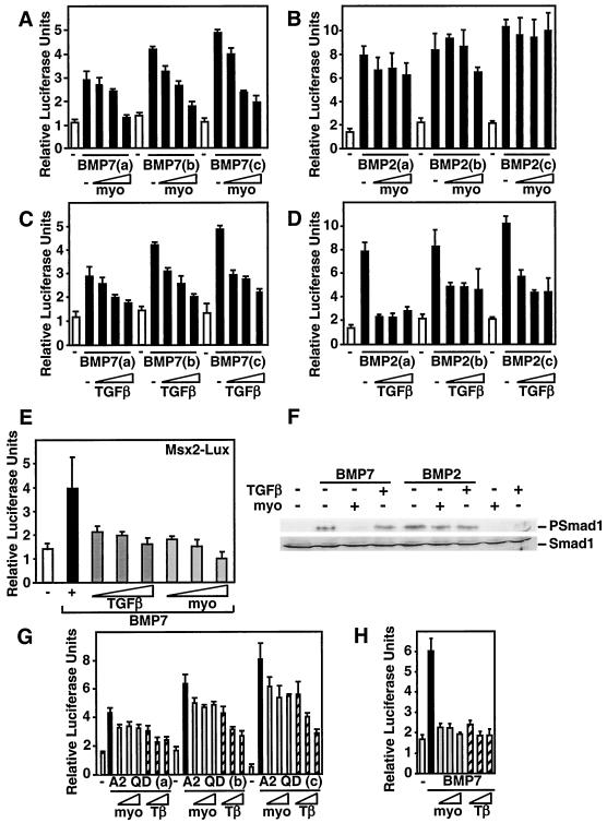
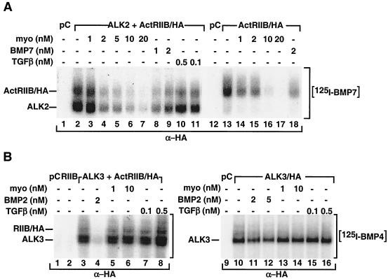
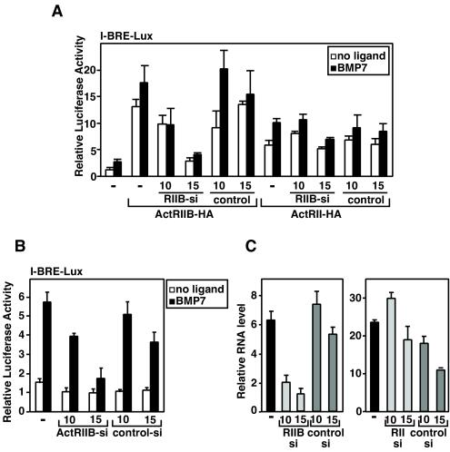
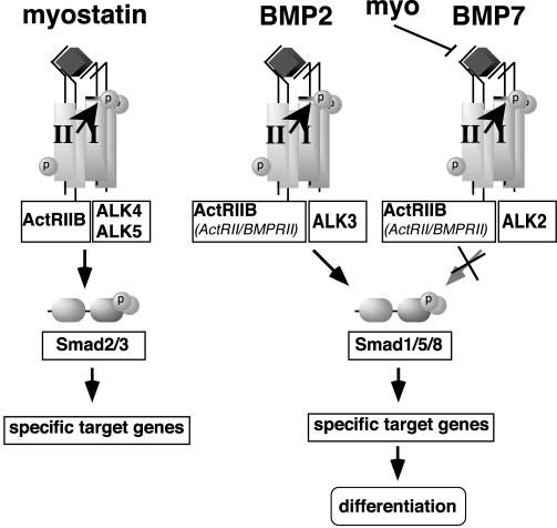
Myostatin, a transforming growth factor beta (TGF-beta) family member, is a potent negative regulator of skeletal muscle growth. In this study we characterized the myostatin signal transduction pathway and examined its effect on bone morphogenetic protein (BMP)-induced adipogenesis. While both BMP7 and BMP2 activated transcription from the BMP-responsive I-BRE-Lux reporter and induced adipogenic differentiation, myostatin inhibited BMP7- but not BMP2-mediated responses. To dissect the molecular mechanism of this antagonism, we characterized the myostatin signal transduction pathway. We showed that myostatin binds the type II Ser/Thr kinase receptor. ActRIIB, and then partners with a type I receptor, either activin receptor-like kinase 4 (ALK4 or ActRIB) or ALK5 (TbetaRI), to induce phosphorylation of Smad2/Smad3 and activate a TGF-beta-like signaling pathway. We demonstrated that myostatin prevents BMP7 but not BMP2 binding to its receptors and that BMP7-induced heteromeric receptor complex formation is blocked by competition for the common type II receptor, ActRIIB. Thus, our results reveal a strikingly specific antagonism of BMP7-mediated processes by myostatin and suggest that myostatin is an important regulator of adipogenesis.


Figures

FIG. 1.
FIG. 1.
Myostatin blocks BMP7-induced adipocyte differentiation. C3H 10T1/2 (A and B) and 3T3-L1 (C and D) cells were incubated for 10 days in the presence of various combinations of BMP2, BMP7, myostatin (10 nM), or no ligand (no TRT) as indicated. (A and C) Lipid accumulation was assessed in fixed cells by Oil Red O staining. (B and D) The effect of myostatin on the BMP2 (3 nM)- and BMP7 (3 nM)-regulated expression of key adipocyte transcription factors and late adipocyte markers was determined by reverse transcription-PCR analysis.
FIG. 2.
FIG. 2.
Myostatin signals through a TGF-β/activin signaling pathway. (A to C) Myostatin (10 nM) activates the TGF-β-responsive reporters 3TP-Lux, A3-Lux, and SBE4-Lux but not the BMP-responsive reporter I-BRE-Lux. HepG2 (A and C), C3H 10T1/2 (B and C), and 3T3-L1 (B) cells were transiently transfected with the indicated reporter constructs, and luciferase activity in cells treated with TGF-β, myostatin, or BMP7 or left untreated was determined. (D to F). Myostatin (10 nM) induces phosphorylation of endogenous Smad2 and Smad3. 3T3-L1 cells were incubated with TGF-β, myostatin, or BMP7 for 30 min, and cell lysates were subjected to anti-Smad2/Smad3 (D and E) or anti-Smad1 (F) immunoprecipitation followed by immunoblotting with anti-phosphospecific Smad2 (PSmad2), anti-phosphospecific Smad2/Smad3 (PSmad2/3), or anti-phosphospecific Smad1 (PSmad 1) antibodies. Total Smad proteins were detected by anti-Smad2 (D and E), anti-Smad3 (E), and anti-Smad1 (F) antibodies (lower panels).
FIG. 3.
FIG. 3.
Myostatin binds to ActRIIB and signals through a heteromeric receptor complex composed of either ALK4 or ALK5. (A to D) COS-1 cells were transiently transfected with various type II receptors (A) or individual type I receptors (B) or the type II receptor ActRIIB together with the type I receptors ALK1 to ALK6 (A1 to A6) (C and D) as indicated. The cells were incubated with [125I]myostatin (0.03 nM [A and C] or 1 nM [B]) in the presence or absence of unlabeled myostatin (15 nM). Total cell lysates were separated by SDS-PAGE, and 125I was detected by autoradiography (A, C, and D) or phosphorimaging (B). Expression of receptor protein in total cell lysates was determined by immunoblotting with anti-HA antibody (B). (E) Mv1Lu cell derivatives, RIB L17 or DR 27 cells, were transiently transfected with the 3TP-Lux reporter either with or without the type I receptors ALK4 or ALK5 (upper panel) or the type II receptor TβRII (lower panel). The fold induction of luciferase activity in cells treated with myostatin, TGF-β, or activin relative to controls was determined.
FIG. 4.
FIG. 4.
Myostatin antagonizes BMP7 but not BMP2 signaling. (A to E) C3H 10T1/2 cells were transiently transfected with the I-BRE-Lux reporter (A to D) or Msx2-Lux (E), and luciferase activity in cells treated with 0.75, 1, or 1.5 nM BMP7 (A, C, and E) or BMP2 (B and D), with or without myostatin (1, 5, or 10 nM) or TGF-β (25, 50, or 100 pM) was determined. (F) Myostatin but not TGF-β blocks BMP7-induced phosphorylation of endogenous Smad1. C3H 10T1/2 cells were incubated for 1 h with 1 nM BMP7 or BMP2 in the presence of 10 nM myostatin or 100 pM TGF-β as indicated. Total cell lysates were immunoblotted with anti-phosphospecific Smad1 or anti-Smad1 antibodies. (G and H) Myostatin does not inhibit signaling by a constitutively activated ALK2 receptor. C3H 10T1/2 cells were transiently transfected with the I-BRE-Lux reporter, and increasing amounts (0.001, 0.002, and 0.01 μg/well) of the activated type I receptor ALK2QD (G) or were treated with 1 nM BMP7 ligand, and myostatin (10, 15, or 20 nM) or TGF-β (50, 75, or 100 pM) (H), and the luciferase activity was measured.
FIG. 5.
FIG. 5.
Myostatin inhibits BMP7 signaling by competing for receptor binding. (A and B) Myostatin blocks BMP7 but not BMP4 receptor binding. COS-1 cells were transiently transfected with various combinations of pCMV5 (pC), ActRIIB/HA (RIIB), and untagged ALK2 (A2) (A) or HA-tagged ALK3 (B) as indicated. The cells were incubated with 2 nM [125I]BMP7 or [125I]BMP4, and various concentrations of unlabeled myostatin, TGF-β, BMP7, or BMP2, as indicated. Cell lysates were subjected to anti-HA immunoprecipitation followed by SDS-PAGE, and bound ligand was visualized by autoradiography.
FIG. 6.
FIG. 6.
RNA interference of ActRIIB abolishes BMP7 signaling in C3H 10T1/2 cells. C3H 10T1/2 cells were transiently transfected with the I-BRE-Lux reporter (A and B) without (B and C) or with (A) ActRII or ActRIIB, as well as various doses (0.8 or 1 μg/well) of ActRIIB-specific siRNA or an irrelevant control/scrambled siRNA (A to C). Luciferase activity of cells treated with BMP7 (1 nM) was assayed (A and B), and changes in ActRIIB and ActRII transcripts were measured by Q-PCR (C). In panel A, ActRIIB siRNA specifically reduces ActRIIB-mediated BMP7 signaling in C3H 10T1/2 cells without affecting ActRII-mediated signaling.
FIG. 7.
FIG. 7.
Model of extracellular and intracellular antagonism of BMP signaling by myostatin and TGF-β. Myostatin signals through ActRIIB and either ALK4 or ALK5 to activate a TGF-β-like signaling pathway. Myostatin potently antagonizes BMP7 but not BMP2 by competing for BMP7 binding to the ActRIIB type II receptor.

References

    1. Aherns, M., T. Ankenbauer, D. Schroder, A. Hollnagel, H. Mayer, and G. Gross. 1993. Expression of human bone morphogenetic proteins-2 or -4 in murine mesenchymal progenitor C3H 10T1/2 cells induces differentiation into distinct mesenchymal cell lineages. DNA Cell Biol. 12:871-880. - PubMed
    1. Alliston, T., L. Choy, P. Ducy, G. Karsenty, and R. Derynck. 2001. TGF-β-induced repression of CBFA1 by Smad3 decreases cbfa1 and osteocalcin expression and inhibits osteoblast differentiation. EMBO J. 20:2254-2272. - PMC - PubMed
    1. Asahina, I., T. K. Sampath, and P. V. Hauschka. 1996. Human osteogenic protein-1 induces chondroblastic, osteoblastic, and/or adipocytic differentiation of clonal murine target cells. Exp. Cell Res. 222:38-47. - PubMed
    1. Attisano, L., J. Cárcamo, F. Ventura, F. M. B. Weis, J. Massagué, and J. L. Wrana. 1993. Identification of human activin and TGF-β type I receptors that form heteromeric kinase complexes with type II receptors. Cell 75:671-680. - PubMed
    1. Attisano, L., and J. L. Wrana. 2002. Signal transduction by the TGF-β superfamily. Science 296:1646-1647. - PubMed

Publication types

MeSH terms

LinkOut - more resources