. 2003 Oct;23(20):7391-402.
doi: 10.1128/MCB.23.20.7391-7402.2003.

Tyrosine phosphorylation of plakoglobin causes contrary effects on its association with desmosomes and adherens junction components and modulates beta-catenin-mediated transcription

Affiliations

Tyrosine phosphorylation of plakoglobin causes contrary effects on its association with desmosomes and adherens junction components and modulates beta-catenin-mediated transcription

Susana Miravet et al. Mol Cell Biol. 2003 Oct.

Abstract

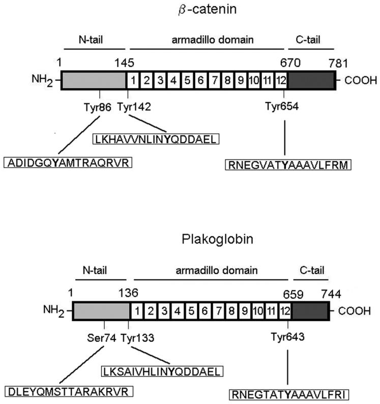
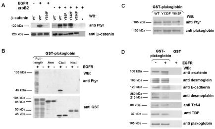
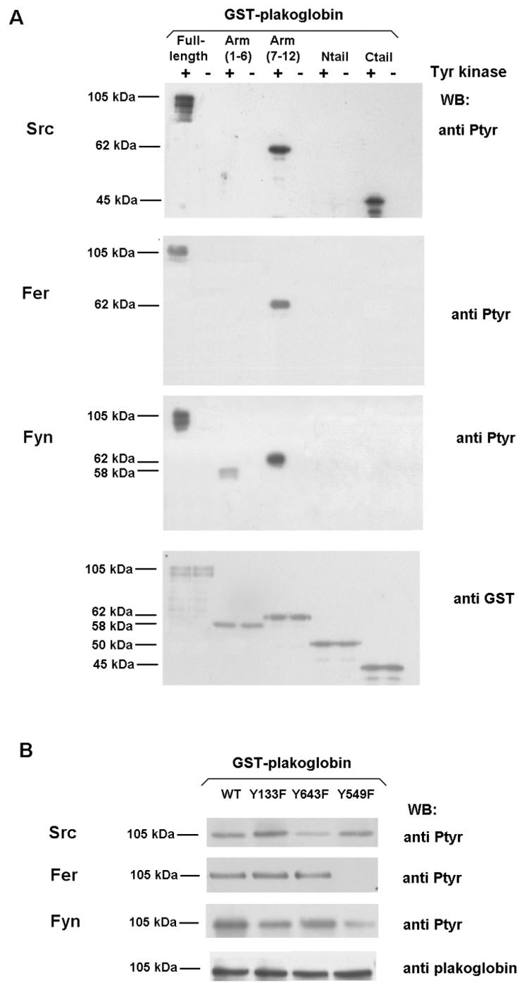
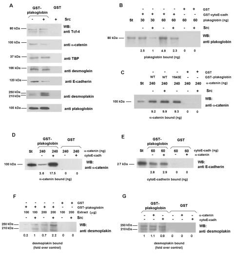
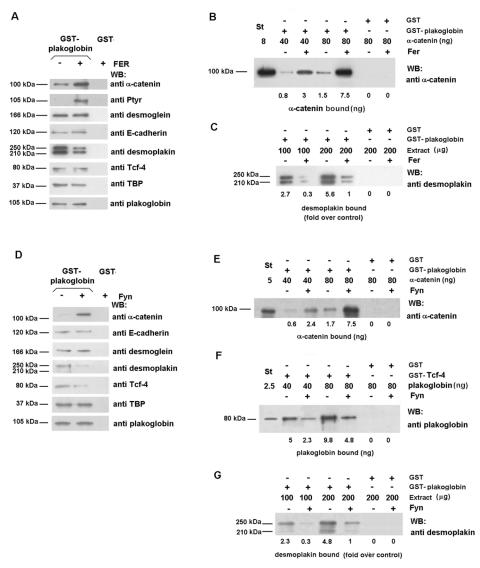
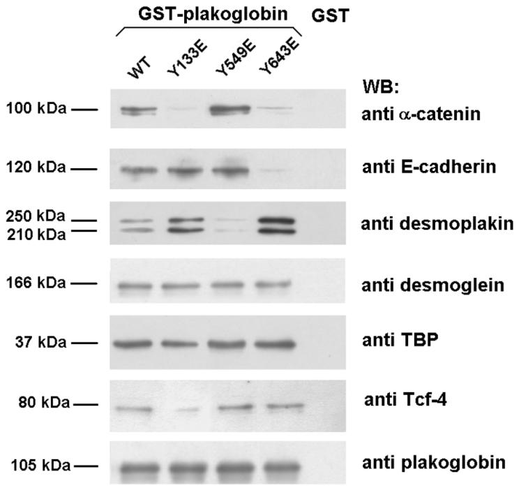
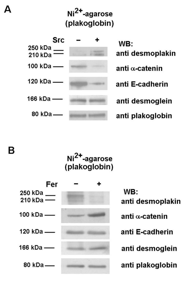
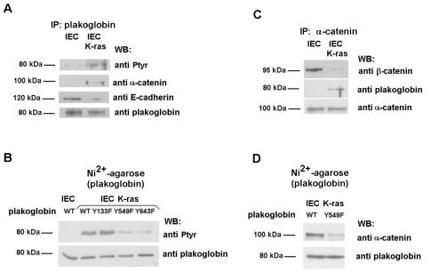
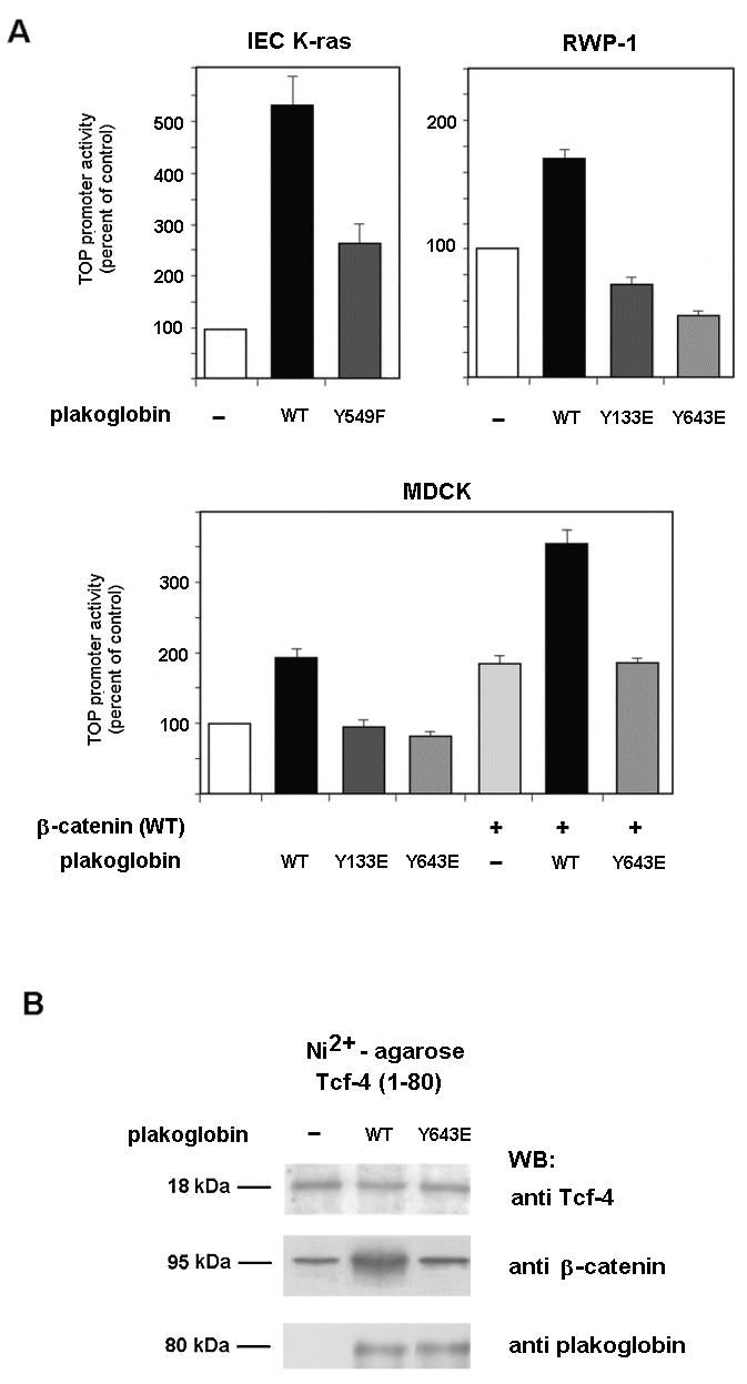
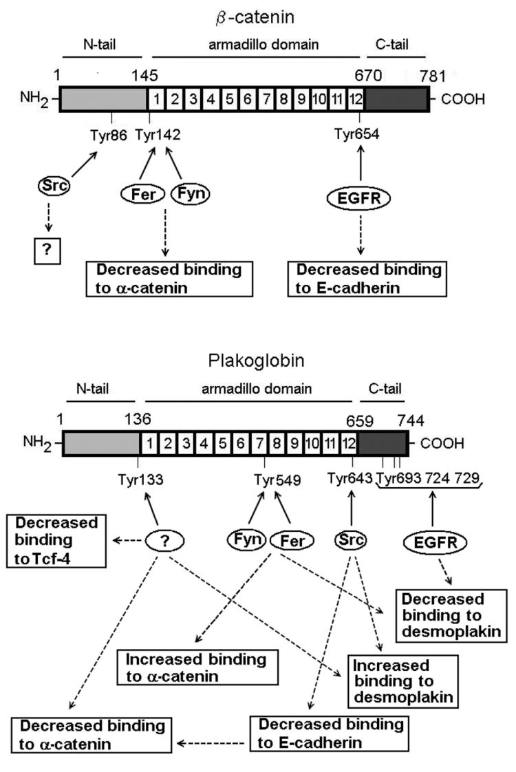
Plakoglobin is a protein closely related to beta-catenin that links desmosomal cadherins to intermediate filaments. Plakoglobin can also substitute for beta-catenin in adherens junctions, providing a connection between E-cadherin and alpha-catenin. Association of beta-catenin with E-cadherin and alpha-catenin is regulated by phosphorylation of specific tyrosine residues; modification of beta-catenin Tyr654 and Tyr142 decreases binding to E-cadherin and alpha-catenin, respectively. We show here that plakoglobin can also be phosphorylated on tyrosine residues, but unlike beta-catenin, this modification is not always associated with disrupted association with junctional components. Protein tyrosine kinases present distinct specificities on beta-catenin and plakoglobin, and phosphorylation of beta-catenin-equivalent Tyr residues of plakoglobin affects its interaction with components of desmosomes or adherens junctions differently. For instance, Src, which mainly phosphorylates Tyr86 in beta-catenin, modifies Tyr643 in plakoglobin, decreasing the interaction with E-cadherin and alpha-catenin and increasing the interaction with the alpha-catenin-equivalent protein in desmosomes, desmoplakin. The tyrosine kinase Fer, which modifies beta-catenin Tyr142, lessening its association with alpha-catenin, phosphorylates plakoglobin Tyr549 and exerts the contrary effect: it raises the binding of plakoglobin to alpha-catenin. These results suggest that tyrosine kinases like Src or Fer modulate desmosomes and adherens junctions differently. Our results also indicate that phosphorylation of Tyr549 and the increased binding of plakoglobin to components of adherens junctions can contribute to the upregulation of the transcriptional activity of the beta-catenin-Tcf-4 complex observed in many epithelial tumor cells.


Figures

FIG. 1.
FIG. 1.
Diagram of β-catenin and plakoglobin. The three different domains that form these two proteins are shown. The amino acid sequences near the three indicated Tyr residues in β-catenin are shown, as are the equivalent sequences in plakoglobin.
FIG. 2.
FIG. 2.
EGFR presents different substrate specificities of phosphorylation on β-catenin and plakoglobin. (A) GST-β-catenin fusion proteins (6.7 pmol) were phosphorylated with 0.5 U of recombinant EGFR kinase or RWP1 cell extracts transfected with erbB2. Phosphorylation was analyzed by Western blotting (WB) with anti-PTyr MAb. The membrane was stripped and reprobed for β-catenin as a control, and similar levels of GST-β-catenin were present. (B and C) Five picomoles of GST-plakoglobin deletion mutants (B) or point mutants (C) was phosphorylated with EGFR under the indicated conditions. Samples were analyzed by Western blotting with anti-PTyr MAb and reblotted against GST (B) or plakoglobin (C). (D) Five picomoles of GST or GST-plakoglobin fusion proteins was phosphorylated with EGFR as described above. Pull-down assays were then performed, and the GST proteins were incubated with the indicated amounts of total cell extracts from SW480. The associated proteins were detected with specific MAbs. WT, wild type; +, present; −, absent. The estimated molecular masses of the bands detected with each antibody are indicated.
FIG. 3.
FIG. 3.
Mapping of residues involved in plakoglobin phosphorylation by Src, Fer, and Fyn kinases. Five picomoles of GST-plakoglobin deletion mutants (A) or the indicated point mutants (B) was phosphorylated with either 0.5 U of recombinant Src (upper panel), Fer kinase purified from transfected RWP1 cells (middle panel), or Fyn kinase immunoprecipitated from transfected RWP1 cells (lower panel) as described in Materials and Methods. Samples were analyzed by Western blotting (WB) with anti-PTyr MAb and reblotted with anti-GST (A) or anti-plakoglobin (B) to ensure that similar levels of GST-plakoglobin forms were present in all cases. +, present; −, absent.
FIG. 4.
FIG. 4.
Phosphorylation of plakoglobin by Src decreases its interaction with α-catenin and E-cadherin and increases plakoglobin-desmoplakin association. (A) Five picomoles of GST or GST-plakoglobin fusion proteins was phosphorylated with 0.5 U of pp60c-src. Pull-down assays were then performed by incubating the GST proteins with 100 μg of total cell extracts from SW480. The amount of associated proteins was determined by using specific MAbs. (B) Control and Src-phosphorylated plakoglobin (0.35 or 0.7 pmol) were incubated with 1.2 pmol of either GST-cytoE-cadh or GST in a final volume of 200 μl. Plakoglobin (0.03 pmol) was included as a reference (St). The numbers below the lanes indicate the amount of bound protein. (C) GST or GST-plakoglobin fusion proteins (control or phosphorylated by Src) (1.8 pmol) were incubated with 2.4 pmol of α-catenin in a final volume of 200 μl. The amount of bound α-catenin was determined with a specific MAb. (D) Modulation of plakoglobin-α-catenin association by E-cadherin. GST or GST-plakoglobin (0.8 pmol) was incubated with 1.4 pmol of α-catenin. When indicated, binding assays were supplemented with 20 pmol of cytoE-cadh. The amount of associated α-catenin was determined by using a specific MAb. (E) Modulation of plakoglobin-E-cadherin association by α-catenin. GST or GST-plakoglobin (0.8 pmol) was incubated with 1.4 pmol of cytoE-cadh. When indicated, binding assays were supplemented with 20 pmol of α-catenin. The amount of associated cytoE-cadh was determined by using a specific MAb. (F) GST or GST-plakoglobin fusion proteins (5 pmol) were phosphorylated with Src, and pull-down assays were then performed by incubating the GST proteins with 100 or 200 μg of total cell extracts from SW480. The amount of associated desmoplakin was determined by using a specific MAb. (G) GST or GST-plakoglobin (5 pmol) was incubated with 200 μg of total cell extracts from SW480 with the presence of 10 pmol of α-catenin or cytoE-cadh when indicated. The amount of associated desmoplakin was determined by using a specific MAb. WB, Western blotting; +, present; −, absent.
FIG. 5.
FIG. 5.
Phosphorylation of plakoglobin by Fer and Fyn kinases decreases plakoglobin-desmoplakin interaction and increases plakoglobin-α-catenin association. (A and D) GST or GST-plakoglobin fusion proteins (5 pmol) were phosphorylated with Fer purified from transfected RWP1 cells (A) or Fyn kinase immunoprecipitated from transfected RWP1 cells (D). Pull-down assays were then performed by incubating the GST proteins with 100 μg of total cell extracts from SW480. The amounts of the associated proteins were determined by using specific MAbs. (B and E) GST or GST-plakoglobin (1.2 pmol) (control and Fer phosphorylated [B] or Fyn phosphorylated [E]) was incubated with 0.4 or 0.8 pmol of α-catenin in a final volume of 200 μl. α-Catenin (0.08 pmol) was included as a reference (St). The numbers below the lanes indicate the amounts of bound α-catenin. (C and G) GST or GST-plakoglobin fusion proteins (5 pmol) were phosphorylated with Fer (C) or Fyn kinases (G), and pull-down assays were then performed by incubating the GST proteins with 100 or 200 μg of total cell extracts from SW480. The amount of associated desmoplakin was determined by using a specific MAb. (F) GST or GST-Tcf-4(1-80) (1.2 pmol) was incubated with 0.5 or 1 pmol of plakoglobin incubated or not with Fyn in a final volume of 200 μl. Plakoglobin (0.03 pmol) was included as a reference (St). The numbers below the lanes indicate the amounts of bound plakoglobin. WB, Western blotting; +, present; −, absent.
FIG. 6.
FIG. 6.
Effect of Tyr-to-Glu point mutants in the association of plakoglobin to its cellular cofactors. Pull-down assays were performed by incubating 8 pmol of GST or GST-plakoglobin fusion proteins with 200 μg of whole-cell extracts from SW480. Protein complexes were affinity purified with glutathione-Sepharose and analyzed by SDS-PAGE and Western blotting (WB). The amounts of the associated proteins were determined by using specific MAbs.
FIG. 7.
FIG. 7.
Src and Fyn modify the association of plakoglobin to its cellular partners. RWP1 cells were cotransfected with 5 μg of pcDNA3.1His-plakoglobin and pCMV-Src (A) or pcDNA3.1His-Fer (B), with empty vectors as controls. After 48 h, cell extracts were prepared, His-tagged plakoglobin was purified by chromatography on nickel-agarose, and the associated proteins were analyzed with specific MAbs against α-catenin, E-cadherin, desmoplakin, desmoglein, and plakoglobin. WB, Western blotting; +, present; −, absent.
FIG. 8.
FIG. 8.
Localization of β-catenin and plakoglobin in IEC and IEC K-ras cells. Cells were grown on glass coverslips as indicated in Materials and Methods and fixed before they reached confluence. The distribution of β-catenin and plakoglobin was analyzed by immunofluorescence with specific MAbs.
FIG. 9.
FIG. 9.
K-ras transfection of IEC epithelial cells induces plakoglobin phosphorylation and upregulates binding to α-catenin. (A and C) Three hundred micrograms of whole-cell extracts from control and K-ras IEC18 cells were immunoprecipitated (IP) with anti-plakoglobin (A) or anti-α-catenin (B) and followed by immunoblotting with the indicated MAbs. (B and D) IEC18 and IEC18 K-ras cells were transfected with 5 μg of pcDNA3.1His-plakoglobin (wild-type [WT] or the indicated mutants). His-tagged plakoglobin was purified by chromatography on nickel-agarose, and the level of tyrosine phosphorylation (B) or α-catenin association (D) was analyzed by Western blotting (WB) with specific antibodies. The membranes were reprobed for plakoglobin to check that similar levels of expression were obtained.
FIG. 10.
FIG. 10.
Tyrosine phosphorylation of plakoglobin affects β-catenin-dependent transcription. (A) The indicated cells were cotransfected with plakoglobin plasmid mutants inserted into pcDNA3.1His (150 ng), TOP-FLASH (20 ng), and pTK-Renilla (20 ng) luciferase plasmids in the presence or absence of wild-type (WT) β-catenin. Relative luciferase activity was determined with a dual-luciferase reporter assay system 48 h after transfection and normalized by using the Renilla luciferase activity for each sample. The percentage of activity was calculated by comparing levels of luciferase activity to those obtained after transfection of the pcDNA3.1His plasmid alone. (B) In vivo association between β-catenin and Tcf-4 in MDCK cells overexpressing plakoglobin. Cells were cotransfected with 5 μg of pcDNA3-plakoglobin (wild-type or Tyr643→Glu mutant) or empty vector as a control and 5 μg of pcDNA3.1His-Tcf-4(1-80). His-tagged Tcf-4(1-80) was purified by nickel-agarose chromatography, and associated β-catenin was analyzed by Western blotting (WB) with anti-β-catenin MAb. To verify that the extent of ectopic expression was similar, blots were reanalyzed with anti-Tcf-4. +, present; −, absent.
FIG. 11.
FIG. 11.
Diagram of β-catenin and plakoglobin tyrosine residues phosphorylated by Src, EGFR, Fer, and Fyn kinases and their corresponding effects. Question marks indicate the points where either an effect of phosphorylation has not been demonstrated (Tyr86 in β-catenin) or the kinase has not been identified (in the case of plakoglobin Tyr133).

Similar articles

Cited by

References

    1. Aberle, H., H. Schwartz, H. Hoschuetzky, and R. Kemler. 1996. Single amino acid substitutions in proteins of the armadillo gene family abolish their binding to α-catenin. J. Biol. Chem. 271:1520-1526. - PubMed
    1. Adams, C., and J. W. Nelson. 1998. Cytomechanics of cadherin-mediated cell-cell adhesion. Curr. Opin. Cell Biol. 10:572-577. - PubMed
    1. Balsamo, J., T. Leung, H. Ernst, M. K. Zanin, S. Hoffman, and J. Lilien. 1996. Regulated binding of PTP1B-like phosphatase to N-cadherin: control of cadherin-mediated adhesion by dephosphorylation of beta-catenin. J. Cell Biol. 134:801-813. - PMC - PubMed
    1. Behrens, J., L. Vakaet, R. Friis, E. Winterhager, F. Van Roy, M. Mareel, and W. Birchmeier. 1993. Loss of epithelial differentiation and gain of invasiveness correlates with tyrosine phosphorylation of the E-cadherin/β-catenin complex in cells transformed with a temperature-sensitive v-SRC gene. J. Cell Biol. 120:757-766. - PMC - PubMed
    1. Bienz, M., and H. Clevers. 2000. Linking colorectal cancer to Wnt signaling. Cell 103:311-320. - PubMed

Publication types

MeSH terms