. 2003 Oct;14(10):4051-66.
doi: 10.1091/mbc.e03-04-0247. Epub 2003 Jul 25.

The role of Cdc42p GTPase-activating proteins in assembly of the septin ring in yeast

Affiliations

The role of Cdc42p GTPase-activating proteins in assembly of the septin ring in yeast

Juliane P Caviston et al. Mol Biol Cell. 2003 Oct.

Abstract

The septins are a conserved family of GTP-binding, filament-forming proteins. In the yeast Saccharomyces cerevisiae, the septins form a ring at the mother-bud neck that appears to function primarily by serving as a scaffold for the recruitment of other proteins to the neck, where they participate in cytokinesis and a variety of other processes. Formation of the septin ring depends on the Rho-type GTPase Cdc42p but appears to be independent of the actin cytoskeleton. In this study, we investigated further the mechanisms of septin-ring formation. Fluorescence-recovery-after-photobleaching (FRAP) experiments indicated that the initial septin structure at the presumptive bud site is labile (exchanges subunits freely) but that it is converted into a stable ring as the bud emerges. Mutants carrying the cdc42V36G allele or lacking two or all three of the known Cdc42p GTPase-activating proteins (GAPs: Bem3p, Rga1p, and Rga2p) could recruit the septins to the cell cortex but were blocked or delayed in forming a normal septin ring and had accompanying morphogenetic defects. These phenotypes were dramatically enhanced in mutants that were also defective in Cla4p or Gin4p, two protein kinases previously shown to be important for normal septin-ring formation. The Cdc42p GAPs colocalized with the septins both early and late in the cell cycle, and overexpression of the GAPs could suppress the septin-organization and morphogenetic defects of temperature-sensitive septin mutants. Taken together, the data suggest that formation of the mature septin ring is a process that consists of at least two distinguishable steps, recruitment of the septin proteins to the presumptive bud site and their assembly into the stable septin ring. Both steps appear to depend on Cdc42p, whereas the Cdc42p GAPs and the other proteins known to promote normal septin-ring formation appear to function in a partially redundant manner in the assembly step. In addition, because the eventual formation of a normal septin ring in a cdc42V36G or GAP mutant was invariably accompanied by a switch from an abnormally elongated to a more normal bud morphology distal to the ring, it appears that the septin ring plays a direct role in determining the pattern of bud growth.


Figures
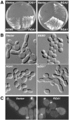
Figure 1.
Figure 1.
Suppression of the growth, morphological, and septin-organization defects of a cdc12-6 mutant by multicopy BEM3, RGA1, or RGA2. ZDS1, a putative non-GAP negative regulator of Cdc42p (Bi and Pringle, 1996), was included as a negative control. (A) Strain M-17 (cdc12-6) was transformed (left panel) with plasmid YEp13 (vector), pPB547 (BEM3 in YEp13), YEp13-RGA1, or YEp13-ZDS1* and (right panel) with plasmid YEp24 (vector), YEp24-RGA1, or pDLB1981 (RGA2 in pRS426). The transformants were streaked and incubated at 30°C on an SC-Leu plate for 3 d (left panel) or an SC-Ura plate for 5 d (right panel). (B) The transformants in A (left panel) were grown to exponential phase in liquid SC-Leu medium at 23°C, diluted 1:10 in prewarmed medium, and incubated at 30°C for 3.4 h. Cells were then sonicated mildly and observed by DIC microscopy. Each image is printed at the same magnification. (C) Strain M-17 carrying plasmid pRS316-CDC3-GFP was further transformed with plasmid YEp13 (left) or YEp13-RGA1 (right). The transformants were grown to exponential phase in liquid SC-Leu-Ura medium at 23°C, shifted to 30°C for 3 h, fixed with 3.7% formaldehyde for 20 min (10 min at 30°C and 10 min at 23°C), and observed by fluorescence microscopy. The images are printed at the same magnification.
Figure 8.
Figure 8.
FRAP studies of septin dynamics in wild-type and mutant cells. All strains carried plasmid pRS316-CDC3-GFP. In some experiments, half the septin structure of interest was bleached, and the subsequent changes in fluorescence of both the bleached and unbleached portions of the structure were monitored; in other experiments, the entire septin structure was bleached before monitoring recovery. In each panel, a representative series of images for a half-bleached structure is shown together with the corresponding quantitation; the quantitative data for a representative total-bleached structure are also shown. b, the cells that were bleached; c, the unbleached control cells used to normalize the fluorescence values from the bleached cells (see MATERIALS AND METHODS). Note that in one case (D, half-bleached), the unbleached septin structure used as control was actually on the same cell as the bleached structure. (A–C) Wild-type strain YEF473A. (A) Unbudded cells. Similar data (i.e., extensive recovery of fluorescence within 2 min) were obtained with seven half-bleached and 12 total-bleached cells. (B) Small-budded cells. Similar data (i.e., very little recovery of fluorescence even after 10 min) were obtained with 10 half-bleached and 7 total-bleached cells. Cells with medium-sized buds behaved similarly (unpublished data). (C) Large-budded cells; all cells examined had septa visible by DIC and thus had already completed cytokinesis but not cell separation. Similar data were obtained with 6 half-bleached (all showed extensive recovery of fluorescence by 4 min) and 15 total-bleached (all showed only limited recovery of fluorescence even after 10 min) cells. (D) cdc42V36G mutant strain YEF2921 and bem3Δ rga1Δ rga2Δ triple-mutant strain YEF2380. Abnormal septin caps and patches were examined; similar data (i.e., extensive recovery of fluorescence within 4 min and further recovery by 10 min) were obtained with five caps (all half-bleached) and five patches (all total-bleached). One half-bleached cap and two total-bleached patches recovered fluorescence more slowly. Structures in the two strains behaved similarly; the data shown were obtained with strain YEF2921.
Figure 2.
Figure 2.
Dependence of cdc12 suppression on the interaction of Rga1p with Cdc42p and/or its GAP activity. Strain M-17 (cdc12-6) was transformed with plasmid YEplac181 (Vector), YEp181-RGA1(WT) (RGA1), or YEp181-RGA1(K872A) (rga1K872A). (A) The transformants were streaked onto a SC-Leu plate and incubated at 30°C for 5 d. (B and C) The transformants were further transformed with plasmid pRS316-CDC3-GFP, grown to exponential phase in liquid SC-Leu-Ura medium at 23°C, and shifted to 30°C for 3 h before fixation (as in Figure 1C) and examination by DIC (B) and fluorescence (C) microscopy.
Figure 3.
Figure 3.
Cell-morphology and septin-organization defects in strains lacking Cdc42p GAPs. (A and B) Wild-type (WT) strain YEF473A, bem3Δ rga1Δ strains YEF2390 (A) and YEF1209 (B), and bem3Δ rga1Δ rga2Δ strain YEF2335 were grown to exponential phase in liquid YM-P (A) or YPD (B) medium at 23°C and then observed by DIC microscopy (A) or processed for immunofluorescence using Cdc11p-specific antibodies (B). (C) bem3Δ rga1Δ rga2Δ strain YEF2380 was transformed with plasmid pRS316-CDC3-GFP, grown overnight on an SC-Ura plate at 24°C, and examined by fluorescence microscopy.
Figure 4.
Figure 4.
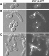
Disorganization of Myo1p in strains lacking Cdc42p GAPs. Wild-type strain YEF473A (A), bem3Δ rga1Δ rga2Δ strain YEF2380 (B), and bem3Δ rga1Δ gin4Δ strain YEF2517 (C) were transformed with plasmid pRS316-N-MYO1-GFP, grown on SC-Ura plates for ∼36 h at 23°C, and examined for cell morphology and Myo1p localization by DIC (left) and fluorescence (right) microscopy.
Figure 5.
Figure 5.
Synthetic effects between Cdc42p-GAP mutations and mutations in protein kinases involved in septin organization. (A) Strains YEF1343 (cla4Δ), YEF1383 (bem3Δ rga1Δ cla4Δ), YEF1341 (ste20Δ), YEF1381 (bem3Δ rga1Δ ste20Δ), YEF1347 (skm1Δ), and YEF1385 (bem3Δ rga1Δ skm1Δ) were observed by DIC microscopy after overnight growth on YPD plates at 23°C. (B) Strain YEF1383 was transformed with plasmid pRS316-CDC3-GFP and observed by fluorescence microscopy after overnight growth on an SC-Ura plate at 24°C. (C) Strains YEF473A (WT), M-267 (gin4Δ), YEF1209 (bem3Δ rga1Δ), and YEF1300 (bem3Δ rga1Δ gin4Δ) were picked from a YPD plate containing 0.9 M KCl (which partially suppresses the gin4Δ mutant phenotype; our unpublished results), streaked onto a YPD plate, and incubated at 23°C for ∼2.5 d. (D) Strains M-267 and YEF1300 were streaked from YPD + 0.9 M KCl plates onto YPD plates, grown overnight at 23°C, and observed by DIC microscopy. (E) Strains M-267 and YEF1300 were grown overnight in liquid YPD + 0.9 M KCl at 23°C, collected by centrifugation, washed twice with liquid YPD medium, diluted 1:50 with fresh YPD, grown for 9 h at 23°C, and processed for immunofluorescence using Cdc11p-specific antibodies. (F) Strain YEF2517 (bem3Δ rga1Δ gin4Δ) was transformed with plasmid pRS316-CDC3-GFP, grown overnight in liquid SC-Ura medium at 23°C, fixed with 3.7% formaldehyde for 10 min at 23°C, and examined by DIC (left) and fluorescence (right) microscopy. (G) Strain YEF1300 was grown as described for panel E and double-stained with anti-Cdc11p antibodies (top) and bisBenzimide to visualize the DNA (bottom).
Figure 6.
Figure 6.
Cell-morphology and septin-organization defects in cdc42V36G cells. Cells of strain YEF2921 (cdc42V36G) carrying plasmid pRS316-CDC3-GFP were grown to exponential phase in liquid SC-Ura medium at 23°C, fixed with 3.7% formaldehyde for 10 min at 23°C, and examined by DIC (A) and fluorescence (B) microscopy. Arrowheads indicate septin caps. Note that the longer bud of cell 1 (A) is out of the plane of focus in B.
Figure 7.
Figure 7.
Localization of Cdc42p GAPs. (A–C) Localization of Bem3p and its dependence on septin function. (A) Strain YEF1423 (3HA:BEM3/3HA:BEM3) was grown to exponential phase in YM-P medium at 23°C and examined by immunofluorescence using anti-HA antibody. (B and C) Strain M-17 (cdc12-6) carrying plasmid YEp13-GFP-BEM3 was grown to exponential phase in liquid SC-Leu medium at 23°C and observed by fluorescence microscopy (B) or shifted to 37°C for 1 h before observation (C). (D) Localization of Rga2p. Strain YEF2291 (RGA2:GFP/RGA2: GFP) was grown overnight on a YPD plate at 23°C and observed by fluorescence microscopy. (E–G) Localization of Rga1p and its dependence on septin function. (E) Strain YEF1026 (bem3Δ rga1Δ) carrying plasmid pRS315-GFP-RGA1 was grown overnight on an SC-Leu plate at 23°C and observed by fluorescence microscopy. (F) Strain YEF1424 (3HA:RGA1/3HA:RGA1) was grown to exponential phase in YM-P medium at 23°C and triple stained using anti-HA antibody (top), anti-Cdc11p antibodies (middle), and bisBenzimide (bottom). (G) Strain YEF1418 (cdc12-6 3HA:RGA1) was grown to exponential phase in liquid SC-Ura medium at 23°C, shifted to 37°C for 1 h, and triple stained as in F.
Figure 9.
Figure 9.
Correlation between the pattern of septin organization and the pattern of bud growth. Time-lapse microscopy was performed at 23°C on cdc42V36G strain YEF2921 (A) and bem3Δ rga1Δ rga2Δ strain YEF2380 (B) carrying plasmid pRS316-CDC3-GFP. Times are indicated in minutes.
Figure 10.
Figure 10.
Two-step model for formation of the mature septin ring. Septin monomers or oligomers are recruited to the presumptive bud site in a process that appears to depend on Cdc42p-GTP but for which the effectors are unknown. It is also not known whether the septins are recruited directly into a ring or first form patches and/or a cap that then converts to a ring before bud emergence. (A) In wild-type cells, the labile (capable of rapid subunit exchange) structure(s) present before bud emergence assembles efficiently into a more stable structure at about the time of bud emergence; this assembly may coincide with the conversion of the initial septin ring into the hourglass-shaped structure of the budded cell. The assembly process also appears to depend on Cdc42p and on multiple factors (the Cdc42p GAPs, Gin4p, Cla4p, Bni5p, Elm1p, Nap1p) that are at least partially redundant in function based on the synthetic phenotypes of double mutants. It is not clear whether all of these factors function at precisely the same stage in the assembly process (see text). (B) In various mutants, the assembly of the stable structure is prevented or delayed, and a labile cap or patches can form and/or persist for an extended period. The eventual formation of a more normal, stable ring is accompanied by a shift to a more normal pattern of bud growth. See text for additional details.

References

    1. Adams, A.E.M., and Pringle, J.R. (1984). Relationship of actin and tubulin distribution to bud growth in wild-type and morphogenetic-mutant Saccharomyces cerevisiae. J. Cell Biol. 98, 934–945. - PMC - PubMed
    1. Ayscough, K.R., Stryker, J., Pokala, N., Sanders, M., Crews, P., and Drubin, D.G. (1997). High rates of actin filament turnover in budding yeast and roles for actin in establishment and maintenance of cell polarity revealed using the actin inhibitor Latrunculin-A. J. Cell Biol. 137, 399–416. - PMC - PubMed
    1. Barral, Y., Parra, M., Bidlingmaier, S., and Snyder, M. (1999). Nim1-related kinases coordinate cell cycle progression with the organization of the peripheral cytoskeleton in yeast. Genes Dev. 13, 176–187. - PMC - PubMed
    1. Barral, Y., Mermall, V., Mooseker, M.S., and Snyder, M. (2000). Compartmentalization of the cell cortex by septins is required for maintenance of cell polarity in yeast. Mol. Cell 5, 841–851. - PubMed
    1. Baudin, A., Ozier-Kalogeropoulos, O., Denouel, A., Lacroute, F., and Cullin, C. (1993). A simple and efficient method for direct gene deletion in Saccharomyces cerevisiae. Nucleic Acids Res. 21, 3329–3330. - PMC - PubMed

Publication types

MeSH terms

Substances