. 2003 Oct;14(10):4207-20.
doi: 10.1091/mbc.e03-03-0147. Epub 2003 Jun 27.

Cross talk between tetanus neurotoxin-insensitive vesicle-associated membrane protein-mediated transport and L1-mediated adhesion

Affiliations

Cross talk between tetanus neurotoxin-insensitive vesicle-associated membrane protein-mediated transport and L1-mediated adhesion

Philipp Alberts et al. Mol Biol Cell. 2003 Oct.

Abstract

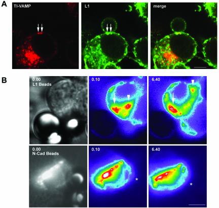
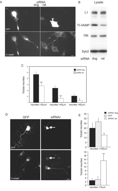
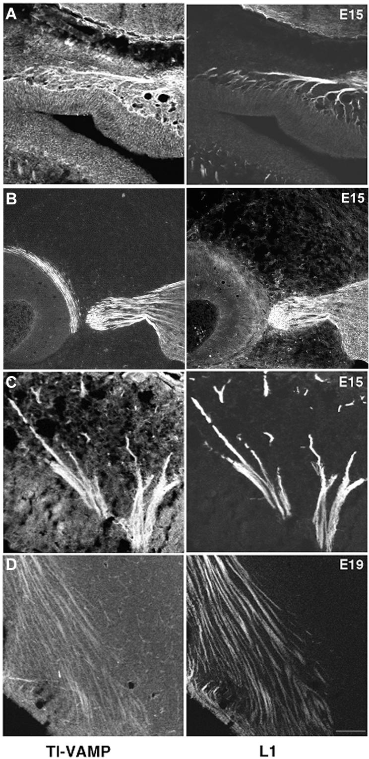
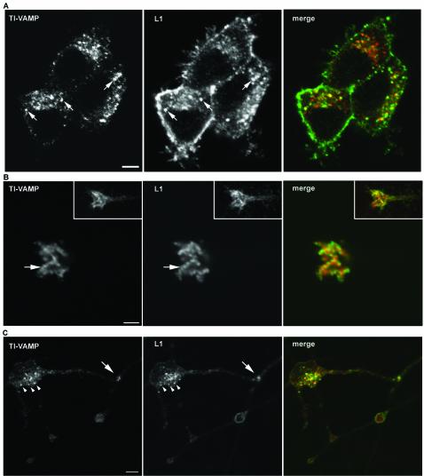
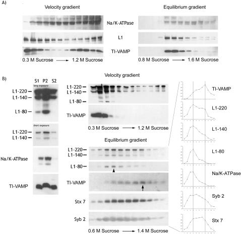
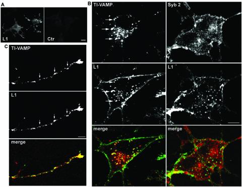
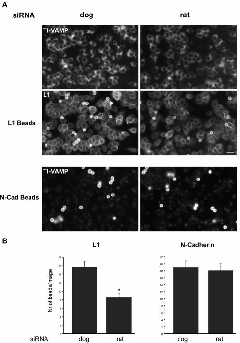
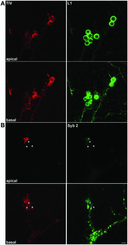
The membrane-trafficking pathway mediated by tetanus neurotoxin-insensitive vesicle-associated membrane protein (TI-VAMP) in neurons is still unknown. We show herein that TI-VAMP expression is necessary for neurite outgrowth in PC12 cells and hippocampal neurons in culture. TI-VAMP interacts with plasma membrane and endosomal target soluble N-ethylmaleimide-sensitive factor attachment protein receptors, suggesting that TI-VAMP mediates a recycling pathway. L1, a cell-cell adhesion molecule involved in axonal outgrowth, colocalized with TI-VAMP in the developing brain, neurons in culture, and PC12 cells. Plasma membrane L1 was internalized into the TI-VAMP-containing compartment. Silencing of TI-VAMP resulted in reduced expression of L1 at the plasma membrane. Finally, using the extracellular domain of L1 and N-cadherin immobilized on beads, we found that the silencing of TI-VAMP led to impaired L1- but not N-cadherin-mediated adhesion. Furthermore, TI-VAMP- but not synaptobrevin 2-containing vesicles accumulated at the site of the L1 bead-cell junction. We conclude that TI-VAMP mediates the intracellular transport of L1 and that L1-mediated adhesion controls this membrane trafficking, thereby suggesting an important cross talk between membrane trafficking and cell-cell adhesion.


Figures

Figure 8.
Figure 8.
L1-dependent adhesive contacts induce clustering of TI-VAMP–positive vesicles at sites of contact in PC12 cells. (A) TI-VAMP vesicles accumulate at sites of L1-bead contact. PC12 cells were incubated with L1-coated beads, processed for immunofluorescence with mAb 158.2 and polyclonal antibody to L1, and analyzed by confocal microscopy. Arrows point at TI-VAMP–positive intracellular structures clustered around the bead cell contact, which are also positive for L1 (bar, 5 μm). (B) L1-beads induce stable clusters of TI-VAMP-GFP. PC12 cells were transfected with TI-VAMP-GFP and incubated with L1-coated beads (top) or N-cadherin–coated beads (bottom). Dynamics of TI-VAMP-GFP were observed by time-lapse videomicroscopy. Shown are snapshots taken with phase contrast (left) and with GFP-filters (middle and right, shown in false color to highlight concentration of TI-VAMP-GFP signal). A stable accumulation of TI-VAMP-GFP close to the site of L1-bead to cell contact could be observed during the recording time (indicated by arrow). A second pool of TI-VAMP-GFP signal indicated by an arrowhead showed a dynamic behavior. N-Cadherin–coated beads did not show any influence on the dynamic behavior of TI-VAMP-GFP (position of N-cadherin bead in false colored images is indicated with asterisk) (bar, 6 μm). See supplemental videos at www.molbiolcell.org .Video 1, dynamics of TI-VAMP-GFP in PC12 cells in contact with a L1-coated bead. PC12 cells were transfected with TI-VAMP-GFP and incubated with L1-coated beads. TI-VAMP-GFP signal was recorded over a time frame of 20 min with exposures of 300 ms every 10 s. Shown is an extract of 77 frames at a display rate of 1 frame per 1/6 s. The starting frame of the video shows the bead contacting the recorded cell in phase contrast. Video 2, dynamics of TI-VAMP-GFP in PC12 cells in contact with a N-cadherin–coated bead. PC12 cells were TI-VAMP-GFP transfected and incubated with N-cadherin–coated beads. Acquisition of GFP signal was performed as above. Shown is an extract of 83 frames at the same display rate as in Video 1. The starting frame of the video shows the bead contacting the recorded cell in phase contrast.
Figure 1.
Figure 1.
Silencing of TI-VAMP expression impairs neurite outgrowth in PC12 cells and neurons. (A) PC12 cells were transfected with siRNAr or siRNAd combined with an expression plasmid encoding GFP. After 72 h, differentiation was induced by treatment with staurosporine for 24 h. SiRNAr silenced the expression of TI-VAMP and inhibited neurite outgrowth (GFP-positive cell, right), whereas siRNAd had no effect (GFP-positive cell, left) (bar, 5 μm). (B) Lysates of cells corresponding to A were analyzed by Western blotting for the expression levels of TI-VAMP, L1, transferrin receptor (TfR), and Syb 2. (C) The neurite length of GFP-positive cells transfected with siRNAr or siRNAd was quantified. Bars represent the percentage of the total number of neurites >30, 45, and 60 μm from two independent experiments (*p < 0.05; **p < 0.01). (D) TI-VAMP expression in hippocampal neurons was silenced by transfecting with siRNA and small amounts of EGFP vector. Three days after plating, TI-VAMP expression in EGFP-positive cells was assessed by immunofluorescence with mAb 158.2. As in PC12 cells, siRNAr decreased expression levels of TI-VAMP and inhibited neurite outgrowth (GFP-positive cell, right) compared with control cells (GFP-positive cell, left) (bar, 10 μm). (E) Quantification of axon lengths in neurons cotransfected with siRNAd or siRNAr and EGFP or transfected with EGFP alone is shown. Bars represent the percentage of total axons >100 μm (top) or <10 μm (bottom) from three independent experiments (*p < 0.02 with respect to GFP control; **p < 0.01 with respect to siRNAd control).
Figure 2.
Figure 2.
TI-VAMP forms SNARE complexes with plasma membrane and endosomal t-SNAREs and colocalizes with Stx 7. (A) SNARE complex formation of TI-VAMP in rat brain. The Triton X-100–soluble fraction of rat brain was incubated with mAb 158.2 or control IgGs (Ctr) covalently coupled to protein G-Sepharose. An aliquot of the starting material (SM) and material bound to immunobeads was analyzed by Western blotting with antibodies to TI-VAMP, Vti1b, Stx7, Stx 13, Stx1, and SNAP25. (B) Localization of TI-VAMP and syntaxin 7 in cortical/striatal neurons in culture. E16 rat cortical-striatal neurons were grown for 1 d in vitro, labeled for TI-VAMP and syntaxin 7, and analyzed by confocal microscopy. Vesicular structures positive for TI-VAMP and Stx 7 are indicated by arrows (bar, 5 μm).
Figure 3.
Figure 3.
TI-VAMP and L1 are coexpressed in growing axonal tracts in the developing rat brain. Expression of L1 and TI-VAMP in the developing brain. E15 and E19 embryos were serially sectioned in the sagittal plane and labeled for TI-VAMP and L1 (right). In all four examples shown there is a high degree of colocalization. (A) Olfactory nerve branching out in the olfactory epithelium. (B and C) Trigeminal ganglion and nerve root. In B, the trigeminal nerve ganglion contains TI-VAMP–labeled neurons. Their labeled axons are seen to converge toward the brainstem, and in C, trigeminal nerve branches fan out toward the facial vibrissae (left). (D) In the forebrain, TI-VAMP–labeled axon tracts are observed that correspond to corticofugal pathways and to cortical afferent pathways (bar, 100 μm).
Figure 4.
Figure 4.
TI-VAMP and L1 colocalize in neurons grown in culture. (A) Localization of TI-VAMP and L1 in PC12 cells. PC12 cells were grown at low density in the absence of NGF and processed for immunofluorescence analysis by confocal microscopy. A considerable pool of L1 immunoreactivity (green) is located on intracellular structures, which largely coincides with TI-VAMP expression (red), as indicated by arrows (bar, 4 μm). (B and C) Localization of TI-VAMP and L1 in neurons grown on L1. Hippocampal neurons were grown for 1 d in vitro on L1 and labeled for TI-VAMP and L1. (B) The confocal sections were taken from the apical part of the growth cone shown in the inset. The arrow points to a row of vesicular structures positive for TI-VAMP (red) and L1 (green); most of L1 (green) at the tip of the growth cone accumulates in clusters at the plasma membrane (bar, 3.5 μm). (C) Two contacting neurons are shown. The arrowheads point at vesicular structures in the cell body of a neuron, which are positive for L1 and TI-VAMP expression. Note also the accumulation of L1 and TI-VAMP immunoreactivity where a neurite extending from the cell body is forming a contact with a passing axon (arrow) (bar, 7 μm).
Figure 5.
Figure 5.
An intracellular pool of L1 cofractionates with TI-VAMP–positive membranes in PC12 cells and adult rat brain. (A) Fractionation of membranes from PC12 cells positive for TI-VAMP and L1 in velocity and equilibrium gradients. PC12 cells were homogenized and membranes were separated on a continuous sucrose gradient by velocity gradient centrifugation. The first four fractions of the gradient were pooled and subjected to equilibrium gradient centrifugation on a continuous sucrose gradient. Equal volumes of each fraction were analyzed by Western blotting with antibodies to TI-VAMP, L1, and Na/K-ATPase. (B) Fractionation of membranes from rat brain positive for TI-VAMP and L1 in velocity and equilibrium gradients. Rat brain was homogenized and the supernatant of a low-speed centrifugation (S1) was subjected to a medium-speed centrifugation to yield a pellet, P2, and a supernatant, S2. Equal amounts of protein were analyzed by Western blotting for TI-VAMP, L1, and Na/K-ATPase expression. An aliquot of S2 was loaded on top of a continuous sucrose gradient, and membranes were separated by velocity gradient centrifugation. Fractions were collected and Western blot analysis of equal volumes of each fraction was performed with antibodies to TI-VAMP, L1, and Na/K-ATPase. The first two fractions were pooled and centrifuged to equilibrium. Analysis was performed as described above with antibodies to TI-VAMP, L1, Na/K-ATPase, Syb 2, and syntaxin 7. The corresponding signals were quantified by densitometry, the peak signal was defined as 1, and the resulting graphs are shown adjacent to the Western blot.
Figure 6.
Figure 6.
Endocytosed L1 reaches the TI-VAMP compartment but is absent from the Syb 2-compartment. (A) L1 is endocytosed in NGF-differentiated PC12-cells. PC12 cells were grown in the presence of NGF for 48 h. Cells were incubated with L1-specific Fab fragments or control IgGs. The cells were “acid stripped” and processed for immunofluorescence analysis (bar, 8 μm). (B) L1 is taken up into the TI-VAMP compartment but not into the Syb 2 compartment. NGF-differentiated PC12 cells were incubated with L1-specific Fab fragments (green) as described above and labeled for TI-VAMP (red) or Syb 2 (red). Arrows highlight intracellular structures positive for TI-VAMP and endocytosed L1-specific Fab fragments (green) (bar, 5 μm). (C) L1 is taken up into the TI-VAMP compartment in neurons in culture. Cortical-striatal neurons grown on collagen were incubated with Fab-fragments directed against L1. After removal of the Fab fragments, the neurons were further incubated at 37°C, fixed, and labeled for TI-VAMP (red) and endocytosed L1 (green). Arrows point at structures in the growing axon positive for TI-VAMP and endocytosed anti-L1 Fab fragments (bar, 3 μm).
Figure 7.
Figure 7.
TI-VAMP–mediated intracellular trafficking is essential for L1-dependent adhesive contacts. (A) Binding of L1-coated beads to PC12 cells is inhibited by silenced TI-VAMP expression. PC12 cells were treated with siRNA dog or rat and incubated with beads coated with L1-Fc or N-cadherin-Fc chimeras. Immunofluorescence was performed with mAb 158.2 and polyclonal L1-antibody. Note that N-cadherin beads are labeled by Cy3-coupled anti-mouse secondary antibody due to the presence of the mouse Fc fragment in N-cadherin chimera (bar, 20 μm). (B) Binding of L1 and N-cadherin–coated beads to siRNA-treated PC12 cells was quantified by counting the number of beads per image (*p < 0.01).
Figure 9.
Figure 9.
L1-dependent adhesive contacts induce clustering of TI-VAMP but not Syb 2-positive vesicles in neuronal growth cones. (A) Accumulation of TI-VAMP at bead/growth cone junctions. Hippocampal neurons grown for 3 d in vitro were incubated with L1-coated beads and processed for confocal microscopy analysis with mAb 158.2 (red) and polyclonal antibody to L1 (green). A basal and an apical section of the same region are shown. Note the formation of bead shaped, TI-VAMP–positive structures in the growth cones. (B) Comparison between TI-VAMP and Syb 2 compartment in a growth cone contacting a bead. Hippocampal neurons were incubated with L1 beads and processed for immunofluorescence with mAb 158.2 and polyclonal antibody to Syb 2. A basal and an apical section of the same region are shown and bead positions are indicated by asterisks. Note the strong TI-VAMP immunoreactivity in the apical confocal section as compared with the basal section (bar, 4 μm).

References

    1. Advani, R.J., Yang, B., Prekeris, R., Lee, K.C., Klumperman, J., and Scheller, R.H. (1999). VAMP-7 mediates vesicular transport from endosomes to lysosomes. J. Cell Biol. 146, 765–775. - PMC - PubMed
    1. Berditchevski, F. (2001). Complexes of tetraspanins with integrins: more than meets the eye. J. Cell Sci. 114, 4143–4151. - PubMed
    1. Bogdanovic, A., Bennett, N., Kieffer, S., Louwagie, M., Morio, T., Garin, J., Satre, M., and Bruckert, F. (2002). Syntaxin 7, Syntaxin 8, Vti1 and VAMP7 form an active SNARE complex for early macropinocytic compartment fusion in Dictyostelium discoideum. Biochem J. 368, 29–39. - PMC - PubMed
    1. Borgonovo, B., Cocucci, E., Racchetti, G., Podini, P., Bachi, A., and Meldolesi, J. (2002). Regulated exocytosis: a novel, widely expressed system. Nat. Cell Biol. 4, 955–962. - PubMed
    1. Brummendorf, T., Kenwrick, S., and Rathjen, F.G. (1998). Neural cell recognition molecule L 1, from cell biology to human hereditary brain malformations. Curr. Opin. Neurobiol. 8, 87–97. - PubMed

Publication types

MeSH terms