. 2003 Oct;14(10):4296-305.
doi: 10.1091/mbc.e03-02-0113. Epub 2003 Jun 27.

Mitogen-activated protein kinase stimulation of Ca(2+) signaling is required for survival of endoplasmic reticulum stress in yeast

Affiliations

Mitogen-activated protein kinase stimulation of Ca(2+) signaling is required for survival of endoplasmic reticulum stress in yeast

Myriam Bonilla et al. Mol Biol Cell. 2003 Oct.

Abstract

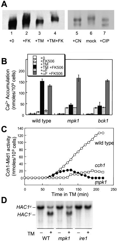
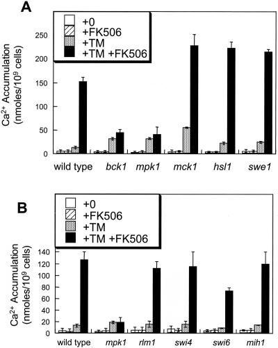
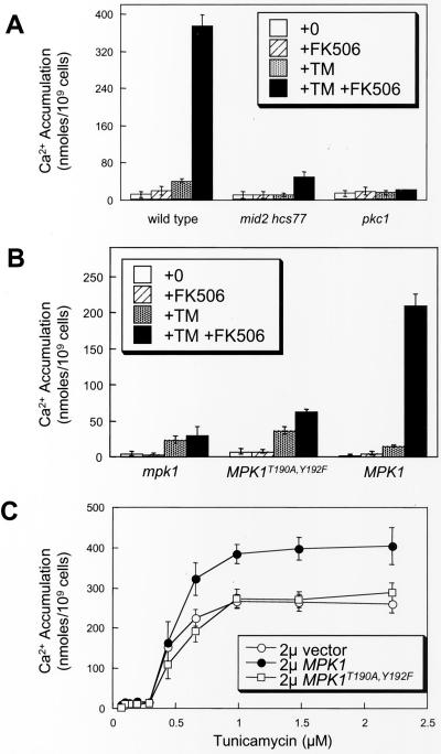
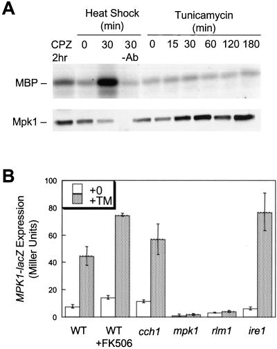
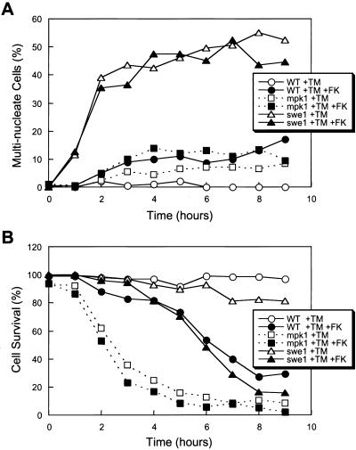
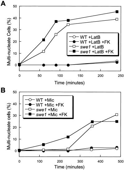
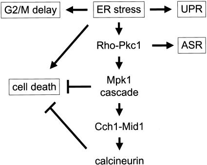
Endoplasmic reticulum (ER) stress in the budding yeast Saccharomyces cerevisiae triggers Ca2+ influx through a plasma membrane channel composed of Cch1 and Mid1. This response activates calcineurin, which helps to prevent cell death during multiple forms of ER stress, including the response to azole-class antifungal drugs. Herein, we show that ER stress activates the cell integrity mitogen-activate protein kinase cascade in yeast and that the activation of Pkc1 and Mpk1 is necessary for stimulation of the Cch1-Mid1 Ca2+ channel independent of many known targets of Mpk1 (Rlm1, Swi4, Swi6, Mih1, Hsl1, and Swe1). ER stress generated in response to miconazole, tunicamycin, or other inhibitors also triggered a transient G2/M arrest that depended upon the Swe1 protein kinase. Calcineurin played little role in the Swe1-dependent cell cycle arrest and Swe1 had little effect on calcineurin-dependent avoidance of cell death. These findings help to clarify the interactions between Mpk1, calcineurin, and Swe1 and suggest that the calcium cell survival pathway promotes drug resistance independent of both the unfolded protein response and the G2/M cell cycle checkpoint.


Figures

Figure 1.
Figure 1.
Identification of kinases required for Cch1-Mid1 Ca2+ channel activation. (A) SDS-urea gradient gel analysis of Cch1-MYC extracted from wild-type cells (strain K1257) treated with tunicamycin (2.5 μg/ml) and FK506 (2.0 μg/ml) as indicated (lanes 1–4). In the right panel (lanes 5–7), Cch1-MYC was first immunoprecipitated from cells grown with tunicamycin and FK506 as described above and treated in vitro with either purified calcineurin, reaction buffer lacking calcineurin, or purified calf intestinal phosphatase. (B) 45Ca2+ accumulation in wild-type cells, mpk1 mutants, and bck1 mutants (strains K1257, RG00993, and RG01328, respectively) was measured after growth for 4 h in SC medium supplemented with tunicamycin, α-factor mating pheromone, and FK506 as indicated. The average of three independent experiments (± SD) is shown. (C) Accumulation of 45Ca2+ in wild-type cells (open circles), cch1 mutants (open squares), and mpk1 mutants (closed circles) (strains K1257, RG04847, and RG00993, respectively) was measured as in A at various times after treatment with tunicamycin plus FK506 and tunicamycin alone. Cch1-dependent Ca2+ influx activity was calculated as the difference between these two measurements at every time point. (D) Northern blot analysis of HAC1 mRNA isolated from wild-type cells, mpk1 mutants, and ire1 mutants (strains K1257, RG00993, and K1259) mutant cells treated with or without tunicamycin for 1 h.
Figure 2.
Figure 2.
Mpk1 stimulates Cch1-Mid1 activity independently of other known targets. (A) 45Ca2+ accumulation was measured in wild-type cells, bck1 mutants, mpk1 mutants, mck1 mutants, hsl1 mutants, and swe1 mutants as described in Figure 1B. (B) 45Ca2+ accumulation in wild-type cells, mpk1 mutants, rlm1 mutants, swi4 mutants, swi6 mutants, and mih1 mutants was measured as in Figure 1B.
Figure 3.
Figure 3.
Mpk1-induced Ca2+ influx through Cch1 requires other components of the cell integrity pathway. (A) 45Ca2+ accumulation was measured in wild-type cells, mid2 hcs77 double mutants, and pkc1 mutants (strains DL1101, DL2282, and DL376) was measured as in Figure 1B except that SC media were supplemented with 0.5 M sorbitol for osmotic support. (B) 45Ca2+ accumulation was measured in mpk1 mutant cells (strain RG00993) transformed with high-dosage plasmids carrying MPK1-HA, mpk1T190A, Y192F-HA, or vector control (pRS425) as in Figure 1B except that cells were grown in SC medium lacking leucine. (C) 45Ca2+ accumulation was measured in wild-type cells (strain K601) transformed with the same plasmids as in B after incubation FK506 and varying concentrations of tunicamycin.
Figure 4.
Figure 4.
Mpk1 activity is stimulated by tunicamycin treatment independently of Ire1 and Ca2+. (A) Immune complex kinase assays were performed using Mpk1-HA isolated from wild-type cells (strain DL1101 transformed with YEp352 MPK1-HA) that had been grown at 23°C and either treated with 250 μM chlorpromazine for 2 h (lane 1), mock-shifted to 23°C (lane 2), heat shocked at 39°C for 30 min (lane 3), or treated with 2.5 μg/ml tunicamycin for the indicated times (lanes 5–10). Immunoprecipitates were also analyzed by SDS-PAGE Western blotting for immunodetection of Mpk1-HA (bottom). (B) Wild-type cells, cch1 mutants, mpk1 mutants, rlm1 mutants, and ire1 mutants (strains K1257, RG04847, RG00993, RG02739, and K1259) bearing the MPK1-lacZ reporter gene were grown to log phase in SC medium lacking uracil and treated with tunicamycin with or without FK506. After 4 h, the cells were harvested and assayed for β-galactosidase activity. The mean of three independent transformants is shown (± SD).
Figure 5.
Figure 5.
Tunicamycin induces Swe1-dependent delay at G2/M phase of the cell cycle and Swe1-independent cell survival pathway (A) Asynchronous log-phase cultures of wild-type cells, mpk1 mutants, and swe1 mutants (strains K1257, RG00993, and RG01238) were treated with tunicamycin or tunicamycin plus FK506 for the indicated periods of time, fixed, stained with DAPI, and analyzed by fluorescence microscopy. The percentage of cells in the population containing two or more nuclei is plotted. (B) Dead cells in the cultures used in A were identified by light microscopy after staining with methylene blue, and the percentage of surviving cells in the population is plotted.
Figure 6.
Figure 6.
Latrunculin B and miconazole trigger the morphogenesis checkpoint with different kinetics. The percentage of multinucleated cells in the population of wild-type and swe1 mutant cultures (strains K1257 and RG01238) is determined as in Figure 5A after treatment with latrunculin B (A) or miconazole (B) with or without K506.
Figure 7.
Figure 7.
Model of the cellular responses to ER stresses in yeast. ER stress can induce the UPR involving Ire1 (1), cell cycle delay at G2/M involving Swe1 (2), a cell death pathway (3), and the cell integrity MAP kinase cascade involving Pkc1 and Mpk1, which stimulates the Cch1-Mid1 Ca2+ channel and calcineurin signaling (4). Calcineurin and Mpk1 also antagonize cell death and Pkc1 regulates transcription of genes encoding ribosomal subunits and other phenomena known as the arrest of secretion response (ASR).

References

    1. Bonilla, M., Nastase, K.K., and Cunningham, K.W. (2002). Essential role of calcineurin in response to endoplasmic reticulum stress. EMBO J. 21, 2343–2353. - PMC - PubMed
    1. Booher, R.N., Deshaies, R.J., and Kirschner, M.W. (1993). Properties of Saccharomyces cerevisiae wee1 and its differential regulation of p34CDC28 in response to G1 and G2 cyclins. EMBO J. 12, 3417–3426. - PMC - PubMed
    1. Costigan, C., Gehrung, S., and Snyder, M. (1992). A synthetic lethal screen identifies SLK1, a novel protein kinase homolog implicated in yeast cell morphogenesis and cell growth. Mol. Cell. Biol. 12, 1162–1178. - PMC - PubMed
    1. Cruz, M.C., Del Poeta, M., Wang, P., Wenger, R., Zenke, G., Quesniaux, V.F., Movva, N.R., Perfect, J.R., Cardenas, M.E., and Heitman, J. (2000). Immunosuppressive and nonimmunosuppressive cyclosporine analogs are toxic to the opportunistic fungal pathogen Cryptococcus neoformans via cyclophilin-dependent inhibition of calcineurin. Antimicrob. Agents Chemother. 44, 143–149. - PMC - PubMed
    1. Cruz, M.C., Goldstein, A.L., Blankenship, J.R., Del Poeta, M., Davis, D., Cardenas, M.E., Perfect, J.R., McCusker, J.H., and Heitman, J. (2002). Calcineurin is essential for survival during membrane stress in Candida albicans. EMBO J. 21, 546–559. - PMC - PubMed

Publication types

MeSH terms

Substances

LinkOut - more resources