. 2003 Oct 14;100(21):12462-7.
doi: 10.1073/pnas.2034101100. Epub 2003 Sep 29.

Four-dimensional multiphoton imaging of brain entry, amyloid binding, and clearance of an amyloid-beta ligand in transgenic mice

Affiliations

Four-dimensional multiphoton imaging of brain entry, amyloid binding, and clearance of an amyloid-beta ligand in transgenic mice

Brian J Bacskai et al. Proc Natl Acad Sci U S A. .

Abstract

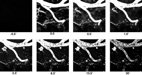
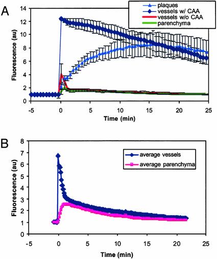
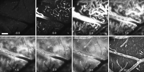
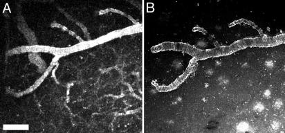
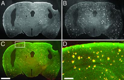
The lack of a specific biomarker makes preclinical diagnosis of Alzheimer's disease (AD) impossible, and it precludes assessment of therapies aimed at preventing or reversing the course of the disease. The development of a tool that enables direct, quantitative detection of the amyloid-beta deposits found in the disease would provide an excellent biomarker. This article demonstrates the real-time biodistribution kinetics of an imaging agent in transgenic mouse models of AD. Using multiphoton microscopy, Pittsburgh compound B (PIB) was imaged with sub-microm resolution in the brains of living transgenic mice during peripheral administration. PIB entered the brain quickly and labeled amyloid deposits within minutes. The nonspecific binding was cleared rapidly, whereas specific labeling was prolonged. WT mice showed rapid brain entry and clearance of PIB without any binding. These results demonstrate that the compound PIB has the properties required for a good amyloid-imaging agent in humans with or at risk for AD.


Figures

Fig. 1.
Fig. 1.
Real-time imaging of fluorescent PIB crossing the BBB, labeling amyloid-β deposits, and clearing from the brain. Each frame is a maximum intensity projection of the fluorescence within a 3D volume of the brain of a live, transgenic, Tg2576 mouse, acquired with a multiphoton microscope. Each 3D volume required 30 sec to obtain. Each frame is 615 × 615 μm wide and ≈150 μm deep. A bolus i.v. injection of 2 mg/kg PIB was made shortly before t = 0 min. PIB is fluorescent, allowing monitoring of its entry into brain (across the BBB), binding to amyloid plaques and CAA, and clearance from brain. Note that brain entry is very rapid (within 30 sec), amyloid targeting occurs within 1 min, and clearance of unbound fluorescence occurs within several minutes. Plaques and CAA are labeled completely within 20 min and remain labeled for the full 30 min.
Fig. 2.
Fig. 2.
Time course of PIB kinetics in Tg2576 mouse brain and cerebral vasculature at the level of single plaques and vessels. (A) These traces are averages from at least three ROIs from the image data shown in Fig. 1. ROIs were drawn within blood vessels containing CAA (⋄) or without CAA (•), plaques (▴) or within the parenchyma (□). For each time point in the experiment, a maximum intensity projection was made of the imaging volume, which was a 615 × 615 × 150-μm deep cortical volume. The mean fluorescence intensity from within each ROI was calculated for each time point, and the average of the ROIs were plotted versus time, for up to 30 min. An i.v. injection of 2 mg/kg of the fluorescent compound PIB was made at t = 0 min. The fluorescence showed up almost immediately in the blood vessels, labeling CAA rapidly. The compound crossed the BBB quickly, entered the parenchyma, and labeled parenchymal amyloid-β deposits (plaques). Complete labeling of plaques was not immediate, because the outer fringes of plaques were labeled first, followed by gradual filling of the cores. Rapid clearance of fluorescent compound occurred in blood vessels without CAA and parenchyma. These kinetics of PIB entry into brain, labeling of amyloid-β deposits, and rapid clearance of unbound tracer demonstrate the utility of this compound for radiolabeled imaging experiments. (B) PIB time courses in WT C57/BL/J6 mice are plotted. These traces are averages from at least three ROIs. The traces are the average of the ROIs from two mice, imaged identically. These animals did not develop CAA or parenchymal amyloid-β deposits. ROIs were drawn within blood vessels (⋄) or within the parenchyma (▪). For each time point in the experiment, a maximum intensity projection was made of the 615 × 615 × 35-μm imaging volume. The mean fluorescence intensity from within each ROI was calculated for each time point, and the average of the ROIs were plotted versus time, for up to 30 min. An i.v. injection of 2 mg/kg of the fluorescent compound PIB was made just before t = 0 min. The fluorescence developed almost immediately in the blood vessels and dissipated rapidly. The compound appeared in the parenchyma with a short delay and was then cleared over time. No structures within the brain were labeled in these mice.
Fig. 3.
Fig. 3.
PIB kinetics in a PDAPP mouse model. Each frame is a maximum intensity projection of the fluorescence within a 3D volume of the brain of a live, transgenic PDAPP mouse, acquired with a Bio-Rad multiphoton microscope. Each 3D volume required 30 sec to obtain. Each frame is 615 × 615 μm wide and ≈150 μm deep. An i.v. injection of 10 mg/kg PIB was made just before t = 0 min. Note that the fluorescent compound appears within blood vessels almost immediately. At t = 0.5 min, the vessels appear larger in diameter and “fuzzy” as the compound crosses the BBB and enters the parenchyma. This is seen in the next time points, t = 1 and 1.5 min. Within 3 min, parenchymal plaques are labeled and remain labeled at t = 30 min (arrows). This transgenic animal had few and small parenchymal plaques and little CAA. The CAA was labeled within 0.5 min and remained labeled for >30 min. By 30 min, the diffuse fluorescence of the blood vessels and parenchyma was markedly reduced compared with the maximal brightness achieved at t = 1–3 min. Some nonspecific fluorescence remains, probably caused by the large dose of PIB given in this animal. (Scale bar: 100 μm.)
Fig. 4.
Fig. 4.
Peripheral injection of thioflavin T labels CAA, but not parenchymal plaques in Tg2576 mice. Eighteen- to 20-month-old transgenic mice were injected with 2 mg/kg thioflavin T in 20% DMSO/80% propylene glycol i.v. Fluorescence was detected with multiphoton microscopy in the living mice, appeared almost instantaneously in the circulation, and remained there for up to 30 min. (A) A maximum intensity projection of a volume of the brain measuring 615 × 615 × 150 μm 10 min after injection. Fluorescence is present in all vessels (fluorescent angiography) and labels CAA to some extent in the large horizontal vessel in this image. Thirty minutes after imaging, the animal was killed, the cranial window was removed, and thioflavin T (0.01% in PBS) was applied directly to the surface of the cortex for 20 min. (B) The same volume (615 × 615 × 150 μm) of brain was reimaged, revealing high-contrast labeling of CAA as in A but without the luminal fluorescence. Numerous parenchymal amyloid-β deposits are now labeled within this volume. These results indicate that thioflavin T has some access to CAA, but does not cross the BBB effectively enough to label parenchymal plaques. (Scale bar: 20 μm.)
Fig. 5.
Fig. 5.
Peripherally administered PIB labels plaques and CAA specifically in transgenic mice. Twelve-month-old PS-APP (3) mice were injected with 2–10 mg/kg PIB in 20% DMSO/80% propylene glycol i.v. The mice were killed 2 h after injection. The brains were removed and fixed in 4% paraformaldehyde for 24 h and cryopreserved in a 15% glycerol solution. Tissue sections (40 μm thick) were cut with a freezing sledge microtome, mounted onto slides without coverslips, and imaged with multiphoton microscopy. The sections were not washed or treated with any organic solvents. Pictures of plaques fluorescently labeled with PIB throughout the coronal sections of brain were obtained by using identical imaging conditions described for in vivo microscopy, but using a ×2 macro objective (numerical aperture = 0.14, Olympus). The tissue sections were then incubated in 0.01% thiazine red R in PBS for 20 min at room temperature. This histochemical stain, like thioflavin S, labels dense-core plaques and CAA but emits red fluorescence (550 nm). The tissue sections were reimaged with two-channel detection. Plaques and CAA labeled with PIB are shown (A) and are pseuodocolored green in C, whereas thiazine red R fluorescence is shown in B and pseuodocolored red in C. Overlapping fluorescence is yellow. (D) A higher-magnification view of the field defined by the rectangle in C. Note that amyloid deposits are stained with very high contrast with PIB, there is no staining of white matter, and the double labeling with thiazine red R is detected 100% of the time. (Scale bars: AC, 1 mm; D, 200 μm.)

References

    1. Hyman, B. T. & Trojanowski, J. Q. (1997) J. Neuropathol. Exp. Neurol. 56, 1095-1097. - PubMed
    1. Markesbery, W. R. (1997) Neurobiol. Aging 18, S13-S19. - PubMed
    1. Klunk, W. E. (1998) Neurobiol. Aging 19, 145-147. - PubMed
    1. Eckelman, W. C. (2002) Nucl. Med. Biol. 29, 777-782. - PubMed
    1. Shoghi-Jadid, K., Small, G. W., Agdeppa, E. D., Kepe, V., Ercoli, L. M., Siddarth, P., Read, S., Satyamurthy, N., Petric, A., Huang, S. C. & Barrio, J. R. (2002) Am. J. Geriatr. Psychiatry 10, 24-35. - PubMed

Publication types

MeSH terms