. 2003 Dec 1;12(23):3075-86.
doi: 10.1093/hmg/ddg332. Epub 2003 Sep 30.

Mouse models of USH1C and DFNB18: phenotypic and molecular analyses of two new spontaneous mutations of the Ush1c gene

Affiliations

Mouse models of USH1C and DFNB18: phenotypic and molecular analyses of two new spontaneous mutations of the Ush1c gene

Kenneth R Johnson et al. Hum Mol Genet. .

Abstract

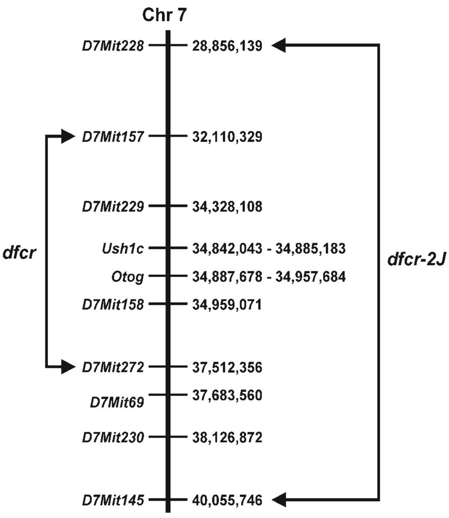
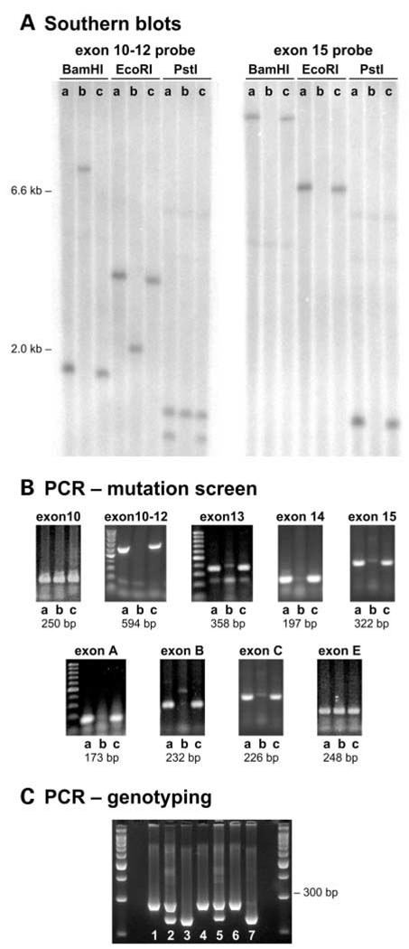
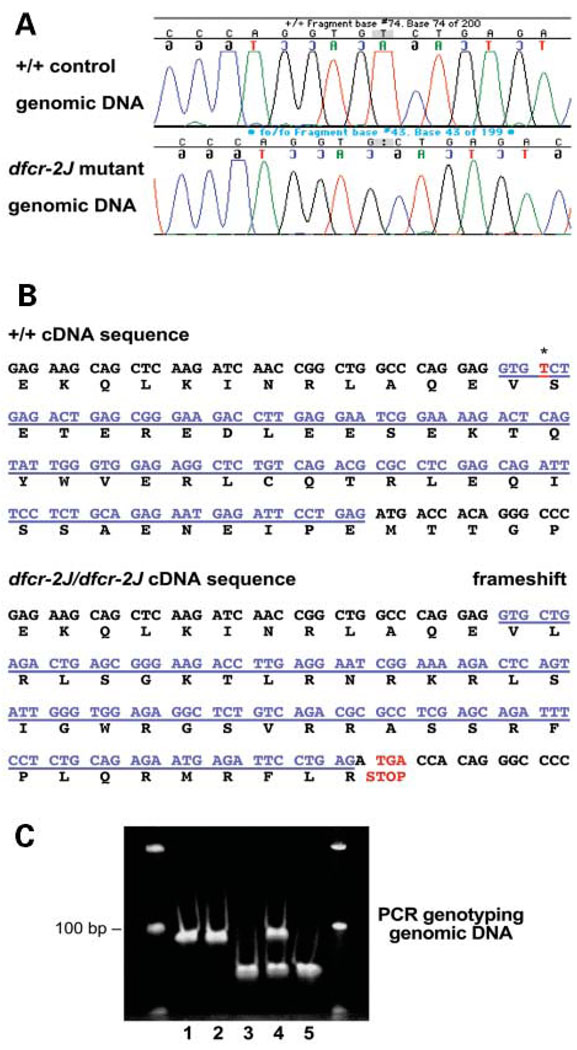
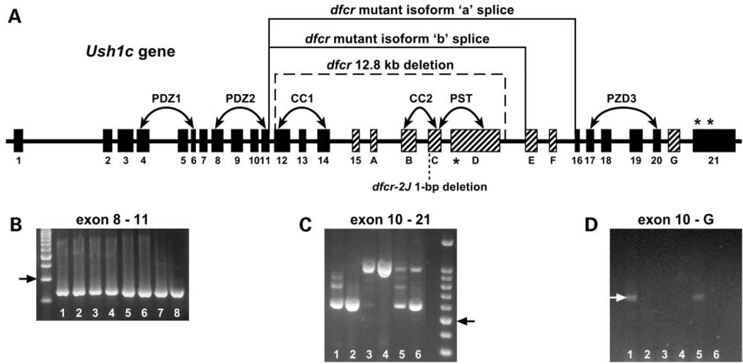
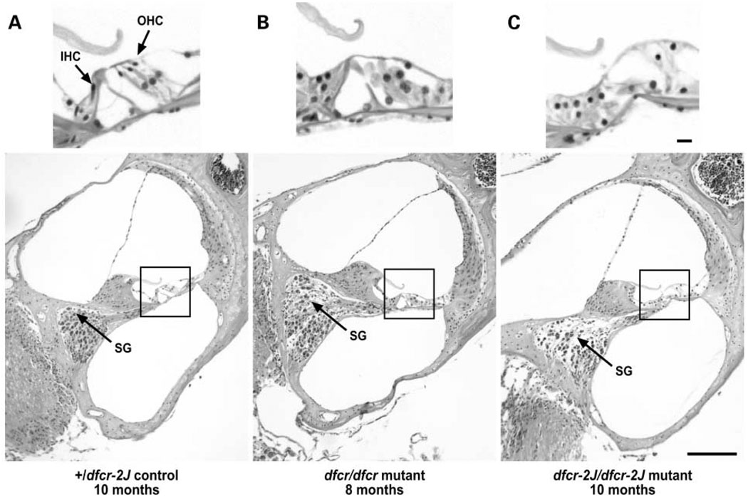
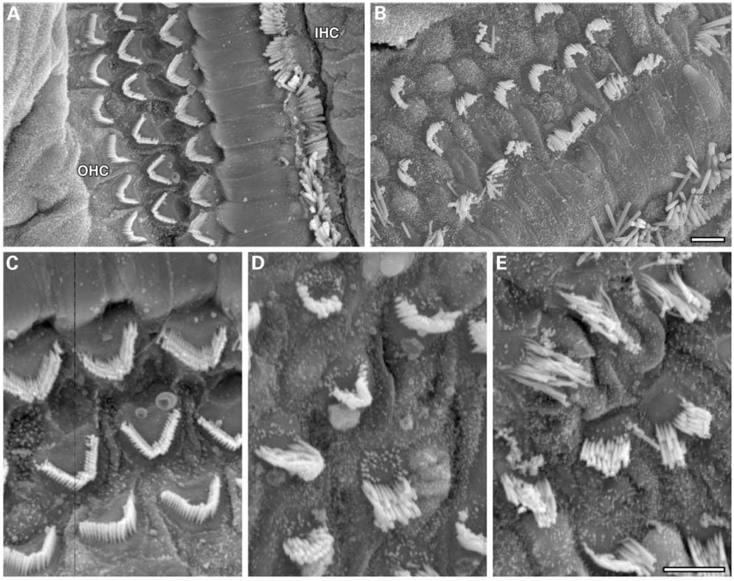
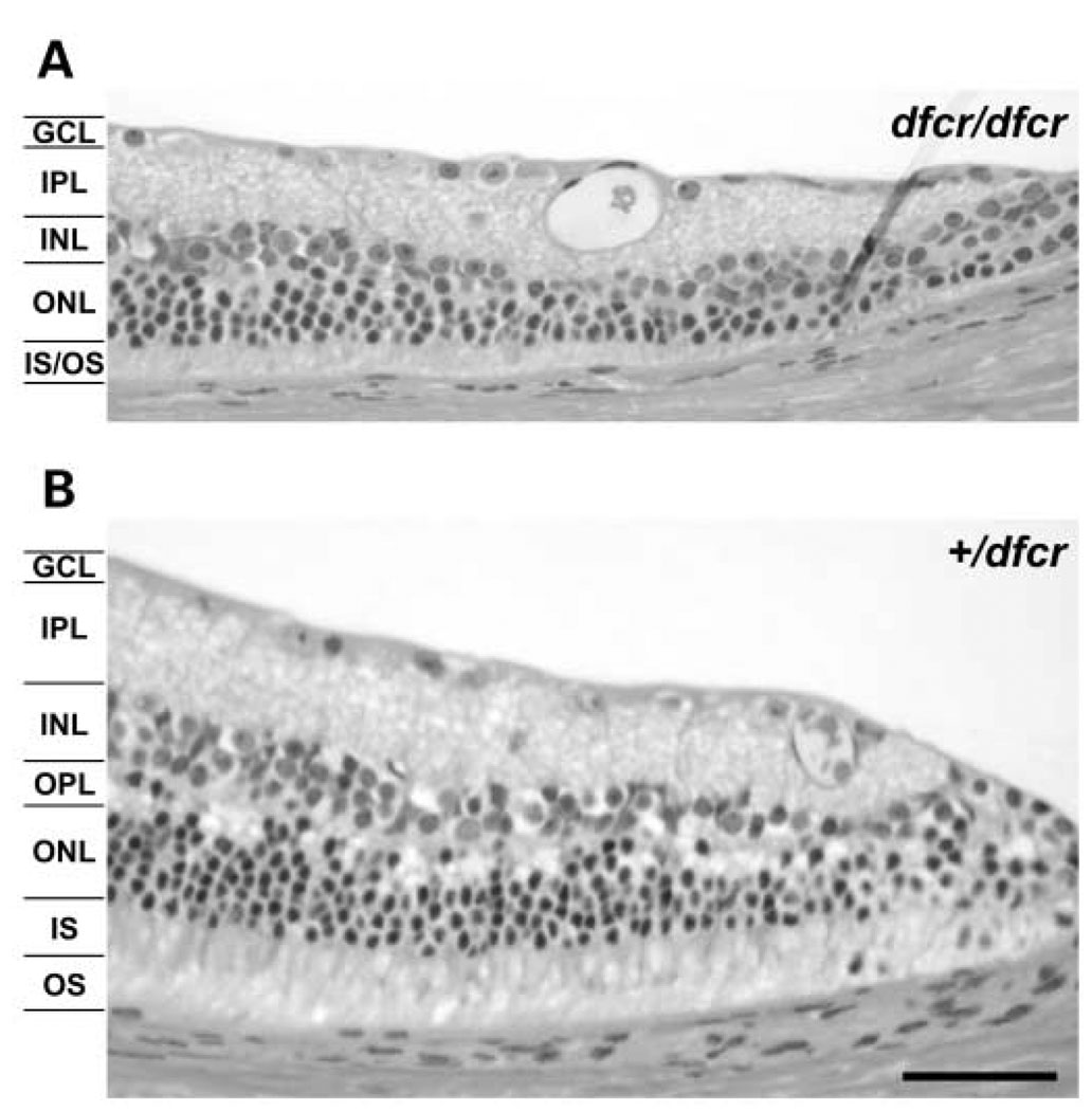
We mapped two new recessive mutations causing circling behavior and deafness to the same region on chromosome 7 and showed they are allelic by complementation analysis. One was named 'deaf circler' (allele symbol dfcr) and the other 'deaf circler 2 Jackson' (allele symbol dfcr-2J). Both were shown to be mutations of the Ush1c gene, the mouse ortholog of the gene responsible for human Usher syndrome type IC and for the non-syndromic deafness disorder DFNB18. The Ush1c gene contains 28 exons, 20 that are constitutive and eight that are alternatively spliced. The dfcr mutation is a 12.8 kb intragenic deletion that eliminates three constitutive and five alternatively spliced exons. The dfcr-2J mutation is a 1 bp deletion in an alternatively spliced exon that creates a transcriptional frame shift, changing 38 amino acid codons before introducing a premature stop codon. Both mutations cause congenital deafness and severe balance deficits due to inner ear dysfunction. The stereocilia of cochlear hair cells are disorganized and splayed in mutant mice, with subsequent degeneration of the hair cells and spiral ganglion cells. Harmonin, the protein encoded by Ush1c, has been shown to bind, by means of its PDZ-domains, with the products of other Usher syndrome genes, including Myo7a, Cdh23 and Sans. The complexes formed by these protein interactions are thought to be essential for maintaining the integrity of hair cell stereocilia. The Ush1c mutant mice described here provide a means to directly investigate these interactions in vivo and to evaluate gene structure-function relationships that affect inner ear and eye phenotypes.


Figures

Figure 1
Figure 1
Genetically derived candidate gene intervals for dfcr and dfcr-2J. The Ensembl base pair locations (from the NCBI 30 Mouse Genome Assembly, May 12, 2003) along Chr 7 are shown to the right of each marker and candidate gene. Arrows connect recombinant flanking markers for each linkage cross, delimiting the candidate gene intervals for each mutation.
Figure 2
Figure 2
The dfcr mutation of Ush1c. Genomic DNA samples from BALB/cByJ +/+control (lane a), dfcr/dfcr (lane b), and dfcr-2J/dfcr-2J (lane c) mice were compared. (A) Southern blot analysis. Restriction fragments from dfcr/dfcr mice are differently sized than those of controls and dfcr-2J/dfcr-2J mice when cDNA corresponding to exons 10–12 of Ush1c is used as a probe and absent when an exon 15 probe is used. (B) RT–PCR analysis. PCR primers flanking Ush1c exons 10–12, 13, 14, 15, A, B, and C amplified products with +/+ and dfcr-2J/dfcr-2J DNA but not with dfcr/dfcr DNA. Primers flanking exons 10 and E amplified products in all three DNA samples, indicating that the dfcr deletion lies between these two exons. (C) Genotyping mice with the dfcr mutation. Two primers flanking the deletion (in11-F and inD-R, Table 2), amplify a 282 bp product only in mutant DNA. The in11-F primer and a primer within the deletion (ex12-R, Table 2), amplify a 234 bp product only in wildtype DNA. The three primers were used together in a single PCR reaction to amplify genomic DNA from +/+ (lanes 3 and 7), +/dfcr (lanes 2 and 5), and dfcr/dfcr (lanes 1, 4, and 6) mice. A 3% agarose gel was used to resolve the products shown here; the two outer lanes contain 100 bp size ladders.
Figure 3
Figure 3
The dfcr-2J mutation of Ush1c. (A) DNA sequence chromatogram of C57BL/6J +/+ control compared with homozygous dfcr-2J mutant, illustrating the deletion of a single bp (T/A) in mutant DNA. (B) The 1 bp deletion is at position 4 (indicated by an asterisk) of exon C (underlined in blue), which causes a frameshift and results in a transcription stop codon (shown in red) at the beginning of exon D. (C) Genotyping mice with the dfcr-2J mutation. Primers mutC-F and mutC-R, flanking the dfcr-2J mutation in exon C (Table 2), were used to amplify genomic DNA from +/+ (lanes 1 and 2), +/dfcr-2J (lane 4), and dfcr-2J/dfcr-2J (lanes 3 and 5) mice, progeny from matings between +/dfcr-2J parents. The reverse primer, mutC-R, ends immediately adjacent to the point of the 1 bp deletion in exon C. Two nucleotides in this primer (underlined in Table 2) were changed (to GT from CA) to create an ApaL1 restriction site (GTGCAC) in mutant but not wildtype DNA. After incubation with the restriction endonuclease ApaL1, the wild-type 93 bp product is unaffected, whereas the mutant product is cleaved into 70 and 22 bp fragments. A 6% acrylamide gel was used to resolve the DNA fragments shown here; the two outer lanes contain 100 bp size ladders.
Figure 4
Figure 4
Transcription of dfcr and dfcr-2J mutations. (A) Ush1c exon–intron structure. Exons are represented as rectangles and introns as lines. Exons (greatly exaggerated in size relative to introns) are designated according to Verpy et al. (4), with the 20 constitutive exons shown in black and the eight alternatively spliced exons in gray. A dashed line indicates the extent of the dfcr deletion and a dotted line marks the position of the dfcr-2J mutation. The dfcr deletion causes an inframe splicing of exon 11 with exon 16 in transcripts encoding the ‘a’ isoform of harmonin and an in-frame splicing of exon 11 with exon E in transcripts encoding the ‘b’ isoform. An asterisk in exon D marks the stop codon created by the dfcr-2J mutation, and asterisks in exon 21 denote the normal stop codons, unchanged by the dfcr mutation. Connected arrows indicate genomic regions encoding the PDZ, CC (coiled-coil), and PST (proline, serine, threonine-rich) domains of the harmonin protein. (B) Ush1c transcripts are detected in both dfcr and dfcr-2J mutant mice. The primers ex8-F (located within exon 8) and ex11-R (located within exon 11) amplified the expected 250 bp product in cDNA derived from inner ear (lanes 1–4) and kidney (lanes 5–8) of +/dfcr mice (lanes1 and 5), dfcr/dfcr mice (lanes 2 and 6), +/dfcr-2J mice (lanes 3 and 7), and dfcr-2J/dfcr-2J mice (lanes 4 and 8). Other tissues examined but not shown here (eye, brain, intestine) gave similar results. The arrow to the left of the figure indicates the position of the 300 bp DNA size marker. (C) Exons12, 13, 14 and 15 (396 nt) are deleted from isoform ‘a’ transcripts in dfcr/dfcr mice. The PCR products amplified with primers ex10-F (located within exon 10) and ex21-R (located within exon 21) from cDNAs derived from inner ear (lanes 1, 3 and 5) and intestine (lanes 2, 4 and 6) were smaller in dfcr/dfcr mice (574 bp; lanes 1 and 2) than those from +/+ mice (970 bp; lanes 3 and 4); both sizes were amplified in heterozygotes (lanes 5 and 6). Sequence analysis of the mutant 574 bp PCR product revealed an in-frame splicing of exon 11 with exon 16. The arrow to the right of the figure indicates the position of the 500 bp DNA size marker. (D) Exons 12, 13, 14, A, B, C and D (1137 nt) are deleted from isoform ‘b’ transcripts in dfcr/dfcr mice. A faint ~700 bp PCR product was amplified from inner ear cDNA with primers ex10-F and exG-R (located within exon G) of dfcr/dfcr (lane 1) and +/dfcr (lane 5) mice, but not from +/+ mice (lane 3) because of the large distance between exons 10 and G (>1800 nt) in wildtype cDNA. No product was amplified from intestine-derived cDNA of any genotype (lanes 2, 4 and 6). Sequence analysis of the mutant product from inner ear (indicated by white arrow) revealed an in-frame splicing of exon 11 with exon E.
Figure 5
Figure 5
Hair cell and spiral ganglion cell loss in cochleae of mutant mice. Cross sections through the basal turn of cochleae of a 10-month-old +/dfcr-2J control (A), an 8-month-old dfcr/dfcr mutant (B), and a 10-month-old dfcr-2J/dfcr-2J mutant (C). The upper panels show magnified images of the organ of Corti corresponding to the boxed areas in the lower panels. IHC, inner hair cell; OHC, outer hair cell; SG, spiral ganglia. Scale bars, 10 µm for upper panels and 100 µm for lower panels.
Figure 6
Figure 6
Stereocilia disorganization in cochlear hair cells of mutant mice. Scanning electron micrographs of hair cell stereocilia in cochleae from 3-week old mice. The three rows of outer hair cells (OHC) and row of inner hair cells (IHC) in a cochlea from a +/dfcr heterozygous mouse (A) show a normal, highly organized pattern of stereocilia, as compared with stereocilia of a dfcr/dfcr mutant mouse (B). Scale bar, 5 µm. Hair cell loss in the mutant cochlea underlies the disrupted appearance of the hair cell pattern (B). Higher magnification of OHCs better illustrates the intricate pattern and rigid morphology of stereocilia of a normal mouse (C) compared with the disorganized and splayed stereocilia of dfcr/dfcr (D) and dfcr-2J/dfcr-2J (E) mutant mice. Scale bar, 5 µm.
Figure 7
Figure 7
Retinal histology of eyes from dfcr mutant mice compared with controls. Histologic sections of retinas from eyes of three BALB/cBy-dfcr/dfcr mutant mice showed evidence of a slight peripheral retinal degeneration at 9 months of age (A), compared with the normal retinal histology seen in eyes from three litter-mate BALB/cBy-+/dfcr controls (B). The overall thickness of cells at the periphery of the retina is reduced in mutant eyes compared with controls. Each layer is indicated as follows: ganglion cell layer (GCL), inner plexiform layer (IPL), inner nuclear layer (INL), outer plexiform layer (OPL), outer nuclear layer (ONL), inner segments (IS) and outer segments (OS). At about 200 µm from the outer edge of the retina, the thickness of the INL-OS layers in mutant eyes was 40–80% that of normal eyes, and mutant eyes appeared to be missing the OPL. Scale bar, 50 µm.

References

    1. Keats BJ, Nouri N, Pelias MZ, Deininger PL, Litt M. Tightly linked flanking microsatellite markers for the Usher syndrome type I locus on the short arm of chromosome 11. Am. J. Hum. Genet. 1994;54:681–686. - PMC - PubMed
    1. Smith RJ, Lee EC, Kimberling WJ, Daiger SP, Pelias MZ, Keats BJ, Jay M, Bird A, Reardon W, Guest M, et al. Localization of two genes for Usher syndrome type I to chromosome 11. Genomics. 1992;14:995–1002. - PubMed
    1. Bitner-Glindzicz M, Lindley KJ, Rutland P, Blaydon D, Smith VV, Milla PJ, Hussain K, Furth-Lavi J, Cosgrove KE, Shepherd RM, et al. A recessive contiguous gene deletion causing infantile hyperinsulinism, enteropathy and deafness identifies the Usher type 1C gene. Nat. Genet. 2000;26:56–60. - PubMed
    1. Verpy E, Leibovici M, Zwaenepoel I, Liu XZ, Gal A, Salem N, Mansour A, Blanchard S, Kobayashi I, Keats BJ, et al. A defect in harmonin, a PDZ domain-containing protein expressed in the inner ear sensory hair cells, underlies Usher syndrome type 1C. Nat. Genet. 2000;26:51–55. - PubMed
    1. Ahmed ZM, Smith TN, Riazuddin S, Makishima T, Ghosh M, Bokhari S, Menon PS, Deshmukh D, Griffith AJ, Friedman TB, et al. Nonsyndromic recessive deafness DFNB18 and Usher syndrome type IC are allelic mutations of USHIC. Hum. Genet. 2002;110:527–531. - PubMed

Publication types